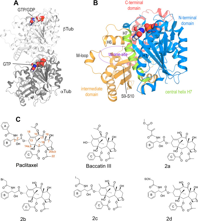
Figure 1. Structures of tubulin and ligands employed in the work.
(A) Tubulin heterodimer (α-tubulin in gray and β-tubulin in white) in ribbon representation, where nucleotide binding sites have been highlighted in sphere representation (B) Structural features of the tubulin β-subunit. (C) Structures of taxanes used in this study.

