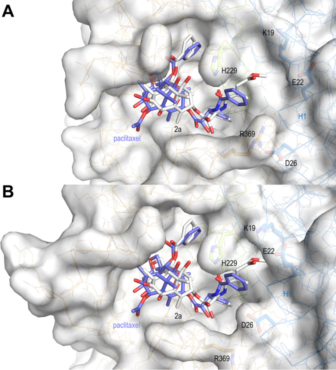
Figure 12. Surface representations of liganded taxane sites in both the curved and straight tubulin conformational states.
(A) Curved tubulin; (B) straight tubulin. The structures of 2a (white) and paclitaxel (slate) bound to microtubules (PDB ID 6WVR) were superimposed onto their central helices βH7. The side chains of the βM loop residue βR278 and of residues surrounding the C13 side chains of the ligands are in stick representation and are labeled. Helix βH1 is highlighted in ribbon representation.

