Abstract
Salt is an essential ingredient in the kimchi fermentation process. Solar salt has antioxidant, anti-cancer, and anti-obesity properties. The aim of this study was to determine the antioxidant and anti-inflammatory effects of solar salt brined kimchi. Purified salt (PS), dehydrated solar salt (DSS), 1-year aged solar salt (SS1), and 3-years aged solar salt (SS3) were investigated. Anti-inflammatory effects were determined by analyzing cytotoxicity, nitric oxide (NO) production, and inflammation-related gene expression in lipopolysaccharide-treated RAW264.7 cells. Antioxidant activities of DSS, SS1, and SS3 were higher than that of PS. Solar salt significantly inhibited NO production with low cytotoxicity and decreased inflammation-related gene expression. Kimchi containing solar salt (DSSK, SS1K, and SS3K) showed higher antioxidant activity than PSK. Additionally, DSSK, SS1K, and SS3K significantly inhibited NO production and decreased the expression of inflammation-related genes. Owing to the antioxidant and anti-inflammatory effects, using solar salt in kimchi preparation could have potential health benefits.
Keywords: Antioxidant, Anti-inflammatory, Solar salt, Kimchi, RAW264.7 cells
Introduction
Solar salt (sea salt) is made by drawing seawater into shallow ponds in the salt farm where the wind and sunlight evaporate its moisture content. Trace salts, such as magnesium chloride, present in the solar salt give it a bitter taste, requiring a draining process over a long period to remove bitterness. Solar salt is widely used in the food and cosmetic industries (Chang et al., 2010; Lee and Lee, 2014). For instance, solar salt-fermented shrimp showed a higher sensory evaluation of overall taste and acceptance than purified salt-fermented shrimp (Lee et al., 2008). Sinan-gun, Yeonggwang-gun, Buan-gun, Taean-gun, and Ongjin-gun are representative solar salt production areas in Korea. Various solar salts have been reported globally, including Celtic sea salt (Sel de Guerande or Sel de Bretagne), fleur de sel (precious flowers of pure sea salt), Khoisan fleur de sel, and bamboo salt, and are reported to have greater health benefits (Amanvida, 2021). The solar salt from Korea has a higher potassium (K) and magnesium (Mg) content than Guerande salt from France, with relatively low levels of sodium (Na) (Kim et al., 2011). Owing to its high mineral content, Korean solar salt is expected to have potential health benefits.
The antioxidant, anti-cancer, and anti-obesity effects of solar salts have been reported previously (Ju et al., 2017, 2016; Lee and Chang, 2011; Park et al., 2021). Oral administration of 1% solar salt exhibited anti-cancer properties by inhibiting the expression of apoptosis- and inflammation-related markers in colon cancer-induced mice (Ju et al., 2017). Solar salt can also suppress serum lipid profiles and adipogenic and lipogenic marker expression and has been reported to have an anti-obesity effect in high-fat diet-fed mice (Ju et al., 2016). In Korea, the National Fishery Products Quality Management Service determines the quality of Korea’s solar salt (Ministry of Oceans and Fisheries, MOF) and fermented food made with solar salt has been reported to have anti-hypertensive and anti-diabetic effects as well as anti-cancer effects (Medical Today, 2019). An in-depth understanding of the effects of solar salt on kimchi would help in its efficient use to promote health benefits.
Salt promotes lactic acid bacteria (LAB) growth and reduces pathogenic and putrefactive bacteria and is often used as a starter during kimchi preparation (Kim and Kim, 2010; Kim et al., 2005). In addition, adding salt to kimchi affects its taste, quality, storage, and its functionality. A high proportion of coccus LAB is observed in kimchi with solar salt, while yeast was not detected. In addition, kimchi with solar salt decreases the Lactobacillus/Leuconostoc ratio and affects kimchi quality by changing in sugars, amino acids, organic acids, lipids, sulfur compounds, and terpenoids (Chang et al., 2011). Preparing kimchi with solar salt is reported to have many health benefits. Kimchi preparation using solar salt having low magnesium and sulfur content showed anti-cancer effects by inhibiting apoptosis and cell cycle arrest in HT-29 colon carcinoma cells (Yu et al., 2020). Even though the anti-cancer effects of solar salts have been widely reported, their anti-inflammatory effects remain largely unidentified.
As mentioned above, solar salt is well known that have higher mineral content than purified salt (refined salt). Based on the fact that minerals have a high effect on health functionality, this study conducted to seek whether there would be a difference in anti-inflammatory effect among purified salt (PS), dehydrated solar salt (DSS), 1-year aged solar salt (SS1), and 3-years aged solar salt (SS3). Antioxidant and anti-inflammatory activities of solar salt and kimchi containing solar salt were investigated. Initially, the antioxidant activities of PS, DSS, SS1, and SS3 were measured. Anti-inflammatory activities were investigated in lipopolysaccharide (LPS)-treated RAW264.7 cells by measuring cytotoxicity, nitric oxide (NO) production, and inflammation-related gene expression. Kimchi containing PS, DSS, SS1, and SS3 was freeze-dried to further analyze their antioxidant and anti-inflammatory properties.
Materials and methods
Chemicals
PS, DSS, SS1, and SS3 were obtained from Sinan, Inc. (Sinan, Korea). Gallic acid, quercetin, lipopolysaccharide, and total antioxidant capacity (TAC) kits were purchased from Sigma-Aldrich (Sigma-Aldrich, St. Louis, MO, USA). Ferric reducing antioxidant power (FRAP) kit were purchased from Ann Arbor (Ann Arbor, MI, USA). Cell Counting kit-8 (CCK-8) was purchased from Dojindo (Dojindo, Tokyo, Japan). Griess Reagent System was purchased from Promega (Promega, USA). TRIzol reagent was purchased from Invitrogen (Invitrogen, Carlsbad, CA). cDNA synthesis kit and SYBR Green premix were purchased from Enzynomics (Enzynomics, Daejeon, Korea).
Preparation of solar salt brined kimchi
Initially, kimchi cabbage was brined with 12% PS, DSS, SS1 and SS3 for 16 h. Then, brined kimchi cabbage was mixed with kimchi seasoning (red pepper, garlic, ginger, onion, radish, and other ingredients). Each kimchi sample, PSK, DSSK, SS1K, and SS3K was prepared with PS, DSS, SS1, and SS3, respectively. Ripened kimchi (pH 4.2) was freeze-dried for in further analysis.
Total flavonoid content analysis
The total flavonoid content (TFC) of the kimchi was measured (Chang et al., 2002). TFC was measured at 415 nm using a microplate reader (BMG LABTECH, Ortenberg, Germany). TFC was expressed as mg QE/g with quercetin standard curve.
Total phenol content analysis
The total phenol content (TPC) of kimchi was measured (Durazzo et al., 2013). TPC was measured at 700 nm and expressed as mg GAE/g with gallic acid standard curve.
Total antioxidant capacity analysis
The antioxidant effect of salt and kimchi were analyzed using a TAC kit. TAC was measured at 570 nm and expressed as μM with Trolox standard curve.
Ferric reducing antioxidant power analysis
The FRAP values of salt and kimchi were measured using a FRAPTM colorimetric detection kit. FRAP was measured at 560 nm and expressed as μM with Fe (II) standard curve.
Cell culture
RAW264.7 cells were obtained from the American Type Culture Collection (ATCC, Manassas, VA, USA) and cultured in Dulbecco’s Modified Eagle’s Medium (DMEM) with 10% fetal bovine serum and 1% penicillin/streptomycin until confluence.
Cytotoxicity analysis
Cytotoxicity was measured using the CCK-8 kit. Cells were seeded at a density of 1 × 10⁴ cells/well and incubated with salt (0, 1, 2, 5, 10, and 15%) or kimchi (0, 0.05, 0.25, 0.5, 2.5, and 5 mg/mL) for 24 h. After washing with phosphate-buffered saline (PBS), cells were incubated with 20 µL of CCK-8 solution for 2 h. Cytotoxicity was measured at 450 nm.
Nitric oxide production analysis
Nitric oxide (NO) production was measured using the Griess Reagent System (Promega, USA). Cells were seeded at a density of 1 × 10⁴ cells/well and incubated with salt (0, 1, 2, 5, 10, and 15%) or kimchi (0, 0.05, 0.25, 0.5, 2.5, and 5 mg/mL) for 24 h with or without LPS (1 μg/mL) treatment. Then, medium was reacted with Griess reagent (1% sulfanilamide and 0.1% naphthylenediamine in 2.5% phosphoric acid). NO was measured at 540 nm.
Quantitative real-time PCR
Cells were seeded at a density of 1 × 10⁴ cells/well and incubated with various salts (10%) or kimchi (2.5 mg/mL) for 24 h with or without LPS (1 µg/mL) treatment. RNA (1 μg/mL) was extracted with TRIzol and cDNA was synthesized with TOPScriptTM cDNA Synthesis Kit. Quantitative real-time PCR (qPCR) was performed with 10 μL of SYBR Green Premix, 0.5 μL of each primer, and 9 μL of cDNA and then amplification was performed with enzyme activation at 94 °C for 10 min, followed by 45 cycles of denaturation at 94 °C for 15 s and annealing and extension at 60 °C for 1 min each. Gene expression were normalized to those for GAPDH (Table 1).
Table 1.
Quantitative real-time PCR primer sequences
| Gene | Sense primer (5′–3′) | Antisense primer (5′–3′) |
|---|---|---|
| GAPDH | TGTCATACTTGGCAGGTTTCT | CGTGTTCCTACCCCCAATGT |
| IL-1β | GCAACTGTTCCTGAACTCAACT | TCTTTTGGGGTCCGTCAACT |
| IL-6 | TAGTCCTTCCTACCCCAATTTCC | TTGGTCCTTAGCCACTCCTTC |
| TNF-α | CCCTCACACTCAGATCATCTTCT | GCTACGACGTGGGCTACAG |
| MCP-1 | GCATCCACGTGTTGGCTCA | CTCCAGCCTACTCATTGGGATCA |
| iNOS | TTCCAGAATCCCTGGACAAG | TGGTCAAACTCTTGGGGTTC |
| NF-κB | ACCGTCCTCTGCCCCAGTAT | GGCTTCCGACAGCGTGCCTT |
Statistical analysis
Data are expressed as means ± standard deviation (SD). Significant difference was analyzed using one-way analysis of variance (ANOVA) followed by Duncan’s multiple range test using GraphPad Prism 7 (GraphPad, Inc., San Diego, CA, USA). P < 0.05 expressed statistical significance.
Results and discussion
Antioxidant effect of solar salt
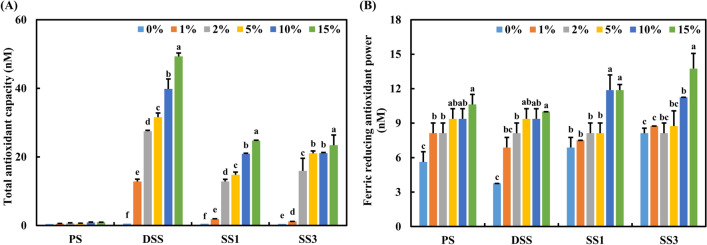
The antioxidant effects of PS, DSS, SS1, and SS3 were measured. As shown in Fig. 1A, the TAC levels of DSS, SS1, and SS3 were dose-dependently and significantly increased (P < 0.05). The DSS showed the highest TAC level, while PS had the lowest. Figure 1B shows FRAP levels of PS, DSS, SS1, and SS3. FRAP levels were dose-dependently increased in all groups. Unlike TAC levels, the FRAP levels of SS1 and SS3 were higher than those of PS and DSS. Similar to our results, solar salt was reported to have higher antioxidant activity in DPPH radical scavenging, ABTS+ radical scavenging, and higher FRAP levels than purified salt (Gao, 2010; Gao et al., 2014, 2015). These results suggest solar salt could also have an antioxidant effect when added to kimchi.
Fig. 1.
Antioxidant activity of purified salt (PS), dehydrated solar salt (DSS), 1-year aged solar salt (SS1), and 3-years aged solar salt (SS3) were analyzed using a total antioxidant capacity (TAC) and ferric reducing antioxidant power (FRAP) analysis. The OD of TAC was measured at 570 nm. FRAP was measured using a FRAP™ colorimetric detection kit. The OD of FRAP was measured at 560 nm (A) Total antioxidant capacity and (B) ferric reducing antioxidant power. Data are expressed as mean ± SD. Different letters indicate significant differences among groups (P < 0.05)
Effect of solar salt on cell viability
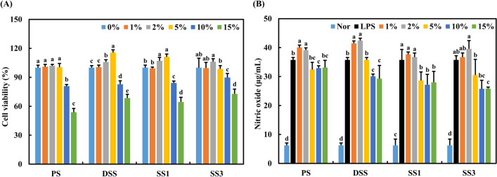
The effect of PS, DSS, SS1, and SS3 at various concentrations (0, 1, 2, 5, 10, and 15%) on cell viability was determined. Salt treatment dose-dependently and significantly decreased the cell viability (Fig. 2A, P < 0.05). The cell viability was over 80% upon treatment with 10% of PS, DSS, SS1, and SS3. Treatment with SS3 did not alter the cells much and showed approximately 90% cell viability at 10% salt concentration. The cells treated with PS showed relatively low cell viability. Previous studies have reported solar salt to have high cell viability in human follicle papilla cells (Choi et al., 2021). Jeong et al., reported that solar salt showed the high cell viability under 9 mg/mL in normal human dermal fibroblasts and macrophages (Jeong, 2009). Conversely, in a previous study, solar salt inhibited cancer cell growth (Yoon and Chang, 2011). Bamboo salt is a traditional Korean solar salt baked several times at high temperatures and is known to have various health benefits (Zhao et al., 2013). Bamboo salt also showed high cell viability in gingival fibroblast, RAW264.7, and human follicle dermal papilla cells (Kim et al., 2016; Lee and Choi, 2015). Based on the cell viability results, the subsequent experiments were performed at a salt concentration of 10%.
Fig. 2.
Cytotoxicity and nitric oxide production of PS, DSS, SS1, and SS3. RAW264.7 cells were incubated with salt (0, 1, 2, 5, 10, and 15%) or kimchi (0, 0.05, 0.25, 0.5, 2.5, and 5 mg/mL) for 24 h with or without LPS (1 μg/mL) treatment. Cytotoxicity was measured using the CCK-8 kit. Nitric oxide production was measured using the Griess reagent method. (A) Cytotoxicity and (B) nitric oxide production. Data were expressed as mean ± SD. Different letters indicate significant differences among groups (P < 0.05)
Inhibitory effect of solar salt on NO production
As shown in Fig. 2B, LPS treatment dramatically increased NO production in RAW26.4 cells compared to the Nor group (without LPS treatment). Previous studies have reported LPS stimulated NO production in RAW264.7 cells (Eo et al., 2021; Li et al., 2021). However, treatment with 5% or more concentrations of PS, DSS, SS1, and SS3 dose-dependently and significantly decreased NO production (P < 0.05). Among the groups, SS1 and SS3 showed potent inhibitory effects on NO production in LPS-induced RAW264.7 cells. In particular, SS3 inhibited NO production by about 28% compared to the LPS group. According to Choi’s study, hot water extract, 30% and 60% ethanol extract of solar salt reduced NO production in 0.1–1 mg/mL concentration (Choi et al., 2021). A similar tendency of NO inhibition was observed with bamboo salt, with no significant difference (Kim et al., 2016). The exact mechanism of salt-mediated NO inhibition remains elusive; however, the inhibitory effects of SS1 and SS3 on NO production could be attributed to the relatively high mineral content in solar salt. Solar salt was found to have abundant mineral content, especially K and Mg, compared to other salts (Jin et al., 2011), leading to health benefits. In a previous study, solar salt with low Na/K ratio showed powerful anti-obesity activity in obese mice (Ju et al., 2016). These results also showed that the inhibitory effect of solar salt (DSS, SS1, and SS3) on NO production was greater than that of PS.
Anti-inflammatory effect of solar salt
The anti-inflammatory effects of PS, DSS, SS1, and SS3, were determined by measuring the expression of inflammation-related genes, such as interleukin (IL)-1 beta (β), IL-6, tumor necrosis factor-alpha (TNF-α), monocyte chemoattractant protein 1 (MCP-1), inducible nitric oxide synthase (iNOS), and nuclear factor kappa-B (NF-κB) using qPCR. IL-1β and IL-6 activates the immune response as major proinflammatory cytokines, and TNF-α is mainly secreted in response to cell damage (Popko et al., 2010). Similarly, iNOS and cyclooxygenase 2 (COX-2) synthesize inflammatory mediators to induce NF-κB activation via the NF-κB pathway (Bak et al., 2012; Hwang et al., 2019; Zhang et al., 2019). In this study, treatment with SS1 and SS3 significantly decreased the expression of inflammation-related genes (Fig. 3; P < 0.05). However, this inhibitory effect was not observed in the PS group. In consistent with our results, solar salt inhibited iNOS and COX-2 genes expression in HCT-116 cells and HT-29 cells, though these inhibitory effects were relatively lower than that of bamboo salt treatment (Zhao, 2011). In another study, bamboo salt was shown to significantly inhibit TNF-α and NF-kB expression in RAW264.7 cells (Kim et al., 2016). Similarly, purple bamboo salt also significantly inhibited TNF-α, IL-1β, and IL-6 secretion in human mast cell -1 (Kim, 2002). Interestingly, beauty salt, composed of solar salt and Impatiens balsamina L. extract, also demonstrated an immunomodulatory effect on human mast cells by suppressing proinflammatory cytokines (Ryu et al., 2017). Our results indicate that SS1 and SS3 suppress inflammation-related markers expression, leading to exerting its anti-inflammatory effect.
Fig. 3.
Anti-inflammatory effect of PS, DSS, SS1, and SS3. RAW264.7 cells were incubated with various salts (10%) or kimchi (2.5 mg/mL) for 24 h with or without LPS (1 µg/mL). Gene expression was determined using qPCR, (A) IL-1β, (B) IL-6, (C) TNF-α, (D) MCP-1, (E) iNOS, and (F) NF-κB. Data were expressed as mean ± SD. Different letters indicate significant differences among groups (P < 0.05)
Antioxidant effect of solar salt brined kimchi
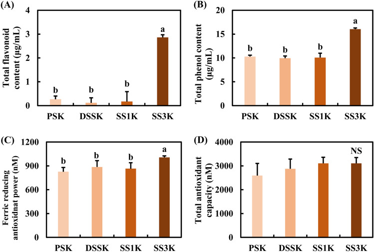
After preparing kimchi with the respective salts, the antioxidant effects of PSK, DSSK, SS1K, and SS3K were measured using total flavonoid, total phenol, TAC, and FRAP assays. Salt alone did not have any detectable flavonoid or phenol content; however, TFC and TPC levels were detected in all solar salt brined kimchi groups. TFC and TPC of SS3K were significantly higher than those of PSK, DSSK, and SS1K (Fig. 4A, B, P < 0.05). The TAC level of SS3K was higher than that of PSK, DSSK, and SS1K, with no significant differences (Fig. 4C). However, the FRAP in SS3K was significantly higher than in the other groups (Fig. 4D, P < 0.05). SS3K showed the highest antioxidant effect among all groups in all antioxidant assays. Even though the method of kimchi preparations differ, kimchi alone was also reported to exhibit antioxidant activity (Yun et al., 2019). Our results suggest that solar salt enhanced the antioxidant effect of kimchi.
Fig. 4.
Antioxidant activities of kimchi with various salts; PSK, DSSK, SS1K, and SS3K. (A) Total flavonoid content (TFC), (B) total phenol content (TPC), (C) TAC, and (D) FRAP. Data are expressed as mean ± SD. Different letters indicate significant differences among groups (P < 0.05)
Effect of solar salt brined kimchi on cell viability
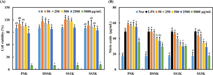
We determined the effect of kimchi with various salts (PSK, DSSK, SS1K, and SS3K) on the cell viability of RAW264.7 cells. As shown in Fig. 5A, treatment with 2.5 mg/mL of solar salt brined kimchi showed cell viability of over 80%. However, cell viability dramatically decreased to less than 10% at 5 mg/mL. No significant difference in the cell viability between the groups was observed. A previous study has reported higher kimchi concentrations showing relatively low cell viability (Yun et al., 2019). In another study, raw mustard leaves, mustard leaf kimchi, and mustard leaf kimchi fermented with Lactobacillus plantarum showed no cytotoxicity in RAW264.7 cells (Lee et al., 2020). These results indicate that kimchi exhibits low cytotoxicity.
Fig. 5.
Cytotoxicity and nitric oxide production of PSK, DSSK, SS1K, and SS3K. (A) Cytotoxicity and (B) nitric oxide production. Data were expressed as mean ± SD. Different letters indicate significant differences among groups (P < 0.05)
NO inhibitory effect of solar salt brined kimchi
The effect of the solar salt on NO production was examined. Figure 5B shows that LPS treatment increased NO production, whereas treatment with solar salt dose-dependently and significantly decreased NO production (P < 0.05). Consistently, SS1K and SS3K showed a high inhibitory effect on NO production. Generally, the health functionality of kimchi is closely related to active components and LAB in kimchi. Therefore, the anti-inflammatory effect of kimchi is also reported on the results of studies on kimchi LAB and kimchi active components rather than kimchi itself. A similar observation was reported in Le’s study, where raw mustard leaves, mustard leaf kimchi, and mustard leaf kimchi fermented with Lactobacillus plantarum significantly suppressed NO production in LPS-treated RAW264.7 cells, although kimchi is not the same sample with solar salt added (Le et al., 2020). In a kimchi LAB study, WiKim39 and WiKim0124 isolated from kimchi significantly inhibited NO production (Yun et al., 2022), In kimchi active component study, HDMPPA isolated from kimchi significantly reduced NO production in LPS-stimulated BV2 microglia in a previous study (Jeong et al., 2015). Our results suggest that the inhibitory effect of kimchi on NO production could be increased using solar salt.
Anti-inflammatory effect of solar salt brined kimchi
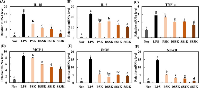
The expression of inflammation-related genes was determined to evaluate the anti-inflammatory effect of solar salt brined kimchi. Even though the inhibitory effect of solar salt brined kimchi was slightly lower than that of solar salt alone, an anti-inflammatory effect was observed in all kimchi groups. SS1K and SS3K significantly decreased the expression of inflammation-related genes (Fig. 6; P < 0.05). Consistent with our results, mustard leaf kimchi significantly and dose-dependently decreased IL-1β and TNF-α mRNA expression levels (Le et al., 2020). Interestingly, optimally and over ripened kimchi significantly reduced iNOS, COX-2, IkB, and NF-kB expression in oxidative stress-induced LLC-PK1 cells (Kim et al., 2014). In kimchi active component study, HDMPPA significantly inhibited proinflammatory mediators and cytokines in LPS-stimulated BV2 microglia (Jeong et al., 2015). Our results suggest that solar salt brined kimchi suppress inflammation-related markers expression, leading to exerting its anti-inflammatory effect.
Fig. 6.
Anti-inflammatory effect of PSK, DSSK, SS1K, and SS3K. Gene expression of (A) IL-1β, (B) IL-6, (C) TNF-α, (D) MCP-1, (E) iNOS, and (F) NF-κB. Data are expressed as mean ± SD. Different letters indicate significant differences among groups (P < 0.05)
We observed that solar salts (DSS, SS1, and SS3) showed a higher antioxidant and anti-inflammatory effect than purified salt. Consequently, kimchi supplemented with solar salt also showed higher antioxidant and anti-inflammatory effects than that supplemented with purified salt. However, further mechanistic and animal studies are essential to understand better the antioxidant and anti-inflammatory effects of solar salt brined kimchi. Based on these results, we will conduct further studies in the future.
Acknowledgements
This research was supported by the World Institute of Kimchi (KE2102-2-2 and KE2201-1), and funded by the Ministry of Sciences and ICT, Republic of Korea.
Declarations
Conflict of interest
The authors declare that they do not have any conflict of interest.
Footnotes
Publisher's Note
Springer Nature remains neutral with regard to jurisdictional claims in published maps and institutional affiliations.
Contributor Information
Ye-Rang Yun, Email: yunyerang@wikim.re.kr.
Yun-Jeong Choi, Email: yjchoi85@wikim.re.kr.
Ye-Sol Kim, Email: rlathf19@wikim.re.kr.
Seo-Young Chon, Email: tjdud1128@wikim.re.kr.
Mi-Ai Lee, Email: leemae@wikim.re.kr.
Young Bae Chung, Email: ybchung@wikim.re.kr.
Sung-Hee Park, Email: shpark@wikim.re.kr.
Sung-Gi Min, Email: skmin@wikim.re.kr.
Ho-Chul Yang, Email: yhc1354@korea.kr.
Hye-Young Seo, Email: hyseo@wikim.re.kr.
References
- Amanvida. Which salt is healthier: Sea salt. Fleur de sel, Himalayan salt or bamboo salt? Available from: https://www.amanvida.eu/en/blog/post/which-salt-is-healthier:-sea-salt-fleur-de-sel-himalayan-salt-or-bamboo-salt
- Bak MJ, Hong SG, Lee JW, Jeong WS. Red ginseng marc oil inhibits iNOS and COX-2 via NF-kB and p38 pathways in LPS-stimulated RAW264.7 macrophages. Molecules. 2012;17:13769–13786. doi: 10.3390/molecules171213769. [DOI] [PMC free article] [PubMed] [Google Scholar]
- Chang CC, Yang MH, Wen HM, Chern JC. Estimation of total flavonoid content in propolis by two complementary colorimetric methods. Journal of Food and Drug Analysis. 2002;10:178–182. doi: 10.38212/2224-6614.2748. [DOI] [Google Scholar]
- Chang M, Kim IC, Chang HC. Effect of solar salt on the quality characteristics of Doenjang. Journal of the Korean Society of Food Science and Nutrition. 2010;39:116–124. doi: 10.3746/jkfn.2010.39.1.116. [DOI] [Google Scholar]
- Chang JY, Kim IC, Chang HC. Effect of solar salt on the fermentation characteristics of kimchi. Korean Journal of Food Preservation. 2011;18:256–265. doi: 10.11002/kjfp.2011.18.2.256. [DOI] [Google Scholar]
- Choi YH, Cho YJ, Kim BL, Han MH, Lee HS, Jeong YG. Functional cosmetic effects of Dendropanax, sea salt, and other extracts to alleviate hair loss symptoms. Asian Journal of Beauty and Cosmetology. 2021;19(1):1–11. doi: 10.20402/ajbc.2020.0100. [DOI] [Google Scholar]
- Durazzo A, Turfani V, Azzini E, Maiani G, Carcea M. Phenols, lignans and antioxidant properties of legume and sweet chestnut flours. Food Chemistry. 2013;140:666–671. doi: 10.1016/j.foodchem.2012.09.062. [DOI] [PubMed] [Google Scholar]
- Eo HJ, Jang JH, Park GH. Anti-inflammatory effects of Berchemia floribunda in LPS-stimulated RAW264.7 cells through stimulation of NF-kappaB and MAPK signaling pathway. Plants (basel) 2021;10:586. doi: 10.3390/plants10030586. [DOI] [PMC free article] [PubMed] [Google Scholar]
- Gao TC. Antioxidant activity of heat-treated solar salts and their effect in rats. Mokpo National University, MS Thesis (2010)
- Gao T, Cho J, Feng L, Chanmuang S, Park S, Ham K, Auh C, Pai T. Mineral-rich solar sea salt generates less oxidative stress in rats than mineral-deficient salt. Food Science and Biotechnology. 2014;23:951–956. doi: 10.1007/s10068-014-0128-y. [DOI] [Google Scholar]
- Gao TC, Cho JY, Feng LY, Chanmuang S, Pongworn S, Jaiswal L, Auh CK, Pai TK, Ham KS. Heat-treated solar sea salt has antioxidant activity in vitro and produces less oxidative stress in rats compared with untreated solar sea salt. Journal of Food Biochemistry. 2015;39:631–641. doi: 10.1111/jfbc.12165. [DOI] [Google Scholar]
- Hwang D, Kang MJ, Jo MJ, Seo YB, Park NG, Kim GD. Anti-inflammatory activity of β-thymosin peptide derived from pacific oyster (Crassostrea gigas) on NO and PGE2 production by down-regulating NF-kB in LPS-induced RAW264.7 macrophage cells. Marine Drugs. 2019;17:129. doi: 10.3390/md17020129. [DOI] [PMC free article] [PubMed] [Google Scholar]
- Jeong JC. The study about the skin regeneration effect of solar salt and natural resource [Mokpo National University MS Thesis]. (2009)
- Jeong JW, Choi IW, Jo GH, Kim GY, Kim J, Suh H, Ryu CH, Kim WJ, Park KY, Choi YH. Anti-inflammatory effect of 3-(4'-hydroxyl-3',5'-dimethoxyphenyl)propionic acid, an active component of Korean cabbage kimchi, in lipopolysaccharide-stimulated BV2 microglia. Journal of Medicinal Food. 2015;18:677–684. doi: 10.1089/jmf.2014.3275. [DOI] [PubMed] [Google Scholar]
- Jin YX, Je JH, Lee YH, Kim JH, Cho YS, Kim SY. Comparison of the mineral contents of sun-dried salt depending on wet digestion and dissolution. Korean Journal of Food Preservation. 2011;18:993–997. doi: 10.11002/kjfp.2011.18.6.993. [DOI] [Google Scholar]
- Ju J, Kim YJ, Park ES, Park KY. Korean solar salt ameliorates colon carcinogenesis in an AOM/DSS-Induced C57BL/6 mouse model. Preventive Nutrition and Food Science. 2017;22:149–155. doi: 10.3746/pnf.2017.22.2.149. [DOI] [PMC free article] [PubMed] [Google Scholar]
- Ju J, Song JL, Park ES, Do MS, Park KY. Korean solar salts reduce obesity and alter its related markers in diet-induced obese mice. Nutrition Research and Practice. 2016;10:629–634. doi: 10.4162/nrp.2016.10.6.629. [DOI] [PMC free article] [PubMed] [Google Scholar]
- Kim B, Choi JM, Kang SA, Park KY, Cho EJ. Antioxidative effect of kimchi under different fermentation stage on radical-induced oxidative stress. Nutrition Research and Practice. 2014;8:638–643. doi: 10.4162/nrp.2014.8.6.638. [DOI] [PMC free article] [PubMed] [Google Scholar]
- Kim HR, Kim MR. Effects of traditional salt on the quality characteristics and growth of microorganisms from Kimchi. Journal of the Korean Society of Food Culture. 2010;25:61–69. [Google Scholar]
- Kim NR, Nam SY, Ryu KJ, Kim HM, Jeong HJ. Effects of bamboo salt and its component, hydrogen sulfide, on enhancing immunity. Molecular Medicine Reports. 2016;14:1673–1680. doi: 10.3892/mmr.2016.5407. [DOI] [PubMed] [Google Scholar]
- Kim SD. A study on the anti-inflammatory activity of Korean folk medicine purple bamboo salt [Wonkwang University Ph.D Thesis]. (2002) [DOI] [PubMed]
- Kim SJ, Kim HL, Han KS. Characterization of kimchi fermentation prepared with various salts. Korean Journal of Food Preservation. 2005;12:395–401. [Google Scholar]
- Kim SY, Kim JB, Kim HW, Kim SN, Kim SY, Cho YS, Kim JH, Weon HY, Ham KS. Variation of fatty acid composition and content in domestic and imported solar-salt by GC-MS. Korean Journal of Environmental Agriculture. 2011;30:419–423. doi: 10.5338/KJEA.2011.30.4.419. [DOI] [Google Scholar]
- Le B, Anh PTN, Yang SH. Enhancement of the anti-inflammatory effect of mustard kimchi on RAW264.7 macrophages by the Lactobacillus plantarum fermentation-mediated generation of phenolic compound derivatives. Foods. 2020;9:181. doi: 10.3390/foods9020181. [DOI] [PMC free article] [PubMed] [Google Scholar]
- Lee HJ, Choi CH. Anti-inflammatory effects of bamboo salt and sodium fluoride in human gingival fibroblasts–An in vitro study. Kaohsiung Journal of Medical Sciences. 2015;31:303–308. doi: 10.1016/j.kjms.2015.03.005. [DOI] [PMC free article] [PubMed] [Google Scholar]
- Lee HJ, Lee JJ. Effects of various kinds of salt on the quality and storage characteristics of tteokgalbi. Korean Journal for Food Science of Animal Resources. 2014;34:604–613. doi: 10.5851/kosfa.2014.34.5.604. [DOI] [PMC free article] [PubMed] [Google Scholar]
- Lee JM, Heo S, Kim YS, Lee JH, Jeong DW. Culture-dependent and -independent investigations of bacterial migration into doenjang from its components meju and solar salt. PLOS ONE. 2020;15:e0239971. doi: 10.1371/journal.pone.0239971. [DOI] [PMC free article] [PubMed] [Google Scholar]
- Lee KD, Choi CR, Cho JY, Kim HL, Ham KS. Physicochemical and sensory properties of salt-fermented shrimp prepared with various salts. Journal of the Korean Society of Food Science and Nutrition. 2008;37:53–59. doi: 10.3746/jkfn.2008.37.1.53. [DOI] [Google Scholar]
- Lee SM, Chang HC. Growth-inhibitory effect of the solar salt-doenjang on cancer cells, AGS and HT-29. Journal of the Korean Society of Food Science and Nutrition. 2011;38:1664–1671. doi: 10.3746/jkfn.2009.38.12.1664. [DOI] [Google Scholar]
- Li M, Ge Q, Du H, Lin S. Tricholoma matsutake-derived peptides ameliorate inflammation and mitochondrial dysfunction in RAW264.7 macrophages by modulating the NF-kB/COX-2 pathway. Foods. 2021;10:2680. doi: 10.3390/foods10112680. [DOI] [PMC free article] [PubMed] [Google Scholar]
- Medical Today (2019). https://mdtoday.co.kr/news/view/179555696602975
- Ministry of Oceans and Fisheries (MOF). Superiority of Korea’s solar salt. https://www.fishtrace.go.kr/Salttrace/en/sub2_2.html
- Park ES, Yu T, Lee HJ, Lim YI, Lee SM, Park KY. Shinan sea salt intake ameliorates colorectal cancer in AOM/DSS with high fat diet-induced C57BL/6N mice. Journal of Medicinal Food. 2021;24:431–435. doi: 10.1089/jmf.2020.4836. [DOI] [PubMed] [Google Scholar]
- Popko K, Gorska E, Stelmaszczyk-Emmel A, Plywaczewski R, Stoklosa A, Gorecka D, Demkow U. Proinflammatory cytokines Il-6 and TNFα and the development of inflammation in obese subjects. European Journal of Medicinal Research. 2010;15(Suppl 2):120–122. doi: 10.1186/2047-783x-15-s2-120. [DOI] [PMC free article] [PubMed] [Google Scholar]
- Ryu KJ, Kim NR, Rah SY, Jeong HJ, Kim HM. Immunomodulatory efficacy of Beauty-salt is mediated by the caspase-1/NF-κB/RIP2/MAP kinase pathway in mast cells. Journal of Food Biochemistry. 2017;41:e12394. doi: 10.1111/jfbc.12394. [DOI] [Google Scholar]
- Yu T, Park ES, Zhao X, Yi RK, Park KY. Lower Mg and S contents in solar salt used in kimchi enhances the taste and anticancer effects on HT-29 colon carcinoma cells. RSC Advances. 2020;10:5351–5360. doi: 10.1039/c9ra09032k. [DOI] [PMC free article] [PubMed] [Google Scholar]
- Yun YR, Park SH, Kim IH. Antioxidant effect of kimchi supplemented with Jeju citrus concentrate and its antiobesity effect on 3T3-L1 adipocytes. Food Science and Nutrition. 2019;7:2740–2746. doi: 10.1002/fsn3.1138. [DOI] [PMC free article] [PubMed] [Google Scholar]
- Yun YR, Lee M, Song JH, Choi EJ, Chang JY. Immunomodulatory effects of Companilactobacillus allii WiKim39 and Lactococcus lactis WiKim0124 isolated from kimchi on lipopolysaccharide-induced RAW264.7 cells and dextran sulfate sodium-induced colitis in mice. Journal of Functional Foods. 2022;90:104969–104978. doi: 10.1016/j.jff.2022.104969. [DOI] [Google Scholar]
- Yoon HH, Chang HC. Growth inhibitory effect of kimchi prepared with four year-old solar salt and topan solar salt on cancer cells. Journal of the Korean Society of Food Science and Nutrition. 2011;40:935–941. doi: 10.3746/jkfn.2011.40.7.935. [DOI] [Google Scholar]
- Zhao X. Anticancer and anti-inflammatory effects of bamboo salt and rubus coreanus miquel bamboo salt [Pusan National University Ph.D Thesis]. (2011)
- Zhao X, Ju J, Kim HM, Park KY. Antimutagenic activity and in vitro anticancer effects of bamboo salt on hepG2 human hepatoma cells. Journal of Environmental Pathology, Toxicology and Oncology. 2013;32:9–20. doi: 10.1615/jenvironpatholtoxicoloncol.2013006370. [DOI] [PubMed] [Google Scholar]
- Zhang Y, Li C, Zhou C, Hong P, Zhang Y, Sun S, Qian ZJ. 2’-Hydroxy-5’-methoxyacetophenone attenuates the inflammatory response in LPS-induced BV-2 and RAW2647 cells via the NF-kB signaling pathway. Journal of Neuroimmunology. 2019;330:143–151. doi: 10.1016/j.jneuroim.2019.03.009. [DOI] [PubMed] [Google Scholar]


