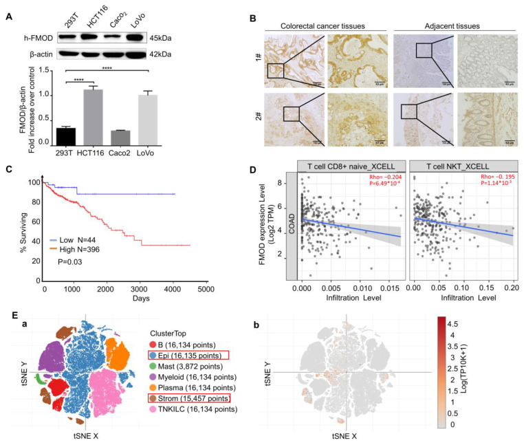
Figure 1.
The expression of FMOD is associated with CRC progression. (A), Protein expression levels of FMOD in CRC cell lines compared with 293T cells were measured by Western blotting. (B), IHC staining of FMOD in CRC and adjacent tissues. Adjacent tissues located more than 2 cm away from the malignant tumor edge and taken out by experienced surgeons. (C), Kaplan–Meier survival analysis and log-rank test showed the overall survival of CRC patients who were FMOD-positive (n = 398) vs. FMOD-negative (n = 44). Data from OncoLnc. (D), Correlation between FMOD expression levels and CD8+ T cell and NKT cell infiltration levels. (E), Single-cell analysis of FMOD in CRC tissues. (a) Tissue clusters in colorectal cancer. (b), FMOD is mostly expressed in the epithelium and stroma. **** means p < 0.0001.

