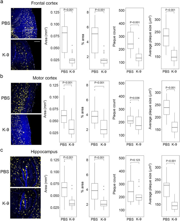
Figure 2.
Kamuvudine-9 treatment leads to decreased amyloid-β deposition in 5xFAD mice.
a–c, Amyloid-β deposition quantification in the brains of 36-week-old 5xFAD mice treated twice daily from 24 weeks of age with Kamuvudine-9 (K-9, i.p. 60 mg/kg twice daily, n = 5) or PBS (i.p. equal volume, n = 5). Amyloid-β quantification performed in five consecutive sections per animal. Left-most panels (a–c) show representative images of amyloid-β deposition (yellow) in the frontal cortex, motor cortex and hippocampus. Nuclei stained with DAPI (blue). Scale bar, 200 μm. Box plots (median, interquartile range (IQR), and outliers defined as greater than 1.5 IQRs from the first or third quartiles shown as dots) show quantification of surface area of amyloid-β (Aβ) plaques, % area, number of plaques, and size of plaques. P values from comparisons conducted using mixed effect models fit to account for repeated samples from each brain region within each mouse. Mixed effect models were fit with nested random effects. A random effect for each mouse and a random effect for each brain region within each mouse were included. Fixed effects include the treatment type (K-9, PBS) as well as the brain region.

