Abstract
The recognized antibacterial properties of silver nanoparticles (AgNPs) characterize them as attractive nanomaterials for developing new bioactive materials less prone to the development of antibiotic resistance. In this work, we developed new composites based on self-assembling Fmoc-Phe3 peptide hydrogels impregnated with in situ prepared AgNPs. Different methodologies, from traditional to innovative and eco-sustainable, were compared. The obtained composites were characterized from a hydrodynamic, structural, and morphological point of view, using different techniques such as DLS, SEM, and rheological measurements to evaluate how the choice of the reducing agent determines the characteristics of AgNPs and how their presence within the hydrogel affects their structure and properties. Moreover, the antibacterial properties of these composites were tested against S. aureus, a major human pathogen responsible for a wide range of clinical infections. Results demonstrated that the hydrogel composites containing AgNPs (hgel@AgNPs) could represent promising biomaterials for treating S. aureus-related infections.
Keywords: peptide hydrogel, composite, silver nanoparticles
1. Introduction
Currently, one of the most interesting objectives from a scientific and biomedical point of view is the search for novel tools to fight bacterial infections that do not involve the use of antibiotics [1,2]. In fact, more and more often, clinicians face pathogenic microorganisms that have developed resistance against currently available antibiotics for treating bacterial infections. Therefore, developing alternative antibiotic agents and treatments to control bacterial infections is critical. A possible answer to this growing issue is the use of nanomaterials with intrinsic antimicrobial properties [3,4,5,6]. Silver nanoparticles (AgNPs) are among the most promising nanostructured materials in the biomedical field thanks to the antibacterial properties of silver. In fact, AgNPs are more reactive and therefore have a greater antibacterial activity as there is an increase in the available surface exposed to microbes (high surface/volume ratio typical of nanomaterials) [7,8,9]. A number of approaches are available for the synthesis of AgNPs, such as physical, electrochemical, and biological methods [10,11]. The role of the functionalizing layer (e.g., polymeric ligands, surfactants, thiols) has also been deeply investigated for surface protection and colloidal stability improvement, also in view of different applications [8,12]. The major problems that could be encountered using traditional methods for the preparation of AgNPs are related to the high cost and use of toxic chemicals that can represent a possible biological and environmental risk. Furthermore, AgNPs used in the biomedical field must meet precise standards of final purity. To overcome these problems, innovative biological synthesis methods, which are more sustainable and less harmful to both humans and the environment, have recently evolved [13,14,15]. These methods, compared to the chemical-physical ones, also allow a simpler control of the size, polydispersion, and shape of the obtained AgNPs. For example, some of the most widely used methods involve bacteria, fungi, and plant extracts [16,17,18].
Another approach being developed in the synthesis of AgNPs involves replacing classical chemical reducing agents, such as NaBH4, with carbohydrates [19]. Among these, the sugars with a greater reducing activity are monosaccharides. For a sugar molecule to have a reducing activity, it must exist in an open chain form with an aldehyde or ketone group, in the case of an aldose or a ketose, respectively. The reducing function is performed by the carbonyl group, which transfers electrons to another species, e.g., to Ag+, producing Ag° clusters [20]. Several works in the literature report the reduction of silver salts using carbohydrates, especially glucose, fructose, and lactose [21]. The AgNPs obtained with this innovative and entirely green methodology have more promising characteristics than those produced with chemical methods: the obtainable dimensions are a few tens of nanometers, the stability of the colloids is several months, and their antibacterial activity is preserved.
It is important to consider that the efficacy of a bioactive species depends not only on its intrinsic characteristics but also on how the substance is administered [22]. Therefore, it becomes crucial to develop new biomaterials capable of entrapping desired molecules, delivering them to the target site, and releasing them in a controlled way over time. In fact, a controlled and prolonged release is essential to ensure a safer and more effective dosage of drugs, preventing the body from being subjected to high drug concentrations that could lead to undesired side effects [23]. Hydrogels are promising biomaterials as controlled release systems. They consist of a water-swollen three-dimensional lattice in which it is possible to entrap a wide variety of bioactive substances, including AgNPs [24]. An interesting class of hydrogels consists of those based on peptides [3]. Self-assembling peptides are a particular class of molecules characterized by the ability to spontaneously organize themselves into ordered and stable structures in conditions of thermodynamic equilibrium, thanks to the formation of non-covalent bonds (hydrogen bonds, ionic, hydrophobic, and Van der Waals interactions) among the side chains of the amino acids of which they are made [25,26,27]. These intra- and inter-molecular interactions allow the peptide to organize into ordered secondary (α-helix and β-sheet) and tertiary (fibers and fibrils) structures. The possibility to obtain stable hydrogels using low molecular weight peptides has emerged in recent years since short peptides with appropriate chemistry can act as hydrogenators in specific pH or temperature conditions; they self-assemble into supramolecular structures, such as nanofibers, which give rise to the three-dimensional lattice which constitutes the solid phase of the hydrogel. During this process, an extremely high amount of water (often more than 90% of the total hydrogel weight) is trapped inside the meshes of the 3D network [28].
In this paper, we worked on the preparation of peptide hydrogel composites containing AgNPs (hgel@AgNPs) using glucose as a green reducing agent. As far as the peptide components are concerned, we used Fmoc-Phe3, a short self-assembling peptide, that we synthesized with a biocatalytic reaction. We have previously reported the optimization and characterization of the native hydrogel [13,28,29]. In this work, we used this system as a platform for the preparation of composite materials with antibacterial properties. The novelty of this work lies in the development of different synthetic methodologies for one-pot preparation of hgel@AgNPs, where AgNP formation occurs in situ and simultaneously with the sol–gel transition. The first synthesis involved the use of sodium 3-mercapto-1-propanesulfonate (3MPS) as a stabilizing agent and NaBH4 as a reducing agent. Then, we moved on to a green approach, exploiting the reducing properties of monosaccharides such as glucose. We compared the effect of the preparation technique of composite hydrogels on the size, monodispersity, and stability of in situ synthesized AgNPs. All the prepared hgel@AgNPs samples were characterized with UV-Vis spectrophotometry, Dynamic Light scattering (DLS) analysis, Scanning Electron Microscopy (SEM), and rheological analysis. Finally, composite hydrogels were tested against Staphylococcus aureus to evaluate their antimicrobial activity.
2. Materials and Methods
2.1. Materials
Fluorenylmethyloxycarbonyl-phenylalanine (FmocPhe, >99%) and diphenylalanine (Phe2, >99%) were from Bachem GmbH (Weil am Rhein, Germany) and used as received. Lipase from Pseudomonas fluorescens (PFL, 20.000 U/mg), silver nitrate (AgNO3, 98%), sodium 3-mercapto-1-propanesulfonate (3MPS), sodium borohydride (NaBH4), sodium dodecyl sulfate (SDS), β-D-glucose, and all other chemicals and solvents were from Sigma Aldrich (St. Louis, MO, USA) and used as received.
2.2. Hydrogel Biosynthesis
Fmoc-Phe3 tripeptide hydrogels were prepared starting from equimolar quantities of Fmoc-Phe and Phe2 according to previously reported procedures [30]. Fmoc-Phe3 is biosynthesized in an aqueous solution by PFL after 30 min of incubation at a controlled temperature. The reaction yield for Fmoc-Phe3 formation was calculated using an HPLC method, as described previously [30].
2.3. Synthesis of AgNPs in a Hydrogel Matrix
AgNPs entrapped within the hydrogel matrix were synthesized using two bottom-up synthetic techniques: a more traditional one requiring NaBH4 as a reducing agent and a green one, in which a monosaccharide (β-D-glucose) is employed (Figure 1). In both cases, a hydrogel precursor solution and a AgNP precursor solution were prepared separately and then mixed. After adjusting pH to neutrality by adding 0.1 M HCl, the enzyme solution was added to start gelation, which occurred after incubation at 30 °C for 30 min.
Figure 1.
Scheme of the preparation of hgel@AgNPs composites.
When using NaBH4, the AgNP precursor solution was prepared as follows: 333 μL of AgNO3 (5 mM) and 333 μL of 3MPS (20 mM) were mixed and bubbled with Ar for 10 min. At the end of bubbling, 333 μL of NaBH4 (25 mM) were added to start the AgNPs nucleation. Regarding the synthesis using β-D-glucose, the AgNP precursor solution was prepared with hydrogel precursors and dissolved in an aqueous phase with the following reagents: 333 μL of AgNO3 (5 mM), 333 μL of SDS (10 mM), and 333 μL of β-D-glucose (75 mM) [31,32,33,34]. The optimized molar ratios among the reagents are equal to 1:2:15 (AgNO3:SDS:β-D-glucose). The mixture was then stirred magnetically for 1 min to start the nucleation of AgNPs.
2.4. UV-Vis Analysis and DLS Measurements
UV-Vis analyses were performed using a UV/Visibile Ultraspec 4000 spectrophotometer (Pharmacia Biotech, Uppsala, Sweden). DLS analyses were performed with a Zetasizer Nano ZS (Malvern Instruments, Malvern, UK). For DLS measurements, hydrogel samples were mechanically broken to obtain a liquid suspension and immediately diluted with deionized water to avoid NPs aggregation. All measurements were performed at room temperature and at least three times, calculating the average value ± standard deviation. Peak intensity analysis was used to determine the average hydrodynamic diameter of the scattering particles.
2.5. Inductively Coupled Plasma-Atomic Emission Spectrometry
The yield of AgNPs in situ formation within the hydrogels was calculated through Inductively Coupled Plasma Atomic Emission Spectrometry (ICP-AES) experiments. Hydrogel samples were prepared as described in Section 2.3 and mechanically broken to obtain a liquid suspension. The suspension was filtered through a 3 kDa Amicon Ultra-100 Centrifugal Filter Unit (Burlington, MA, USA) inside a centrifuge operating at 15,000× g rpm for 30 min at 20 °C in order to separate AgNPs from unreacted silver ions. The separated AgNPs were digested in HNO3. Ag concentration in the digestate was analyzed using a Varian Vista RL CCD Simultaneous ICP-AES spectrometer (338.289 nm Ag emission line was taken into account). The AgNPs formation yield was calculated with Equation (1):
| yield% = (experimental Ag mass)/(theoretical Ag mass) × 100 | (1) |
2.6. SEM Measurements
AgNPs morphology was studied with Scanning Electron Microscopy at the CNIS laboratory of Sapienza University. A drop of each sample was deposited on aluminum stabs and air-dried. Analyses were carried out using a Zeiss Auriga 405 microscope (Oberkochen, Germany) at a low extracting voltage and current.
2.7. Rheological Measurements
An Anton Paar MCR 302 rotational rheometer was used to record the elastic (G′) and viscous (G″) moduli of hydrogel samples. The instrument, equipped with a temperature control unit, used a plate-plate geometry. The gelation process was followed at 30 °C, applying a deformation of 1% and a constant frequency of 1 Hz in time sweep experiments. The elastic and viscous moduli of preformed hydrogels were measured in frequency sweep experiments, varying the applied frequency between 0.01 and 20 Hz at 30 °C.
2.8. Evaluation of Hydrogel Stability in Physiological Conditions
The degradation kinetics of the native hydrogels as well as hydrogel composites containing AgNPs, under physiological conditions, were evaluated using the following stability assay. Briefly, 8.5 mL of Ringer’s solution, containing NaCl (8.6 mg/mL), KCl (0.3 mg/mL), and CaCl2 (0.33 mg/mL), were added to each hydrogel sample. Samples were incubated at 37 °C for 30 days. After incubation, the supernatant was removed. The hydrogels were weighed before adding Ringer’s solution (W0) and after its removal (Wt). The percentage of weight loss (ΔW%) was calculated using Equation (2):
| ΔW% = (W0 − Wt)/Wt × 100 | (2) |
To obtain the daily degradation rate (T), the percentage of weight loss (ΔW%) was then divided by the total incubation time (30 days) using Equation (3):
| T = (ΔW%)/30 | (3) |
2.9. Antibacterial Activity Studies
The antimicrobial activity of hgel@AgNPs composites was evaluated using the S. aureus strain from the American Type Culture Collection (ATCC) 25923. MICs (minimum inhibitory concentrations) were calculated using the broth microdilution method. The laboratory strain S. aureus ATCC 25923 was collected from a Blood Agar plate (Oxoid, Basingstoke, Hampshire, UK) and inoculated in 2 mL of 0.45% saline solution (Air Life, Fresno, CA, USA) to obtain turbidity of 0.5 ± 0.1 McFarland (McF). Subsequently, 100 µL of bacterial suspension, diluted 1:100 in cation-adjusted Mueller–Hinton broth (MHB), corresponding to 1 × 10⁶ CFU/mL, was used to inoculate a 96-well polystyrene plate (Corning Inc., Corning, NY, USA). The bacteria were incubated at 37 °C for 24 h in the presence of different concentrations of antimicrobial compounds.
After treatment with the different antimicrobial compounds, viable cells were determined with plate counting for the CFU/mL determination [34]. All experiments were in triplicate and repeated three times.
3. Results and Discussion
3.1. Synthesis of hgel@AgNPs Composites
Our research group has extensively studied the lipase-mediated biosynthesis of self-assembling peptide hydrogels [29,35]. Such systems can be prepared in mild reaction conditions and are highly versatile, allowing the incorporation of bioactive molecules, nanoparticles, or other molecules, serving as a tailorable platform for the preparation of composites with desired features for a wide variety of applications [13]. In this work, starting from our previously optimized protocols, we attempted to prepare antibacterial hydrogel composites containing AgNPs. We developed two different synthetic protocols, both based on the reduction of AgNO3, as described in Section 2.3: one was based on the use of chemical reducing agents such as NaBH4, while the other relied on environmentally friendly natural reducing agents such as β-D-glucose. It was necessary to optimize the amounts of reducing agents for in situ AgNP formation. Both synthetic protocols successfully yielded self-supporting hydrogels after approximately 30 min of incubation at 37 °C, with a tripeptide reaction yield of about 30%. Comparing these results with our previously published data on the native hydrogel, we can observe that AgNP precursors did not interfere with tripeptide formation and self-assembling [30].
3.2. UV-Vis Analysis
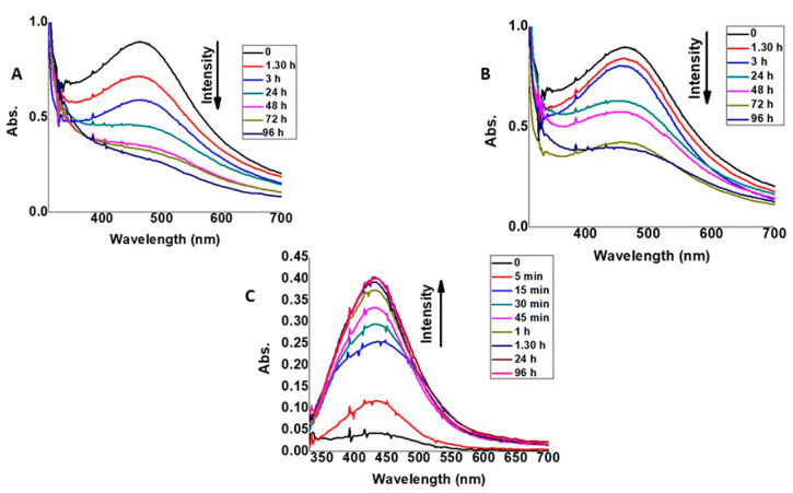
Aiming to characterize in situ formed AgNPs, their optical features such as the presence of SPR (surface plasmon resonance) band as a function of time were studied. Different experiments were performed to investigate the influence of storage temperature and duration on AgNP surface plasmon resonance values. Firstly, the evolution of AgNPs absorbance in the presence of NaBH4 as a function of reaction time was investigated. Samples stored at room temperature (RT) and 4 °C were examined. Figure 2A,B show the time evolution of the adsorption spectra of AgNPs synthesized in the hydrogel with NaBH4 and 3MPS at different temperatures. For both temperatures, the SPR band of AgNPs, centered at 475 nm, tends to decrease, and the plasmonic peak tends to widen over time. This is most likely due to the poor stability of AgNPs, which tend to aggregate. The reported data show that sample storage at 4 °C partially prevents aggregation, probably due to a reduction in diffusion phenomena within the hydrogel matrix.
Figure 2.
UV-Vis analysis in water. (A,B) The absorbance of hgel@AgNPs-3MPS at RT and 4 °C, respectively. (C) The absorbance of hgel@AgNPs-SDS at RT.
As can be seen from the spectra (Figure 2C), the SPR band of AgNPs-SDS synthesized with β-D-glucose at RT increases over time, while in the case of NaBH4-mediated AgNPs synthesis, the SPR band decreases with time. In addition, for AgNPs synthesized with glucose, the SPR band is present at a smaller wavelength (425 nm), and it is narrower than the SPR peak of AgNPs synthesized with NaBH4. These experimental observations could be due to the different AgNP formation kinetics obtained with the two different reducing agents. NaBH4 is a strong reducing agent, used in combination with 3MPS thiol. AgNPs could be subjected to an oxidation process with the formation of an Ag2O shell [36]. As a result, aggregation equilibria occur, and the band gradually widens and decreases over time.
However, the use of a milder reducing agent such as β-D-glucose may account for a slower AgNP formation that increases with time. In addition, the presence of SDS as the capping agent seems to prevent AgNPs aggregation, resulting in a smaller and narrower SPR band.
3.3. Inductively Coupled Plasma-Atomic Emission Spectrometry
The yield of AgNPs formed from Ag+ ions, using both β-D-glucose and NaBH4 as reducing agents, was calculated on the basis of ICP-AES experiments. According to ICP data, the amount of formed AgNPs in the presence of β-D-glucose and NaBH4 is equal to 1.2 ± 0.14 and 4.2 ± 0.11 ppm, respectively. Dividing these amounts by the theoretical value indicates that the reaction yields are 23.5% and 82.3% for AgNPs formed using β-D-glucose and NaBH4, respectively. As expected, in the optimized reaction conditions, the use of NaBH4 leads to higher yields of formed AgNPs, given that this compound is a strong reducing agent.
3.4. DLS Measurements
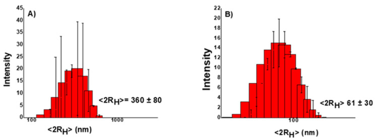
DLS measurements were performed to estimate AgNP size and polydispersion degree. The size of the AgNPs is a fundamental parameter to be considered in the realization of antimicrobial nanosystems. In fact, many of their chemical and physical properties, such as stability, reactivity (proportional to their surface/volume ratio), and release kinetics of Ag+ ions could be affected by size. The interaction processes of AgNPs with biological systems are also size dependent. Figure 3A,B shows the distributions of the hydrodynamic diameters of AgNPs synthesized using NaBH4 and β-D-glucose as reducing agents.
Figure 3.
Hydrodynamic distribution of (A) hgel@AgNPs-3MPS and (B) hgel@AgNPs-SDS.
As can be seen, AgNPs synthesized using NaBH4 as a reducing agent have larger dimensions and polydispersion (~360 nm, PDI: 0.475), compared to those obtained using β-D-glucose (~60 nm, PDI: 0.233). Certainly, different factors may be responsible for such differences in AgNP dimensions and polydispersion. Higher AgNP concentrations may enhance particle aggregation within the hydrogel structure (as when using NaBH4). In addition, the structure of the capping agent could affect the final dimensions and/or particle aggregation.
3.5. SEM Measurements
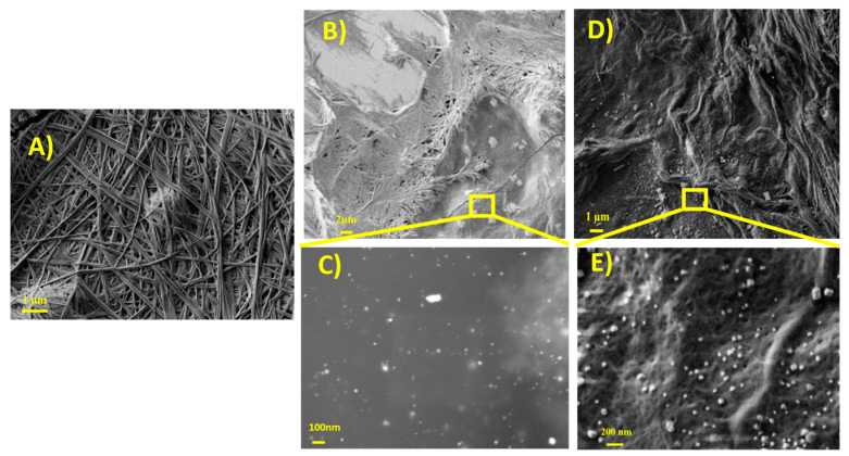
The morphological features of the native hydrogel and hgel@AgNPs were investigated using SEM. Figure 4A shows a micrograph of the native hydrogel in which the fibrous nature of the highly reticulated three-dimensional network can be seen. Figure 4B–E is related to hgel@AgNPs-SDS and hgel@AgNPs-3MPS, respectively, and their high-resolution images. It can be seen that the presence of AgNPs does not affect the ability of the Fmoc-Phe3 tripeptide to self-assemble into supramolecular structures giving rise to composite gels with a highly cross-linked fibrillar morphology. Moreover, in both composites, AgNPs appear as well dispersed inside the hydrogel matrix.
Figure 4.
SEM images of (A) native hydrogel; (B) hgel@AgNPs-SDS; (C) hgel@AgNPs-SDS at high magnification; (D) hgel@AgNPs-3MPS; and (E) hgel@AgNPs-3MPS at high magnification.
3.6. Rheological Studies
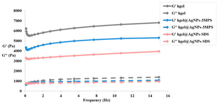
Studying the rheological features of hydrogels is pivotal for evaluating their application potential. With the aim to analyze the viscoelastic behavior of hydrogel composites, we used a dynamo-mechanical analysis, and the measurements were conducted in an oscillatory mode, at different application frequencies of the stimulus under constant strain. Both hydrogel samples containing AgNPs, formed in situ with the two different protocols described in Section 2.3, as well as the native control hydrogel, were analyzed. The aim of this study was to quantify the influence of in situ formed AgNPs on the viscoelastic behavior of the composites, and therefore how the elastic and viscous moduli of the hydrogels changed. The results for all samples are reported in Figure 5.
Figure 5.
Frequency sweep of the hydrogel; hgel@AgNPs-3MPS; and hgel@AgNPs-SDS.
Regarding all types of hydrogels, the elastic modulus G′ and the viscous one G” do not vary according to the oscillation frequency but remain almost constant for the entire frequency range analyzed. On this basis, a solid-like behavior is found (G′ > G″), whatever the frequency of the stimulus to which the sample is subjected. This indicates that there is a gel or a three-dimensional network in which chains, in this case, peptides, are held together thanks to physical and chemical crosslinks that prevent their sliding (typical of a liquid system) even at low frequencies. In fact, in the gel, there are only conformational variations in the peptide chains and translational motions of these chains are absent. Furthermore, from the frequency sweep experiments conducted, it is possible to obtain information also regarding the strength of the gel. Since the elastic modulus G′ is much higher than the viscous one G″ for all samples, it is possible to state that they are in the presence of strong and stable hydrogels.
In particular, with hgel@AgNPs-3MPS, we can observe a reduction in the mechanical properties compared to the hydrogel alone. In this case, the 3MPS stabilizer and NaBH4 reducing agent could negatively influence the hydrogel gelling ability. In fact, physical crosslinks are present in the peptide hydrogel, such as hydrophobic interactions and aromatic π–π stacking (due to the presence of benzyl groups in the side chain of the amino acid phenylalanine), which can be reduced in the presence of polar compounds such as 3MPS and NaBH4. As far as the hgel@AgNPs-SDS samples are concerned, we obtained a significant reduction in the viscous modulus G″ and, in particular, of the elastic one (G′). This indicates that a weaker gel is present, probably due to the presence of the amphiphilic surfactant SDS. In fact, the latter has a polar head that could affect the hydrophobic interactions that hold together the peptide lattice of the hydrogel.
3.7. Study of Hydrogel Stability in Physiological Conditions
The stability of hydrogel composites in physiological conditions, being an important feature for biological applications, was evaluated. The obtained results are summarized in Table 1.
Table 1.
Percentage of weight loss (ΔW %) after 30 days of incubation in Ringer solution and daily degradation rate (T) of native and composite hydrogels.
| Sample | ΔW % | T |
|---|---|---|
| Native hydrogel | 9.11 ± 2.34 | 0.31 ± 0.08 |
| hgel@AgNPs-SDS | 10.89 ± 1.76 | 0.36 ± 0.06 |
| hgel@AgNPs-3MPS | 12.60 ± 2.65 | 0.49 ± 0.09 |
Overall, all samples demonstrated good stability, accounting for a weight loss of approximately 10% after 1 month. The reported values of weight loss percentage (ΔW%) and degradation rate (T) of the analyzed composites do not differ much from those obtained for the native hydrogel. Therefore, it is possible to state that the in situ synthesis of AgNPs, both through the use of NaBH4 and glucose, does not significantly compromise the stability of the peptide hydrogel in physiological conditions.
3.8. Antibacterial Properties of AgNPs and hgel@AgNPs Composites
To test the antibacterial effects of silver and the application of these nanocomposites in the biomedical field, it was decided to carry out in vitro inhibition tests against S. aureus, which is a major human pathogen and the most common Gram-positive bacteria isolated from skin ulcers [36].
The tests were carried out on the hgel@AgNPs composites made with the three different synthesis strategies and with a Ag concentration equal to 0.094 mg/mL. In addition, the three types of AgNPs in solution with [Ag] = 0.094 mg/mL and the hydrogel as it is in the absence of AgNPs were tested as controls.
A viability test was then carried out on S. aureus in the presence of these biomaterials at different concentrations to determine the MIC (minimum inhibitory concentration). It was evidenced that the hydrogel exhibited limited antibacterial activity even in the absence of AgNPs. Indeed, it is known that the peptides of which it is made up have antimicrobial properties [13]. In particular, the hydrogel was effective against S. aureus at the following concentrations: Fmoc-Phe: 0.9 mg/mL, Phe2: 0.725 mg/mL, and Fmoc-Phe3: 0.7 mg/mL. The results of the microbiological analyzes obtained for the AgNPs in solution and for the hgel@AgNPs composites prepared under the different experimental conditions are reported in Table 2.
Table 2.
Minimum inhibitory concentration (MIC) for S. aureus ATCC 25923 for AgNPs in solution and hgel@AgNPs samples.
| Sample | Reducing Agent | MIC (mg/mL) |
|---|---|---|
| AgNPs-3MPS | NaBH4 | 0.008 |
| hgel@AgNPs-3MPS | NaBH4 | 0.006 |
| AgNPs-SDS | β-D-glucose | 0.004 |
| hgel@AgNPs-SDS | β-D-glucose | 0.006 |
Comparing the MIC data, it can be seen that there are no substantial differences either with the variation in the synthesis technique of AgNPs (using NaBH4 or β-D-glucose) or as a function of sample type (AgNPs colloidal suspension or hgel@AgNPs composites). In each of the analyzed cases, the MIC ranged between 0.004 and 0.008 mg/mL. Such values are comparable with those reported in the literature for similar systems. Thus, all the systems studied are promising candidates for treating S. aureus infections [13]. In particular, hgel@AgNPs samples have the advantage of being able to release silver ions in a controlled way over time; therefore, the bacteria, in the presence of these composites, will be subjected to the antimicrobial agent for a prolonged time and the duration of the antibacterial effect will be greater. Moreover, the green synthesis we propose, which uses glucose as a reducing agent, avoids the use of chemicals that could represent a biological risk.
4. Conclusions
In this study, peptide hydrogel composites containing AgNPs were synthesized. The AgNps were obtained with an in situ reduction of Ag+ using both traditional chemical reducing agents such as NaBH4 as well as natural reducers such as β-D-glucose. The use of β-D-glucose yielded composites containing AgNPs with improved physical features, e.g., smaller dimensions and greater stability over time, while allowing a more sustainable synthesis. The AgNPs synthesized in situ did not affect either the highly cross-linked fibrillar morphology of the hydrogel or its stability.
From the MIC data collected with the microbiological analyses, it appears that, in each of the experimental conditions used in the synthesis of AgNPs, the composites exhibit antibacterial activity against S. aureus. Therefore, it would be interesting to evaluate the bactericidal activity of these composites against other pathogenic bacteria such as the Gram-negative Pseudomonas aeruginosa.
Overall, the composite material prepared using β-D-glucose exhibits promising features and can easily be obtained using a sustainable approach. Therefore, such a composite will be further investigated to analyze the release kinetics of silver ions from the composite, with the prospect of its application in the biomedical field.
Author Contributions
Conceptualization, C.P. and L.C.; methodology, E.G.D.D., R.B. and S.C.; validation, L.C. and S.C.; investigation, L.C., R.B., S.M. and F.S.; resources, C.P., S.M., E.G.D.D. and I.F.; data curation, R.B. and F.H.H.; writing—original draft preparation, R.B. and C.P.; writing—review and editing, L.C. and I.F.; visualization, R.B. and F.H.H.; funding acquisition, C.P. and L.C. All authors have read and agreed to the published version of the manuscript.
Institutional Review Board Statement
Not applicable.
Informed Consent Statement
Not applicable.
Data Availability Statement
Data are contained within the article.
Conflicts of Interest
The authors declare no conflict of interest.
Funding Statement
This research received funding from Ateneo Sapienza (Progetti di Avvio alla Ricerca 2021, grant number AR22117A80326A88 and Progetti di Ricerca Ateneo Sapienza 2022, grant number RM12218167B480E0).
Footnotes
Disclaimer/Publisher’s Note: The statements, opinions and data contained in all publications are solely those of the individual author(s) and contributor(s) and not of MDPI and/or the editor(s). MDPI and/or the editor(s) disclaim responsibility for any injury to people or property resulting from any ideas, methods, instructions or products referred to in the content.
References
- 1.Łojewska E., Sakowicz T. An Alternative to Antibiotics: Selected Methods to Combat Zoonotic Foodborne Bacterial Infections. Curr. Microbiol. 2021;78:4037–4049. doi: 10.1007/s00284-021-02665-9. [DOI] [PMC free article] [PubMed] [Google Scholar]
- 2.Kumar M., Sarma D.K., Shubham S., Kumawat M., Verma V., Nina P.B., Jp D., Kumar S., Singh B., Tiwari R.R. Futuristic Non-Antibiotic Therapies to Combat Antibiotic Resistance: A Review. Front. Microbiol. 2021;12:609459. doi: 10.3389/fmicb.2021.609459. [DOI] [PMC free article] [PubMed] [Google Scholar]
- 3.Binaymotlagh R., Chronopoulou L., Haghighi F.H., Fratoddi I., Palocci C. Peptide-Based Hydrogels: New Materials for Biosensing and Biomedical Applications. Materials. 2022;15:5871. doi: 10.3390/ma15175871. [DOI] [PMC free article] [PubMed] [Google Scholar]
- 4.da Silva M.R.P., Matos R.S., Monteiro M.D.S., Santos S.B., Filho H.D.F., Andrade G.R.S., Salerno M., Almeida L.E. Exploiting the Physicochemical and Antimicrobial Properties of PHB/PEG and PHB/PEG/ALG-e Blends Loaded with Ag Nanoparticles. Materials. 2022;15:7544. doi: 10.3390/ma15217544. [DOI] [PMC free article] [PubMed] [Google Scholar]
- 5.Fierascu I.C., Fierascu I., Baroi A.M., Ungureanu C., Ortan A., Avramescu S.M., Somoghi R., Fierascu R.C., Dinu-Parvu C.E. Phytosynthesis of Biological Active Silver Nanoparticles Using Echinacea purpurea L. Extracts. Materials. 2022;15:7327. doi: 10.3390/ma15207327. [DOI] [PMC free article] [PubMed] [Google Scholar]
- 6.Toro R.G., Adel A.M., de Caro T., Brunetti B., Al-Shemy M.T., Caschera D. A Facile One-Pot Approach to the Fabrication of Nanocellulose–Titanium Dioxide Nanocomposites with Promising Photocatalytic and Antimicrobial Activity. Materials. 2022;15:5789. doi: 10.3390/ma15165789. [DOI] [PMC free article] [PubMed] [Google Scholar]
- 7.Yang X., Wu J.-Y. Synthetic Conditions, Physical Properties, and Antibacterial Activities of Silver Nanoparticles with Exopolysaccharides of a Medicinal Fungus. Materials. 2022;15:5620. doi: 10.3390/ma15165620. [DOI] [PMC free article] [PubMed] [Google Scholar]
- 8.Gherasim O., Puiu R.A., Bîrcă A.C., Burdușel A.-C., Grumezescu A.M. An Updated Review on Silver Nanoparticles in Biomedicine. Nanomaterials. 2020;10:2318. doi: 10.3390/nano10112318. [DOI] [PMC free article] [PubMed] [Google Scholar]
- 9.Romdoni Y., Kadja G.T.M., Kitamoto Y., Khalil M. Synthesis of Multifunctional Fe3O4@SiO2-Ag Nanocomposite for Antibacterial and Anticancer Drug Delivery. Appl. Surf. Sci. 2023;610:155610. doi: 10.1016/j.apsusc.2022.155610. [DOI] [Google Scholar]
- 10.Islam M.A., Jacob M.V., Antunes E. A Critical Review on Silver Nanoparticles: From Synthesis and Applications to Its Mitigation through Low-Cost Adsorption by Biochar. J. Environ. Manag. 2021;281:111918. doi: 10.1016/j.jenvman.2020.111918. [DOI] [PubMed] [Google Scholar]
- 11.Zharkova M.S., Golubeva O.Y., Orlov D.S., Vladimirova E.V., Dmitriev A.V., Tossi A., Shamova O.V. Silver Nanoparticles Functionalized With Antimicrobial Polypeptides: Benefits and Possible Pitfalls of a Novel Anti-Infective Tool. Front. Microbiol. 2021;12:750556. doi: 10.3389/fmicb.2021.750556. [DOI] [PMC free article] [PubMed] [Google Scholar]
- 12.Desireddy A., Conn B.E., Guo J., Yoon B., Barnett R.N., Monahan B.M., Kirschbaum K., Griffith W.P., Whetten R.L., Landman U., et al. Ultrastable Silver Nanoparticles. Nature. 2013;501:399–402. doi: 10.1038/nature12523. [DOI] [PubMed] [Google Scholar]
- 13.Binaymotlagh R., Del Giudice A., Mignardi S., Amato F., Marrani A.G., Sivori F., Cavallo I., Di Domenico E.G., Palocci C., Chronopoulou L. Green In Situ Synthesis of Silver Nanoparticles-Peptide Hydrogel Composites: Investigation of Their Antibacterial Activities. Gels. 2022;8:700. doi: 10.3390/gels8110700. [DOI] [PMC free article] [PubMed] [Google Scholar]
- 14.Roy A., Bulut O., Some S., Mandal A.K., Yilmaz M.D. Green Synthesis of Silver Nanoparticles: Biomolecule-Nanoparticle Organizations Targeting Antimicrobial Activity. RSC Adv. 2019;9:2673–2702. doi: 10.1039/C8RA08982E. [DOI] [PMC free article] [PubMed] [Google Scholar]
- 15.Kakakhel M.A., Sajjad W., Wu F., Bibi N., Shah K., Yali Z., Wang W. Green Synthesis of Silver Nanoparticles and Their Shortcomings, Animal Blood a Potential Source for Silver Nanoparticles: A Review. J. Hazard. Mater. Adv. 2021;1:100005. doi: 10.1016/j.hazadv.2021.100005. [DOI] [Google Scholar]
- 16.Gong D., Sun L., Li X., Zhang W., Zhang D., Cai J. Micro/Nanofabrication, Assembly, and Actuation Based on Microorganisms: Recent Advances and Perspectives. Small Struct. 2023 doi: 10.1002/sstr.202200356. [DOI] [Google Scholar]
- 17.Arif R., Uddin R. A Review on Recent Developments in the Biosynthesis of Silver Nanoparticles and Its Biomedical Applications. Med. Devices Sens. 2021;4:e10158. doi: 10.1002/mds3.10158. [DOI] [Google Scholar]
- 18.Gong D., Celi N., Zhang D., Cai J. Magnetic Biohybrid Microrobot Multimers Based on Chlorella Cells for Enhanced Targeted Drug Delivery. ACS Appl. Mater. Interfaces. 2022;14:6320–6330. doi: 10.1021/acsami.1c16859. [DOI] [PubMed] [Google Scholar]
- 19.Yazgan I., Gümüş A., Gökkuş K., Demir M.A., Evecen S., Sönmez H.A., Miller R.M., Bakar F., Oral A., Popov S., et al. On the Effect of Modified Carbohydrates on the Size and Shape of Gold and Silver Nanostructures. Nanomaterials. 2020;10:1417. doi: 10.3390/nano10071417. [DOI] [PMC free article] [PubMed] [Google Scholar]
- 20.Durmazel S., Üzer A., Erbil B., Sayın B., Apak R. Silver Nanoparticle Formation-Based Colorimetric Determination of Reducing Sugars in Food Extracts via Tollens’ Reagent. ACS Omega. 2019;4:7596–7604. doi: 10.1021/acsomega.9b00761. [DOI] [PMC free article] [PubMed] [Google Scholar]
- 21.Sidhu A.K., Verma N., Kaushal P. Role of Biogenic Capping Agents in the Synthesis of Metallic Nanoparticles and Evaluation of Their Therapeutic Potential. Front. Nanotechnol. 2022;3:801620. doi: 10.3389/fnano.2021.801620. [DOI] [Google Scholar]
- 22.Nocca G., D’Avenio G., Amalfitano A., Chronopoulou L., Mordente A., Palocci C., Grigioni M. Controlled Release of 18-β-Glycyrrhetinic Acid from Core-Shell Nanoparticles: Effects on Cytotoxicity and Intracellular Concentration in HepG2 Cell Line. Materials. 2021;14:3893. doi: 10.3390/ma14143893. [DOI] [PMC free article] [PubMed] [Google Scholar]
- 23.Orekhova A., Palocci C., Chronopoulou L., De Angelis G., Badiali C., Petruccelli V., D’Angeli S., Pasqua G., Simonetti G. Poly-(Lactic-Co-Glycolic) Acid Nanoparticles Entrapping Pterostilbene for Targeting Aspergillus Section Nigri. Molecules. 2022;27:5424. doi: 10.3390/molecules27175424. [DOI] [PMC free article] [PubMed] [Google Scholar]
- 24.Dong Q., Zu D., Kong L., Chen S., Yao J., Lin J., Lu L., Wu B., Fang B. Construction of Antibacterial Nano-Silver Embedded Bioactive Hydrogel to Repair Infectious Skin Defects. Biomater. Res. 2022;26:36. doi: 10.1186/s40824-022-00281-7. [DOI] [PMC free article] [PubMed] [Google Scholar]
- 25.Chen H., Zhang T., Tian Y., You L., Huang Y., Wang S. Novel Self-Assembling Peptide Hydrogel with PH-Tunable Assembly Microstructure, Gel Mechanics and the Entrapment of Curcumin. Food Hydrocoll. 2022;124:107338. doi: 10.1016/j.foodhyd.2021.107338. [DOI] [Google Scholar]
- 26.Das S., Das D. Rational Design of Peptide-Based Smart Hydrogels for Therapeutic Applications. Front. Chem. 2021;9:770102. doi: 10.3389/fchem.2021.770102. [DOI] [PMC free article] [PubMed] [Google Scholar]
- 27.Liu C., Zhang Q., Zhu S., Liu H., Chen J. Preparation and Applications of Peptide-Based Injectable Hydrogels. RSC Adv. 2019;9:28299–28311. doi: 10.1039/C9RA05934B. [DOI] [PMC free article] [PubMed] [Google Scholar]
- 28.Chronopoulou L., Di Nitto A., Papi M., Parolini O., Falconi M., Teti G., Muttini A., Lattanzi W., Palmieri V., Ciasca G. Biosynthesis and Physico-Chemical Characterization of High Performing Peptide Hydrogels@ Graphene Oxide Composites. Colloids Surf. B Biointerfaces. 2021;207:111989. doi: 10.1016/j.colsurfb.2021.111989. [DOI] [PubMed] [Google Scholar]
- 29.Chronopoulou L., Margheritelli S., Toumia Y., Paradossi G., Bordi F., Sennato S., Palocci C. Biosynthesis and Characterization of Cross-Linked Fmoc Peptide-Based Hydrogels for Drug Delivery Applications. Gels. 2015;1:179–193. doi: 10.3390/gels1020179. [DOI] [PMC free article] [PubMed] [Google Scholar]
- 30.Mehta S.K., Chaudhary S., Gradzielski M. Time Dependence of Nucleation and Growth of Silver Nanoparticles Generated by Sugar Reduction in Micellar Media. J. Colloid Interface Sci. 2010;343:447–453. doi: 10.1016/j.jcis.2009.11.053. [DOI] [PubMed] [Google Scholar]
- 31.Antony J.J., Sivalingam P., Siva D., Kamalakkannan S., Anbarasu K., Sukirtha R., Krishnan M., Achiraman S. Comparative Evaluation of Antibacterial Activity of Silver Nanoparticles Synthesized Using Rhizophora Apiculata and Glucose. Colloids Surf. B Biointerfaces. 2011;88:134–140. doi: 10.1016/j.colsurfb.2011.06.022. [DOI] [PubMed] [Google Scholar]
- 32.Peng H., Yang A., Xiong J. Green, Microwave-Assisted Synthesis of Silver Nanoparticles Using Bamboo Hemicelluloses and Glucose in an Aqueous Medium. Carbohydr. Polym. 2013;91:348–355. doi: 10.1016/j.carbpol.2012.08.073. [DOI] [PubMed] [Google Scholar]
- 33.Dong Y.-Y., Li S.-M., Ma M.-G., Yao K., Sun R.-C. Compare Study Cellulose/Ag Hybrids Using Fructose and Glucose as Reducing Reagents by Hydrothermal Method. Carbohydr. Polym. 2014;106:14–21. doi: 10.1016/j.carbpol.2014.02.023. [DOI] [PubMed] [Google Scholar]
- 34.Sivori F., Cavallo I., Kovacs D., Guembe M., Sperduti I., Truglio M., Pasqua M., Prignano G., Mastrofrancesco A., Toma L., et al. Role of Extracellular DNA in Dalbavancin Activity against Methicillin-Resistant Staphylococcus aureus (MRSA) Biofilms in Patients with Skin and Soft Tissue Infections. Microbiol. Spectr. 2022;10:e0035122. doi: 10.1128/spectrum.00351-22. [DOI] [PMC free article] [PubMed] [Google Scholar]
- 35.Tong S.Y.C., Davis J.S., Emily E., Holland T.L., Fowler V.G., Jr. Staphylococcus Aureus Infections: Epidemiology, Pathophysiology, Clinical Manifestations, and Management. Clin. Microbiol. Rev. 2015;28:603–661. doi: 10.1128/CMR.00134-14. [DOI] [PMC free article] [PubMed] [Google Scholar]
- 36.Elyamny S., Eltarahony M., Abu-Serie M., Nabil M.M., Kashyout A.E.-H.B. One-Pot Fabrication of Ag @Ag2O Core–Shell Nanostructures for Biosafe Antimicrobial and Antibiofilm Applications. Sci. Rep. 2021;11:22543. doi: 10.1038/s41598-021-01687-4. [DOI] [PMC free article] [PubMed] [Google Scholar]
Associated Data
This section collects any data citations, data availability statements, or supplementary materials included in this article.
Data Availability Statement
Data are contained within the article.


