Abstract
Nanotechnology is used in a variety of scientific, medical, and research domains. It is significant to mention that there are negative and severe repercussions of nanotechnology on both individuals and the environment. The toxic effect of nanoparticles exerted on living beings is termed as nanotoxicity. Nanoparticles are synthesized by various methods such as chemical, biological, physical, etc. These nanoparticles’ nanotoxicity has been observed to vary depending on the synthesis process, precursors, size of the particles, etc. Nanoparticles can enter the cell in different ways and can cause cytotoxic effects. In this review, the toxicity caused in the reproductive system and the role of the antioxidants against the nanotoxicity are briefly explained.
Keywords: antioxidant, reproductive system, nanotoxicity
1. Introduction
Nanotechnology is an emerging field that focuses on the design, characterization, manufacturing of devices, and systems by manipulating shape and size at the nanoscale [1]. Recent developments in nanotechnology and nanoscience find its application in various fields such as food technology, medicine, transportation, drug delivery, cosmetics, etc. [1,2,3,4,5,6]. Nanomaterials can interact with cells and the milieu in a way that their chemically identical, larger biological counterparts cannot, and this is because of their exceedingly small size [7]. When nanoparticles are discharged into the environment, they may readily enter the cells by receptor-mediated endocytosis or by passive diffusion, where they interact with the cellular proteins, lipids, and genomic DNA [8,9]. This results in oxidative stress caused by Reactive Oxidative Species which is considered to be the most significant contributor to nanotoxicity [10,11]. Despite the antioxidant property of a few nanoparticles like gold, silver, copper, iron nanoparticles, etc., most of them are involved in the formation of intracellular ROS depending on the cellular absorption of nanoparticles, intracellular response, and intracellular metal ion release [12,13,14] and are produced during several cellular signalling processes as well as they are a part of the immune system’s defense mechanism. ROS damages biological macromolecules such as proteins, lipids, and DNA, causing negative effects on cells and causing mitochondrial dysfunction [15]. A high level of ROS can also produce a range of physiopathological effects, such as apoptosis, necrosis, hypertrophy, genotoxicity, inflammation, fibrosis, and even cancer. It also increases the production of pro-inflammatory cytokines and activates inflammatory cells like macrophages, which further increases the production of ROS [16]. These nanoparticles could also affect the reproductive system by breaching the guard tissues of the reproductive system like the epithelial, blood-testis, placenta and placental barriers and reproductive cells like germ cells, Leydig cells, and Sertoli cells [17]. Particularly, in the male reproductive system, nanoparticles could cause toxic effects by creating ROS which acts as molecular mediators in signal transduction pathways of spermatogenesis, steroidogenesis, and hypothalamic-pituitary-gonadal axis regulation affecting sperm maturation, such as DNA compaction and flagellar modification; whereas, in female reproduction, nanoparticles affect the physiological processes, including maturation of the oocyte, fertilization, development of the embryo, and pregnancy [18,19]. The prevalence of interspecies extrapolation factors between humans and test species helps in computing the toxicology in the reproductive system after exposure to drugs or chemicals [18]. Thus, this review briefs readers on the impacts of nanoparticles on the reproductive system, its mechanism, quantitative test, and potential nanotoxicity, using animal models.
2. Nanoparticles and Reactive Oxygen Species (ROS)
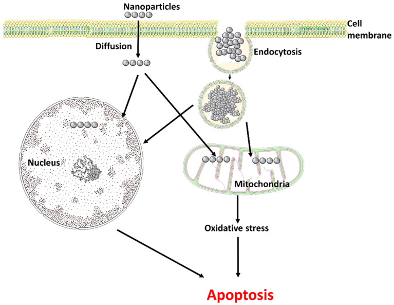
The unique characteristics of nanoparticles make them suitable for a variety of applications, such as biomedical applications, chemical and biological sensors, drug delivery, and various other fields [20]. The physical features of nanoparticles have a significant impact on cellular interactions. Nanoparticles can enter cells via endocytosis, diffusion, or via interactions with phospholipids. They can disrupt cell membranes by physicochemical interactions with the surface of the membrane, thereby impairing transport processes. Additionally, they can also trigger oxidative stress by releasing ions. Also, the function of cell organelles, particularly the mitochondria and peroxisomes, can be impacted by nanoparticles, which can disrupt intracellular transport and cause oxidative stress [21] (Figure 1).
Figure 1.
Entry of nanoparticles into the cell (Figure was drawn using the images and illustrations available on Smart Servier Medical Art).
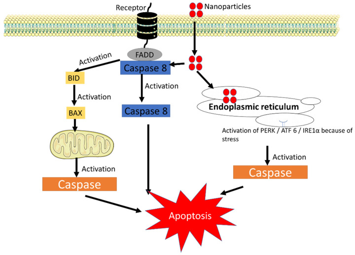
Silver, platinum, cerium, and zinc nanoparticles are some of the metal-based nanoparticles that can cause membrane lipid peroxidation and oxidative stress leading to mitochondrial dysfunction, DNA damage, and a number of other adverse effects [22]. Hydrogen peroxide, hydroxyl radicals, singlet oxygen, and superoxide anion radicals are some examples of biologically relevant ROS produced by normal cellular metabolism or by any antigenic/mitogenic responses or as a by-product in mitochondrial respiration [23]. Also, ROS are generated when immune cells like neutrophils and macrophages react against environmental toxins/microorganisms/any other antigens/any internal stimuli [24]. Oxidative stress occurs when ROS are abundant in the body and cause potentially harmful biological reactions. This is because ROS produces an imbalance between the amount produced and the biological system’s ability to quickly detoxify reactive intermediates or to repair the damage by activating antioxidant enzymes and non-enzymatic antioxidants [24,25]. When the ROS level exceeds the level because of nanoparticle entry, immune cell activation and activated cell signalling pathways lead to additional pathological effects such as inflammation, genotoxicity, and fibrosis [24]. Most nanoparticles could interfere with the cell signalling pathway through three primary routes resulting in the apoptotic process (Figure 2). The direct nanoparticle occupancy of the FADD receptor is the first mechanism. The death receptor including TNFR1 (TNF receptor superfamily member 1A), FAS/CD95 (FAS cell surface death receptor) etc. could initiate death signalling, where the ligands bind to these receptors and activate FADD (FAS associated via death domain) and then activate proCASP8 to CASP8 and end with activating apoptosis [26]. The second pathway involves the location of nanoparticles pacing in the endoplasmic reticulum; while the endoplasmic reticulum is facing high stress, there is the activation of Protein kinase R-like ER kinase (PERK), inositol-requiring enzyme 1α (IRE1α), and activating transcription factor 6 (ATF6), which are important transmembrane proteins that cascade various cellular signalling pathways, ending with activation of caspase and leading to apoptosis [27,28,29]. The third pathway involves modulating the mitochondrial function in the presence of nanoparticles. All these mechanisms eventually lead to caspase activation, which causes the mitochondria to produce more ROS, to make more BH3 Interacting Domain Death Agonist (BID protein), and to activate Bax or Bak1 proteins, all of which can cause cell death, DNA cleavage, and organelle damage [30]. The mechanism by which each nanoparticle produces ROS is unique; to date, there is no exact evidence that shows how the ROS are produced. Through Fenton-type reactions, metal-containing nanoparticles cause toxicity by producing free radicals, whereas carbon nanotube-mediated ROS production is significantly influenced by mitochondrial injury [31]. Also, nanoparticles alter electron transfer, elevating the ratio of NADP+/NADPH, and interfering with mitochondrial function, resulting in intracellular ROS formation. Additionally, nanoparticles are known to affect genes associated with oxidative stress, such as oxyR, ahpC, soxR, and soxS antioxidant genes such as gpx, sod1, and NADPH synthesis genes such as met9. Nanoparticles are associated with an increase in intracellular ROS because of their instability in oxidative and antioxidant defence genes [16]. Based on the mechanism, nanotoxicity is classified into two types: (i) primary oxidative stress, and (ii) secondary oxidative stress. Primary oxidative stress is defined as the photocatalytic activity of metal nanoparticles, for example titanium oxide could directly induce oxidative stress by ROS production, whereas secondary oxidative stress causes mitochondrial malfunction by the exposure to nanoparticles that results in the formation of ROS [12].
Figure 2.
Nanoparticles interfering with the cell signalling pathway to induce apoptosis. ATF6, activating transcription factor 6; PERK, protein kinase R-like ER kinase IRE1α, inositol-requiring enzyme 1α (Figure was drawn using the images and illustrations available on Smart Servier Medical Art).
3. Entry of Nanoparticles into the Reproductive System
Since the 20th century, experiments in consumer items, industry, and medicine have significantly increased, which also increases the number of nanosized particles in the air. This increase in the air holds the chance of nanoparticle-induced toxicity in humans [32,33,34]. Generally, the entry of nanoparticles into the human body is comprised of four types: (i) ingestion; (ii) inhalation; (iii) dermal penetration, and (iv) blood circulation. Since nanoparticles are smaller, it is easier for them to quickly penetrate deeper into the respiratory system after being inhaled. The process of absorption also allows nanoparticles to enter the dermal system through wound or abrasion skin where particles smaller than 5 nm diffuse into the skin easily. Thus, entered nanoparticles travel throughout the bloodstream, interact with the systems/organs and accumulate, which could cause severe impact on organs including the lung, liver, reproductive system, and kidneys [21]. Numerous nanoparticles have been shown to have the potential to overcome biological boundaries and injure important organs like the kidney, brain, and liver [33,34]. These nanoparticles could even reach the reproductive system where there are more investigations done on in vitro and in vivo models for studying the medical and molecular effects of nanotoxicity on genital organs [33]. As discussed in the previous section, these nanoparticles tend to cause cytotoxicity in the genetic and molecular levels by promoting inflammation, apoptosis, and oxidative stress through ROS [34,35]. Nanoparticles reach and accumulate in reproductive organs by breaching the guard reproductive tissues including epithelial, placental, and blood-testis barriers and by destroying Leydig cells, germ cells, and Sertoli cells. This accumulation does harm the male including epididymis and testes and impairs the quality, quantity, motility, and shape of sperm. Female reproductive organs like the ovary and uterus showed a reduction in the number of mature oocytes and interfered with the growth of primary and secondary follicles on the entry of nanoparticles [17,34].
In the male reproductive system, nanomaterials trigger cell death after penetrating through the blood testis barrier (BTB). It results in the lowering of mobility of the sperm due to mitochondrial dysfunction. Its transmission depends upon the polarity, shape, and size of the nanoparticle [33,34,35,36]. According to recent studies, nanoparticles can be endocytosed by granulosa and thecal cells, which stops oocyte development in vivo and causes aberrant hormone release [37]. The most common nanoparticles involved in studies assessing their toxicity to female reproductive systems are carbon nanoparticles, metal, metal oxides, and quantum dots. Studies conducted in both in vivo and in vitro conditions revealed that specific sizes of nanoparticles could enter and accumulate in different female germ cells, triggering a variety of cellular reactions including oxidative stress, signal transduction inhibition, apoptosis, DNA damage, and inflammation [38].
4. Impact on the Male Reproductive System
Both oxidative stress and inflammation are believed to be sensitive to the male reproductive system; where nanoparticles’ exposure readily increases oxidative stress and causes cell death and poor spermatogenesis, oxidative stress is reported to be the primary cause of 30–80% of infertility problems in male [39,40]. Studies suggest that nanoparticles react to distinct germ cells and damage cells differently. The changes of the cytoskeleton on the entry of nanoparticles can affect the production of sperm flagella and their ability to migrate, as well as the formation of Sertoli cell tight junctions and the tight junctions between them; all these changes can affect spermatogenesis [41]. Liu et al. [42] have proposed that ZnO nanoparticles caused down-regulation of tight junction proteins in Sertoli cells resulting in BTB dysfunction and no changes in cytoskeleton dynamics were identified [42]. The various nanoparticles involved in the toxicity of the male reproductive system are tabulated in Table 1.
The buildup of nanoparticles destroys germ cells, Leydig cells, and Sertoli cells, impairing the motility, shape, quality, and quantity of the sperm and limiting the number of mature oocytes or preventing the growth of primary and secondary follicles. Further, nanoparticles can alter the number of hormones released, altering sexual behavior [34]. In a study, Mathias et al. [43] found there were no differences in any of the sexual activities in silver nanoparticles on the following sexual behaviors like number of mounts, intromissions, ejaculatory intervals, attempted mounts, and ejaculations, but the exposure to nanoparticles was reducing sperm quality. Nanoparticles such as silica nanoparticles were found to increase the levels of micronucleus frequencies, malondialdehyde levels, and lower activity of catalase and glutathione content in testicular tissues at a higher concentration treated group, pointing to mechanisms of DNA damage and oxidative stress. Significant testicular histological changes were also seen in this group along with inflammation, testicular apoptosis, and oxidative stress by enhancing the gene expression corresponding to the pro-inflammatory activity, apoptotic activity, and oxidative stress caspase 3 and iNOS [44].
Metal nanoparticles like zinc oxide nanoparticles (ZnO) are observed to be breaking the cell membrane and outer membrane of mitochondria in Sertoli cells and down-regulating the production of gap junction proteins, thus damaging the BTB, further compromising its integrity. Additionally, ROS and cytokine release play substantial roles in BTB disruption, markedly raising the oxidative stress status by causing elevated ROS and malondialdehyde levels and lowered glutathione levels and raising the TNF-α levels in Sertoli cells [42]. Similarly, Bara and Kaul [45] have observed that ZnO nanoparticles considerably lowered the expression of the antioxidant enzyme gene (SOD) and significantly enhanced the expression of genes associated with steroidogenesis by the up-regulation of steroidogenic acute regulatory protein and cytochrome P450 side-chain cleavage enzyme in the mouse model. In contrast, exposure to ZnO nanoparticles markedly boosted testosterone synthesis at a concentration level of 2 mg/mL [45].
Likewise, the subcutaneous injection of titanium oxide nanoparticles in pregnant mouse found to have reduced sperm production, disordered seminiferous tubules and olfactory bulb apoptosis was observed [46]. Some of the studies have been done on the estimation of the levels of endocrine and reproductive hormones such as follicle-stimulating hormone, testosterone, luteinizing hormone, estradiol, and gonadotropin-releasing hormone that can be related to the rise in ROS and the concurrent decline in antioxidant enzymes. The sex hormone profile might also be disturbed by titanium oxide nanoparticles’ administration, as seen by lower blood testosterone levels and higher serum levels of estradiol, luteinizing hormone, and follicle-stimulating hormone [47,48]. According to prior research using mouse, rat, and porcine Leydig cells, the inhibitory effect of TNF-α on testosterone production may be caused by a decrease in the 17 α-hydroxylase/C17-20 lyase gene and protein expression and cholesterol side-chain cleavage enzyme, two essential enzymes in testosterone biosynthesis [49]. Contradictory reports are also there, which are as follows: Lauvås et al. [50] and Ogunsuyi et al. [51] found titanium oxide nanoparticles do not cause changes in testosterone levels but according to Miura et al. [52], titanium oxide nanoparticles did not impact sex hormones related to spermatogenesis but they do report that titanium nanoparticles tend to reduce testosterone levels and sperm mobility.
Cerium oxide nanoparticles were found to influence prepubertal spermatogenesis and harm Sertoli cells. Also, lowered expression of the steroidogenic enzyme’s genes Cyp17-1 and HSD3b1 is associated with decreased expression of Insl3, a gene that specifically marks Leydig cells [53]. There are studies where the Superparamagnetic Iron Oxide Nanoparticles (SPIONs), gold and silver nanoparticles reduce the earthworm in number, but the direct interaction with the reproductive system is not clear [54,55,56,57,58].
Table 1.
Various nanoparticles are involved in the toxicity of the male reproductive system.
| S. No | Nanoparticles | Animal Model | Toxicity Effect | References |
|---|---|---|---|---|
| 1. | Silica nanoparticles | Male Albino Rats | Higher levels of micronucleus frequencies and malondialdehyde levels, and lesser catalase and glutathione activity in testicular tissues. | [44] |
| 2. | ZnO nanoparticles | TM-4 Sertoli cell line and GC2-spd spermatocyte cell line of mouse | Breakdown of the cell membrane and outer membrane of mitochondria in Sertoli cells; down-regulating the production of gap junction proteins; disruption of BTB disruption. | [42] |
| Mouse testis Leydig cells | Decreased antioxidant enzyme gene expression (SOD) and increased steroidogenesis-related gene expression. | [45] | ||
| 3. | Titanium oxide nanoparticles | Pregnant mouse model | Apoptosis of the olfactory bulb occurs with decreased sperm production and motility; disordered and disrupted seminiferous tubules. | [46] |
| Mouse, rat, and porcine Leydig cells | 17–α hydroxylase/C17-20 lyase and cholesterol side-chain cleavage enzyme gene and protein expression are affected by TNF- α to decrease testosterone synthesis. | [49] | ||
| 4. | Cerium oxide nanoparticles | Pregnant mouse model | Involvement in the prepubertal spermatogenesis and germ cell; reduction of germ cells, deformation of Sertoli cells; impairment steroidogenesis. | [53] |
Spermatogenesis and Toxicity
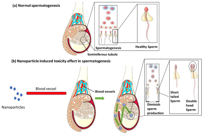
According to some of the research, nanoparticles are found to have the ability to quickly pass the BTB, after building up in the testis and having a negative impact on spermatogenesis. ROS generation in the seminiferous tubule, the place where spermatogenesis takes place, can cause DNA damage to spermatogenic cells [59]. While the testicular tissues are exposed to nanoparticles, seminiferous tubules undergo histological changes, which damage the testicles and diminish sperm production [40] (Figure 3).
Figure 3.
Comparison between normal spermatogenesis and nanotoxicity effect induced spermatogenesis in the male reproductive system (Figure was drawn using the images and illustrations available on Smart Servier Medical Art).
Nanoparticles are proven to enter the male rat reproductive organs or tissues in various ways; the testes and the epididymis are considered to be the most vulnerable to damage. In a study, it has been proposed that nanoparticles may affect the testes in a variety of ways, resulting in differences in the production of sperm quantity and quality production. Male mouse testes exposed to water-soluble carbon nanotubes showed oxidative stress, which reduced spermatogenic epithelium thickness [8]. Subcutaneous exposure of silver nanoparticles in male rats displayed abnormalities in the testes as well as changes in sperm quantity and motility, as well as levels of testosterone, luteinizing hormone, and follicle-stimulating hormone. While some studies claim that nanoparticles cannot permeate the skin, others have found evidence of metallic nanoparticles, especially iron nanoparticles, penetrating the skin through hair follicles [60]. Mice exposed to iron oxide nanoparticles caused histopathological changes in seminiferous tubules of the testes including sloughing, detachment, and vacuolization [61]. Exposure to silver nanoparticles resulted in various sperm cell irregularities, such as multiple heads, lengthy tails, and hook attachment errors [62]. Additionally, ultrastructural changes in spermatogonia, spermatogenic cells, and Sertoli cells, as well as atrophy in seminiferous tubules, necrosis, and cell disintegration with abnormal development of spermatids were observed [63].
Also, non-metal nanoparticles such as carbon nanotubes were reported to disrupt the Leydig and Sertoli cell function in the testes, which may lead to a variety of problems in steroidogenesis and germ cell differentiation. Carbon nanotubes might also prevent BTB and hemato-testicular barrier (HTB) from functioning properly [64]. These barriers are permeable to carbon nanotubes, which therefore have a direct impact on the neuroendocrine pathways, spermatogenesis, and reproductive organs [65]. They may have an impact on the level of sex hormones in the blood and the serum or tissue level of endocrine hormones in carbon nanotubes [64]. In a study, ROS levels and biomarkers indicative of oxidative stress in the testes are measured to better understand the mechanism underlying the oligospermia and teratozoospermia brought on by silica nanoparticles. The results have revealed that silica nanoparticles significantly increased the ROS level and malondialdehyde as well as the activity of superoxide dismutases. As a result, it has been hypothesized that alterations in the redox system and sex hormones might be the reason for the lower sperm quality and quantity [66].
Administration of quantum dots to mice was found to decrease the follicle-stimulating hormone and testosterone and a dose-dependent increase in the toxicity of quantum dots on spermatogenesis occurred, and it lasted for about 60 days. Additionally, approximately 60% of pachytene spermatocytes showed unrepaired double-strand break at days 14 and 30 after injection and were found to be vanished at day 60 indicating that double-strand break repair was impaired after exposure to quantum dots. The quantum dots-treated groups were still observed to have a lot of H2AX foci in the pachytene stage, and during meiosis I, the fraction of spermatocytes in the pachytene stage was much higher than expected on exposure [67]. Hong et al. [68] have demonstrated that mice exposed to titanium nanoparticles had lesions of the testes and epididymis, experienced reductions in the concentration and motility of sperm, and produced a greater number of defective sperm. Additionally, in mice’s testes, exposure to titanium nanoparticles increased the activities of testicular-marked enzymes such as alkaline phosphatase, acid phosphatase, and total nitric oxide synthase while decreasing the activities of succinate dehydrogenase, sorbitol dehydrogenase, lactate dehydrogenase, and glucose-6-phosphate dehydrogenase. Additionally, exposure to titanium nanoparticles increased the generation of ROS, malondialdehyde, a product of lipid peroxidation, carbonyl, a product of protein oxidation, and 8-hydroxydeoxyguanosine, a product of DNA oxidation in the testes. Also, a reduction in sperm count and lesions induced by titanium nanoparticles was observed [68].
5. Impact on the Female Reproductive System
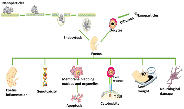
Nanoparticles alter sex hormone levels by triggering secretory cells such as thecal cells, follicle cells, granule cells, and corpus luteum via the hypothalamic pituitary gonadal axis, or by directly stimulating secretory cells such as granule cells, thecal cells, follicle cells, and the corpus luteum [38]. Some nanoparticles can reach the foetus by passive diffusion or endocytosis, causing fetal inflammation, apoptosis, genotoxicity, reproductive deficit, lower weight, cytotoxicity, immunodeficiency, and neurological damage, among other effects [38] (Figure 4). The primary female sex hormones in females are estrogen and progesterone, which are mostly produced in the ovaries or placenta during pregnancy in humans. Some data suggest that certain nanoparticles can change the gene expression that encodes proteins involved in steroidogenesis, such as ovarian genes essential for the synthesis of estrogen and/or progesterone [32]. The various nanoparticles involved in the toxicity of the female reproductive system are given in Table 2.
Figure 4.
Entry of nanoparticles into foetus and its adverse effect (Figure was drawn using the images and illustrations available on Smart Servier Medical Art).
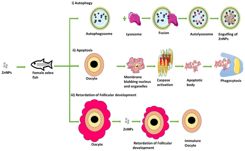
In a study, it has been proposed that ZnO nanoparticles have triggered autophagy and apoptosis in a caspase-dependent manner and induced oxidative stress by raising the level of the mutant ovarian p53 protein in maturing oocytes. Necroptosis, having the characteristics of both necrosis and apoptosis, have also been discovered where ZnO nanoparticles produced a necrotic environment that was conducive to retardation of follicular development, changed ovulation of oocyte, and decreased female zebrafish fertility [69] (Figure 5). In a pregnant mouse model, metal ions such as cadmium oxide nanoparticles are found to delay weight gain, decrease the weight of the placenta, and increase the weight of the uterus. Additionally, noticeable changes in the number of estrogen receptors α and β expression in uterine tissues finally led to a decrease in implantations. The release of cadmium ions from cadmium oxide nanoparticles has the potential to disrupt and unbalance the blastocyst before implantation as well as cause endocrine disturbance to prevent it [70,71].
Figure 5.
ZnO nanoparticles inducing oxidative stress by autophagy, apoptosis, and retardation of follicular development (Figure was drawn using the images and illustrations available on Smart Servier Medical Art).
The toxicity of the titanium oxide nanoparticles was found to be associated with Cyp17a1, a gene that regulates the secretion of the hormone, which was up-regulated, indicating enhanced estradiol production. Genes, including bmf, were up-regulated while some of the genes that control apoptosis were down-regulated. Changes in the ovary’s immunological and inflammatory responses, oxidoreductase activity, oxidative stress, transcription, cell proliferation, and ion transport were also noted [72]. The Zona Pellucida (ZP) of oocytes was found to be accumulated by cerium oxide nanoparticles in follicular cells through endocytosis. Follicular cell endocytosis and zona pellucida trapping could not able to shield mature oocytes from oxidative stress and DNA damage when exposed to high concentrations [73]. Mouse oocytes cultivated in media containing cerium oxide nanoparticles at a lower concentration (0.01 mg/L) during in vitro fertilization (IVF) had a considerably decreased fertilization rate compared to the control group. Low fertility rates could be caused by gamete genotoxicity and oxidative stress brought on by cerium oxide [38,74]. Silver nanoparticles have decreased the primary oocytes which resulted in the influence and inhibition of ovulation by entering the ovaries after entering the circulation and penetrating cells to cause oxidative stress, which activates oxidative stress factors in ovarian cells and causes apoptosis. Hence, it can be concluded that silver nanoparticles, at various concentrations, could cause oxidative stress by increasing degeneration in primary oocytes associated with lower antioxidant status [75].
Table 2.
Various nanoparticles are involved in the toxicity of the female reproductive system.
| S. No. | Nanoparticles | Animal Model | Toxicity Effect | References |
|---|---|---|---|---|
| 1. | ZnO nanoparticles | Female zebrafish | Autophagy and apoptosis occurring in a caspase-dependent manner; increased oxidative stress by inducing mutant ovarian p53 protein; necroptosis; follicular developmental retardation; deformation of oocyte ovulation, and decreased female zebrafish fertility. | [69] |
| 2. | Cadmium oxide nanoparticles | Pregnant mouse model | Weight gain, increased uterus weight, and decreased weight of placenta; decreased quantity of estrogen receptors. | [70] |
| 3. | Titanium oxide nanoparticles | Female mice | Up-regulation of Cyp17a1 resulted in enhanced estradiol production; up-regulation of bmf genes; apoptotic genes were down-regulated. | [72] |
| 4. | Cerium oxide nanoparticles | Mouse oocytes | Accumulation in the zona pellucida (ZP) of oocytes; DNA damage due to follicular cell endocytosis and zona pellucida trapping. | [73] |
| 5. | Silver nanoparticles | Ovaries of female albino rats | Inhibition of the ovulation; activation of oxidative stress factors in ovarian cells resulting in apoptosis. | [75] |
Nanotoxicity on the Steroidogenic Pathway
The potential harmful effects of nanoparticles such as induction of cellular states of oxidative stress, modulation of enzyme activity, inflammation, and cell death include disruption of gonadal steroidogenesis-related biochemical and physiologic processes [16]. Larson et al. [76] reported that gold nanoparticles could be a new class of ovarian endocrine-inhibiting compounds if exposed for extended periods or at high concentrations as they disrupt the production of sex-steroid hormones, resulting in reproductive problems in humans and animals [76]. By contrast, Gallus domesticus treated with silver nanoparticles showed no significant differences in progesterone levels between the granulosa and theca layers, but a progressive reduction in estradiol and testosterone levels was observed in theca cells. [77]. In preovulatory rat granulosa cells, multi-walled carbon nanotubes (MWCNTs) prevented the synthesis of progesterone. Particularly, at concentrations of 10 and 50 g/mL/48 h, the production was drastically reduced. Expression of Steroidogenic Acute Regulatory protein (StAR), a steroidogenic protein that helps in the movement of cholesterol from the outer to the inner mitochondrial membrane, where it is processed by P450scc to pregnenolone, was also altered by MWCNTs [78,79]. Other non-metal nanoparticles such as calcium phosphate nanoparticles have induced apoptosis and have no effect on S phase cell cycle arrest, progesterone level, estradiol levels, and the mRNA levels of P450scc, and StAR was observed in human ovarian granulosa cells cultured in-vitro [80]. Similarly, nano-silica particles caused fetotoxicity and placental dysfunction in foetus placenta of pregnant mice [81]. The decreased activity of the steroidogenic enzymes 3β and 17β-hydroxysteroid dehydrogenase affected gonadal steroidogenesis in fullerene-treated female Anabas testudineus. Serum cortisol levels significantly increased whereas estradiol levels in female fish significantly decreased. There was a significant difference in alkali-labile phosphate levels, plasma calcium, and total protein among female and male fish, possibly due to C60 fullerene antiestrogenic properties. In the ovaries and brain of female fish, aromatase enzyme activity considerably decreased [80].
6. Nanotoxicity Quantification Tests
The presence of nanotoxicity in the reproductive system can be identified by the collection of test samples and subjecting them to the following tests. These include tests for genotoxicity, proliferation, and mutated gene expression in cell culture as well as tests for cytotoxicity-altered metabolism, reduced growth, or lytic or apoptotic cell death [82].
6.1. Assay for the Determination of ROS Production Due to Oxidative Stress
Nanoparticles entering the cell may cause oxidative stress to the cell leading to the production of reactive oxidative species such as superoxide, α-oxygen, hydroxyl radical, peroxides, singlet oxygen, etc. The defense mechanisms of ROS production are of two types: enzymatic and non-enzymatic scavengers. Enzymatic scavengers include catalase, glutathione peroxidase, and superoxide dismutase, whereas non-enzymatic scavengers are glutathione, melatonin and vitamin A, C, and E [83]. These biomarkers act as indicators for biological, pathogenic processes, therapeutic, or pharmacological responses [84].
6.1.1. Superoxide Dismutase (SOD) Assay
Superoxide dismutases (SODs) are a crucial antioxidant defense system in the body that protects against oxidative damage. Reactive oxygen species-related disorders can be treated effectively with the enzyme. [85]. It is involved in the conversion of hydrogen peroxide and molecular oxygen from superoxide radicals [86]. SOD is the first line of defense mechanism against ROS in live cells by accelerating this conversion by redox disproportionation [87]. The assay measures the activity of superoxide dismutase in the mitochondria of the cell helping in the quantification of stress formed in the cell. In order to identify the presence of SOD by reaction with INT (2-(4-iodophenyl)-3-(4-nitrophenol)-5-phenyltetrazolium chloride) and formation of red formazan. SOD present in the sample prevents the conversion of superoxide radicals into oxygen. Further, the absorbance was observed at 505 nm. The SOD activity of the sample was estimated using the standard curve drawn using SOD enzyme as the standard solution [88].
6.1.2. Catalase Assay
Hydrogen peroxide (H2O2), a non-radical ROS, is the primary substrate for catalase, an essential enzyme. It is responsible for breaking down hydrogen peroxide and neutralizing it, as well as maintaining the required level of the molecules in the cell for cellular signalling [89]. The catalase activity can be determined by the conversion of cobalt (II) to cobalt (III) using H2O2 in the presence of bicarbonate solution. The formation of the complex carbonato-cobaltate (III) complex ([Co(CO3)3]Co) is used for the determination of catalase enzyme activity in the sample, where sample is added with 10 mM hydrogen peroxide and incubated at the temperature of 37 °C for 2 min. Then the mixture solution containing cobalt (II), sodium bicarbonate, Graham salt solution is added. This was then vortexed for 5 sec and then kept at room temperature for 10 min [90] the absorbance of the standard solution (mostly 10mM hydrogen peroxide and not added with sample) and sample are taken at 440 nm and compared [90].
6.1.3. Glutathione Peroxidase Assay
Glutathione peroxidase (GPx) is a cytosolic enzyme that is responsible for reducing hydrogen peroxide to water and oxygen as well as reducing peroxide radicals to alcohols and oxygen [91]. It is a qualitative reaction of tert-butyl and cumene hydroperoxides and glutathione transferase which helps in the evaluation of peroxidase activity. It is estimated by measuring the reduction of H2O2 by glutathione peroxidase to alcohol through NADPH loss. The gel containing glutathione is run with the sample, further incubated in 0.008% of cumene hydroperoxide for 10 min and then stained using 1% ferric chloride (FeCl3) and 1% potassium ferricyanide (C6N6FeK3). The formation of achromatic bands indicates the presence of glutathione peroxidase [86].
6.2. Other Methods
Various biological specimens (serum, urine, plasma, follicular/peritoneal/seminal fluid) can be used to assess oxidative stress, enabling accurate evaluation of redox status and the planning of therapeutic antioxidant supplements, if necessary [92]. Oxidative stress can be measured by the identification of the modification of protein, lipid, and DNA. The lipid modification can be identified by the byproduct of malondialdehyde, 4-hydroxy 2-nonenal, 4-oxonon-2-enal, and acrolein; for protein, dityrosine is used as biomarker, and for DNA, 8-hydroxy-2′-deoxyguanosine (8-oxodG) is used as biomarker, which is measured using ELISA [93]. Numerous techniques are used to quantify ROS in semen, including (i) chemiluminescence, (ii) cytochrome c reduction test, (iii) nitro blue tetrazolium test, and (iv) electron spin resonance. Various other methods include (i) fluorescent anisotropy which assesses membrane fluidity sperm motility defects, (ii) oxidation-reduction potential (ORP) measurement, which detects oxidative stress in seminal fluid, and (iii) Oxygen Radical Absorbance Capacity (ORAC) Assay, (iv) ALDETECT Assay [92]. Oxidative stress in the female reproductive system can be evaluated using common biomarkers like SOD, GPx, oxidative DNA adducts, conjugated dienes, thiobarbituric acid reactive substances, lipid peroxides, reverse transcription-polymerase chain reaction, enhanced chemiluminescence assay, immunocytochemical staining, etc [94].
7. Role of Antioxidants in Nanoparticle-Induced Stress
Any agent or compound that could prevent the oxidation of a suitable substrate even at low concentrations is referred to as an antioxidant. These are stable compounds that can give an undesired free radical species an electron, neutralize it, and reduce its destructive potential. Generally, the scavenging abilities of these antioxidants either prevent or delay cellular damage. These antioxidants’ low molecular weights make it simple for them to engage with ROS (i.e., free radicals) and stop their chain reaction before they damage important molecules involved in the normal functioning of the cells [95]. Some of the naturally occurring antioxidants mostly obtained from plants [96,97,98,99] have the ability to strengthen antioxidant defense mechanisms and prevent and reduce organism damage brought on by oxidative stress produced by nanoparticles. In particular, they play a key role in the prevention, treatment, and control of nanoparticle-induced toxicity and oxidative stress [100]. Vitamin C, sometimes referred to as ascorbic acid, is an antioxidant that can scavenge free radicals. The formation of ROS by the silver nanoparticles is completely reduced by the addition of ascorbic acid. It concurrently reduces DNA damage, apoptosis, and mitochondrial damage brought on by silver nanoparticles, lessening their harmful effects [15]. One naturally occurring flavonoid found in numerous plants and foods, quercetin, is an antioxidant with the capacity to scavenge free radicals. By promoting bad phosphorylation and translocation of Nrf2 through PI3-K/Akt-dependent pathways, quercetin has been demonstrated to lessen oxidative damage and inflammation brought on by Fe2O3 nanoparticles [101].
Lycopene has been shown to control the activity of redox-sensitive molecular targets, including the mitogen-activated protein kinases and Nrf2 activation [102]. In a study, rats receiving lycopene supplements had higher levels of expression of Nrf2, HO-1, glutathione, and antioxidant enzymes such as catalase, superoxide dismutase, and glutathione peroxidase, thus increasing antioxidant activity against oxidative stress [103]. Also, diterpenes from the leaves of Stevia rebaudiana reduce the proinflammatory cytokines production (TNF-α, IL-1β, and IL-6) by altering the I- κB/NF- κB pathway [104]. The ROS- mediated lethal toxicity of the ZnO nanoparticles and titanium oxide nanoparticles was found to be scavenged by curcumin and vitamin C in Caenorhabditis elegans (C. elegans) [105]. Some of the other findings have reported that the antioxidants such as vitamin E and anti-amyloid compounds glycyrrhizic acid significantly reduce the effects of nano-aluminum oxide-induced oxidative stress, graphene oxide nanoparticles-induced toxicity, and nano-silica-induced inhibition of serotonin neurotransmission in C. elegans [106,107,108].
Recently, antioxidants in nanoparticle form have been suggested as an original way to enhance their properties [23]. Several nanoparticles made of biologically derived compounds with antioxidant properties have been discovered where loading or tagging of bioactive compounds is possible [23,96,97]. An innovative and potent replacement could be phyto-antioxidant functionalized nanoparticles. In addition to serving their original purpose, they can offer oxidative damage protection [109]. Copper nanoparticles synthesized using Hibiscus rosa-sinensis showed great scavenging of H2O2 and Ferric-Reducing Antioxidant Power (FRAP), demonstrating a strong antioxidant activity [110]. Similarly, copper nanoparticles made from Dioscorea bulbifera tubers (DBTE) revealed scavenging activity against nitric oxide, superoxide radicals, and 1,1-diphenyl-2-picrylhydrazyl (DPPH). This demonstrated the importance of copper nanoparticles in the reduction of oxidative stress [111]. Silver nanoparticles made from Costus leaves had antioxidant activity comparable to that of ascorbic acid with an IC50 value of less than 50 mg/L, making them more potent DPPH scavengers than leaf extract of Costus [112]. Similarly, silver nanoparticles made from Cestrum nocturnum leaves tested for antioxidant activity were discovered to have effective DPPH scavengers with a percentage of 29.55% at the concentration of 100 μg/mL rather than ascorbic acid with a percentage of 24.28% at the same dosage [113]. Also, the recent development of ROS-responsive drug delivery systems such as sulfur-containing polymers, thioether-containing polymers, poly(thioketal), selenium-containing polymers, etc., act by breaking the chemical bonds and/or transitions from hydrophobic to hydrophilic phases, favoring the release of carrier medicines helping in the treatment of ROS [114].
8. Conclusions
The reproductive system is now being exposed to more nanoparticles due to the increased advancement in nanotechnology. Male and female reproductive systems have been reported to be negatively affected by a number of nanoparticles. The transmission of genetic and epigenetic information to subsequent generations is carried out by germ cells, which serve as a link between generations. It has been addressed how different nanoparticles can be hazardous to the reproductive system. Even though it is unavoidable and has been determined to have a slower effect on people, animals, and the environment, careful consideration of the effects and toxicity of nanoparticles is essential.
Acknowledgments
The authors thank MAHSA University, Malaysia and Sathyabama Institute of Science and Technology, India for providing support to complete this review.
Author Contributions
Conceptualization, A.V.S.; Methodology, A.V.S.; Writing—original draft, L.X.N.R.P.; Writing—review & editing, A.V.S. and L.X.N.R.P.; Visualization, A.V.S.; Supervision, A.V.S. All authors have read and agreed to the published version of the manuscript.
Institutional Review Board Statement
Not applicable.
Informed Consent Statement
Not applicable.
Data Availability Statement
The data used to support the findings of this study are included in the article. Should further data or information be required, these are available from the corresponding author upon request.
Conflicts of Interest
The authors declare that there are no conflict of interest regarding the publication of this paper.
Funding Statement
This research received no external funding.
Footnotes
Disclaimer/Publisher’s Note: The statements, opinions and data contained in all publications are solely those of the individual author(s) and contributor(s) and not of MDPI and/or the editor(s). MDPI and/or the editor(s) disclaim responsibility for any injury to people or property resulting from any ideas, methods, instructions or products referred to in the content.
References
- 1.Aziz Z.A.A., Mohd-Nasir H., Ahmad A., Setapar S.H.M., Peng W.L., Chuo S.C., Khatoon A., Umar K., Yaqoob A.A., Ibrahim M.N.M. Role of nanotechnology for design and development of cosmeceutical: Application in makeup and skin care. Front. Chem. 2019;7:739. doi: 10.3389/fchem.2019.00739. [DOI] [PMC free article] [PubMed] [Google Scholar]
- 2.Thiruvengadam M., Rajakumar G., Chung I.M. Nanotechnology: Current uses and future applications in the food industry. 3 Biotech. 2018;8:74. doi: 10.1007/s13205-018-1104-7. [DOI] [PMC free article] [PubMed] [Google Scholar]
- 3.Abiodun-Solanke I., Ajayi D., Arigbede A. Nanotechnology and its application in dentistry. Ann. Med. Health Sci. Res. 2014;4:171–177. doi: 10.4103/2141-9248.141951. [DOI] [PMC free article] [PubMed] [Google Scholar]
- 4.Mathew J., Joy J., George S.C. Potential applications of nanotechnology in transportation: A review. J. King Saud Univ. Sci. 2019;31:586–594. doi: 10.1016/j.jksus.2018.03.015. [DOI] [Google Scholar]
- 5.Samrot A.V., Angalene J.L.A., Roshini S.M., Stefi S.M., Preethi R., Raji P. Purification, characterization and exploitation of Azadirachta indica gum for the production of drug loaded nanocarrier. Mater. Res. Express. 2020;7:055007. doi: 10.1088/2053-1591/ab8b16. [DOI] [Google Scholar]
- 6.Samrot A.V., Angalene J.L.A., Roshini S.M., Stefi S.M., Preethi R., Raji P., Kumar A.M., Paulraj P., Kumar S.S. Purification, characterization and utilization of polysaccharide of Araucaria heterophylla gum for the synthesis of curcumin loaded nanocarrier. Int. J. Biol. Macromol. 2019;1:93–400. doi: 10.1016/j.ijbiomac.2019.08.121. [DOI] [PubMed] [Google Scholar]
- 7.Akçan R., Aydogan H.C., Yildirim M.Ş., Taştekin B., Sağlam N. Nanotoxicity: A challenge for future medicine. Turk. J. Med. Sci. 2020;50:1180–1196. doi: 10.3906/sag-1912-209. [DOI] [PMC free article] [PubMed] [Google Scholar]
- 8.Habas K., Demir E., Guo C., Brinkworth M.H., Anderson D. Toxicity mechanisms of nanoparticles in the male reproductive system. Drug Metab. Rev. 2014;53:604–617. doi: 10.1080/03602532.2021.1917597. [DOI] [PubMed] [Google Scholar]
- 9.Shalviri A., Cai P., Rauth A.M., Henderson J.T., Wu X.Y. Evaluation of new bi-functional terpolymeric nanoparticles for simultaneous in vivo optical imaging and chemotherapy of breast cancer. Drug Deliv. Transl. Res. 2012;2:437–453. doi: 10.1007/s13346-012-0103-1. [DOI] [PubMed] [Google Scholar]
- 10.Cypriyana P.J.J., Saigeetha S., Samrot A.V., Ponniah P., Chakravarthi S. Overview on toxicity of nanoparticles, it’s mechanism, models used in toxicity studies and disposal methods–A review. Biocatal. Agric. Biotechnol. 2021;36:102117. doi: 10.1016/j.bcab.2021.102117. [DOI] [Google Scholar]
- 11.Samrot A.V., Singh S.P.R., Deenadhayalan R., Rajesh V.V., Padmanaban S., Radhakrishnan K. Nanoparticles, a Double-Edged Sword with Oxidant as Well as Antioxidant Properties—A Review. Oxygen. 2022;2:591–604. doi: 10.3390/oxygen2040039. [DOI] [Google Scholar]
- 12.Ge X., Cao Z., Chu L. The Antioxidant Effect of the Metal and Metal-Oxide Nanoparticles. Antioxidants. 2022;11:791. doi: 10.3390/antiox11040791. [DOI] [PMC free article] [PubMed] [Google Scholar]
- 13.Valgimigli L., Baschieri A., Amorati R. Antioxidant activity of nanomaterials. J. Mater. Chem. B. 2018;6:2036–2051. doi: 10.1039/C8TB00107C. [DOI] [PubMed] [Google Scholar]
- 14.Horie M., Tabei Y. Role of oxidative stress in nanoparticles toxicity. Free Radic. Res. 2021;55:331–342. doi: 10.1080/10715762.2020.1859108. [DOI] [PubMed] [Google Scholar]
- 15.Khanna P., Ong C., Bay B.H., Baeg G.H. Nanotoxicity: An Interplay of Oxidative Stress, Inflammation and Cell Death. Nanomaterials. 2015;5:1163–1180. doi: 10.3390/nano5031163. [DOI] [PMC free article] [PubMed] [Google Scholar]
- 16.Yu Z., Li Q., Wang J., Yu Y., Wang Y., Zhou Q., Li P. Reactive Oxygen Species-Related Nanoparticle Toxicity in the Biomedical Field. Nanoscale Res. Lett. 2020;15:115. doi: 10.1186/s11671-020-03344-7. [DOI] [PMC free article] [PubMed] [Google Scholar]
- 17.Wang R., Song B., Wu J., Zhang Y., Chen A., Shao L. Potential adverse effects of nanoparticles on the reproductive system. Int. J. Nanomed. 2018;13:8487–8506. doi: 10.2147/IJN.S170723. [DOI] [PMC free article] [PubMed] [Google Scholar]
- 18.Working P.K. Male reproductive toxicology: Comparison of the human to animal models. Environ. Health Perspect. 1988;77:37–44. doi: 10.1289/ehp.887737. [DOI] [PMC free article] [PubMed] [Google Scholar]
- 19.Baskaran S., Finelli R., Agarwal A., Henkel R. Reactive oxygen species in male reproduction: A boon or a bane? Andrologia. 2021;53:e13577. doi: 10.1111/and.13577. [DOI] [PubMed] [Google Scholar]
- 20.Derakhshi M., Daemi S., Shahini P., Habibzadeh A., Mostafavi E., Ashkarran A.A. Two-dimensional nanomaterials beyond graphene for biomedical applications. J. Funct. Biomater. 2022;13:27. doi: 10.3390/jfb13010027. [DOI] [PMC free article] [PubMed] [Google Scholar]
- 21.Mihailovic V., Stankovic J.S.K., Selakovic D., Rosic G. An overview of the beneficial role of antioxidants in the treatment of nanoparticle-induced toxicities. Oxidative Med. Cell. Longev. 2021;2021:7244677. doi: 10.1155/2021/7244677. [DOI] [PMC free article] [PubMed] [Google Scholar]
- 22.Ganguly R., Singh A.K., Kumar R., Gupta A., Pandey A.K., Pandey A.K. Nanoparticles as modulators of oxidative stress. Nanotechnol. Mod. Anim. Biotechnol. 2019:29–35. doi: 10.1016/B978-0-12-818823-1.00003-X. [DOI] [Google Scholar]
- 23.Larson J.K., Carvan M.J., III, Hutz R.J. Engineered nanomaterials: An emerging class of novel endocrine disruptors. Biol. Reprod. 2014;91:20–21. doi: 10.1095/biolreprod.113.116244. [DOI] [PMC free article] [PubMed] [Google Scholar]
- 24.Manke A., Wang L., Rojanasakul Y. Mechanisms of nanoparticle-induced oxidative stress and toxicity. Biomed Res. Int. 2013;2013:942916. doi: 10.1155/2013/942916. [DOI] [PMC free article] [PubMed] [Google Scholar]
- 25.Sies H., editor. Oxidative Stress Oxidants and Antioxidants. Volume 15e22 Academic Press; London, UK: 1991. Oxidative Stress: Introduction. [Google Scholar]
- 26.Mohammadinejad R., Moosavi M.A., Tavakol S., Vardar D.Ö., Hosseini A., Rahmati M., Dini L., Hussain S., Mandegary A., Klionsky D.J. Necrotic, apoptotic and autophagic cell fates triggered by nanoparticles. Autophagy. 2019;15:4–33. doi: 10.1080/15548627.2018.1509171. [DOI] [PMC free article] [PubMed] [Google Scholar]
- 27.Mao J., Hu Y., Ruan L., Ji Y., Lou Z. Role of endoplasmic reticulum stress in depression (Review) Mol. Med. Rep. 2019;20:4774–4780. doi: 10.3892/mmr.2019.10789. [DOI] [PMC free article] [PubMed] [Google Scholar]
- 28.Bettigole S.E., Glimcher L.H. Endoplasmic reticulum stress in immunity. Annu. Rev. Immunol. 2015;33:107–138. doi: 10.1146/annurev-immunol-032414-112116. [DOI] [PubMed] [Google Scholar]
- 29.Carrara M., Prischi F., Nowak P.R., Kopp M.C., Ali M.M. Noncanonical binding of BiP ATPase domain to Ire1 and Perk is dissociated by unfolded protein CH1 to initiate ER stress signaling. Elife. 2015;4:e03522. doi: 10.7554/eLife.03522. [DOI] [PMC free article] [PubMed] [Google Scholar]
- 30.Čapek J., Roušar T. Detection of Oxidative Stress Induced by Nanomaterials in Cells-The Roles of Reactive Oxygen Species and Glutathione. Molecules. 2021;26:4710. doi: 10.3390/molecules26164710. [DOI] [PMC free article] [PubMed] [Google Scholar]
- 31.Huang Y., Wu C., Aronstam R. Toxicity of transition metal oxide nanoparticles: Recent insights from in vitro Studies. Materials. 2010;3:4842–4859. doi: 10.3390/ma3104842. [DOI] [PMC free article] [PubMed] [Google Scholar]
- 32.Brohi R.D., Wang L., Talpur H.S., Wu D., Khan F.A., Bhattarai D., Rehman Z.U., Farmanullah F., Huo L.J. Toxicity of nanoparticles on the reproductive system in animal models: A review. Front. Pharmacol. 2017;8:606. doi: 10.3389/fphar.2017.00606. [DOI] [PMC free article] [PubMed] [Google Scholar]
- 33.Marziyeh A., Keyhanfar F., Moosavi M.A., Shabani R., Mehdizadeh M., Varma R.S. Potential toxicity of nanoparticles on the reproductive system animal models: A review. J. Reprod. Immunol. 2021;148:103384. doi: 10.1016/j.jri.2021.103384. [DOI] [PubMed] [Google Scholar]
- 34.Dianová L., Tirpák F., Halo M., Slanina T., Massányi M., Stawarz R., Formicki G., Madeddu R., Massányi P. Effects of Selected Metal Nanoparticles (Ag, ZnO, TiO2) on the Structure and Function of Reproductive Organs. Toxics. 2022;10:459. doi: 10.3390/toxics10080459. [DOI] [PMC free article] [PubMed] [Google Scholar]
- 35.Mitrea D., Toader A., Hoteiuc O. Oxidative Stress Produced by Urban Atmospheric Nanoparticles. In: Clichici S., Filip A., do Nascimento G.M., editors. Nanomaterials—Toxicity, Human Health and Environment. IntechOpen; London, UK: 2019. [DOI] [Google Scholar]
- 36.Zhou Q., Yue Z., Li Q., Zhou R., Liu L. Exposure to PbSe nanoparticles and male reproductive damage in a rat model. Environ. Sci. Technol. 2019;53:13408–13416. doi: 10.1021/acs.est.9b03581. [DOI] [PubMed] [Google Scholar]
- 37.Jamnongjit M., Gill A., Hammes S.R. Epidermal growth factor receptor signaling is required for normal ovarian steroidogenesis and oocyte maturation. Proc. Natl. Acad. Sci. USA. 2005;102:16257–16262. doi: 10.1073/pnas.0508521102. [DOI] [PMC free article] [PubMed] [Google Scholar]
- 38.Hou C.C., Zhu J.Q. Nanoparticles and female reproductive system: How do nanoparticles affect oogenesis and embryonic development. Oncotarget. 2017;8:109799–109817. doi: 10.18632/oncotarget.19087. [DOI] [PMC free article] [PubMed] [Google Scholar]
- 39.Bisht S., Faiq M., Tolahunase M., Dada R. Oxidative stress and male infertility. Nat. Rev. Urol. 2017;14:470–485. doi: 10.1038/nrurol.2017.69. [DOI] [PubMed] [Google Scholar]
- 40.Iftikhar M., Noureen A., Uzair M., Jabeen F., Daim M.A., Cappello T. Perspectives of Nanoparticles in Male Infertility: Evidence for Induced Abnormalities in Sperm Production. Int. J. Environ. Res. Public Health. 2021;18:1758. doi: 10.3390/ijerph18041758. [DOI] [PMC free article] [PubMed] [Google Scholar]
- 41.Vassal M., Rebelo S., de Lourdes Pereira M. Metal Oxide Nanoparticles: Evidence of Adverse Effects on the Male Reproductive System. Int. J. Mol. Sci. 2021;22:8061. doi: 10.3390/ijms22158061. [DOI] [PMC free article] [PubMed] [Google Scholar]
- 42.Liu Q., Xu C., Ji G., Liu H., Mo Y., Tollerud D.J., Gu A., Zhang Q. Sublethal effects of ZnOnanoparticles on male reproductive cells. Toxicol. In Vitro. 2016;35:131–138. doi: 10.1016/j.tiv.2016.05.017. [DOI] [PubMed] [Google Scholar]
- 43.Mathias F.T., Romano R.M., Kizys M.M., Kasamatsu T., Giannocco G. Daily exposure to silver nanoparticles during prepubertal development decreases adult sperm and reproductive parameters. Nanotoxicology. 2015;9:64–70. doi: 10.3109/17435390.2014.889237. [DOI] [PubMed] [Google Scholar]
- 44.Azouz R.A., Korany R.M.S., Noshy A. Silica Nanoparticle–Induced Reproductive Toxicity in Male Albino Rats via Testicular Apoptosis and Oxidative Stress. Biol. Trace Elem. Res. 2022;201:1816–1824. doi: 10.1007/s12011-022-03280-w. [DOI] [PMC free article] [PubMed] [Google Scholar]
- 45.Bara N., Kaul G. Enhanced steroidogenic and altered antioxidant response by ZnO nanoparticles in mouse testis Leydig cells. Toxicol. Ind. Health. 2018;34:571–588. doi: 10.1177/0748233718774220. [DOI] [PubMed] [Google Scholar]
- 46.Takeda K., Suzuki K., Ishihara A., Kubo-Irie M., Fujimoto R., Tabata M., Oshio S., Nihei Y., Ihara T., Sugamata M. Nanoparticles transferred from pregnant mice to their offspring can damage the genital and cranial nerve systems. J. Health Sci. 2009;55:95–102. doi: 10.1248/jhs.55.95. [DOI] [Google Scholar]
- 47.Meena R., Kajal K., Paulraj R. Cytotoxic and genotoxic effects of titanium dioxide nanoparticles in testicular cells of male Wistar rat. Appl. Biochem. Biotechnol. 2015;175:825–840. doi: 10.1007/s12010-014-1299-y. [DOI] [PubMed] [Google Scholar]
- 48.Shahin N.N., Mohamed M.M. Nano-sized titanium dioxide toxicity in rat prostate and testis: Possible ameliorative effect of morin. Toxicol. Appl. Pharmacol. 2017;334:129–141. doi: 10.1016/j.taap.2017.08.014. [DOI] [PubMed] [Google Scholar]
- 49.Xiong Y., Hales D.B. The role of tumor necrosis factor-alpha in the regulation of mouse Leydig cell steroidogenesis. Endocrinology. 1993;132:2438–2444. doi: 10.1210/endo.132.6.8504748. [DOI] [PubMed] [Google Scholar]
- 50.Lauvås A.J., Skovmand A., Poulsen M.S., Kyjovska Z.O., Roursgaard M., Goericke-Pesch S., Vogel U., Hougaard K.S. Airway exposure to TiO2 nanoparticles and quartz and effects on sperm counts and testosterone levels in male mice. Reprod. Toxicol. 2019;90:134–140. doi: 10.1016/j.reprotox.2019.07.023. [DOI] [PubMed] [Google Scholar]
- 51.Ogunsuyi O.M., Ogunsuyi O.I., Akanni O., Alabi O.A., Alimba C.G., Adaramoye O.A., Cambier S., Eswara S., Gutleb A.C., Bakare A.A. Alteration of sperm parameters and reproductive hormones in Swiss mice via oxidative stress after co-exposure to titanium dioxide and ZnOnanoparticles. Andrologia. 2020;52:e13758. doi: 10.1111/and.13758. [DOI] [PubMed] [Google Scholar]
- 52.Miura N., Ohtani K., Hasegawa T., Yoshioka H., Hwang G.W. High sensitivity of testicular function to titanium nanoparticles. J. Toxicol. Sci. 2017;42:359–366. doi: 10.2131/jts.42.359. [DOI] [PubMed] [Google Scholar]
- 53.Lee W.Y., Park H.J. Toxicity of Cerium Oxide Nanoparticles on Neonatal Testicular Development in Mouse Organ Culture. Reprod. Toxicol. 2022;111:120–128. doi: 10.1016/j.reprotox.2022.05.014. [DOI] [PubMed] [Google Scholar]
- 54.Samrot A.V., Bhavya K.S., Sahithya C.S., Sowmya N. Evaluation of toxicity of chemically synthesised gold nanoparticles against Eudrilus eugeniae. J. Clust. Sci. 2018;29:1217–1225. doi: 10.1007/s10876-018-1440-0. [DOI] [Google Scholar]
- 55.Samrot A.V., Saipriya C., Agnes L.A., Roshini S.M., Cypriyana J., Saigeetha S., Raji P., Kumar S. Evaluation of nanotoxicity of Araucaria heterophylla gum derived green synthesized silver nanoparticles on Eudrilus eugeniae and Danio rerio. J. Clust. Sci. 2019;30:1017–1024. doi: 10.1007/s10876-019-01561-4. [DOI] [Google Scholar]
- 56.Samrot A.V., Ujjala B., Padmanaban S., Yamini P., Rabel A.M. A study on toxicity of chemically synthesised silver nanoparticle on Eudriluseugeniae. Toxicol. Environ. Health Sci. 2018;10:162–167. doi: 10.1007/s13530-018-0360-6. [DOI] [Google Scholar]
- 57.Shobana N., Prakash P., Samrot A.V., Saigeetha S., Sathiyasree M., Thirugnanasambandam R., Sridevi V., Kumar M.B., Shankar S.G., Dhiva S., et al. Nanotoxicity studies of Azadirachta indica mediated silver nanoparticles against Eudrilus eugeniae, Danio rerio and its embryos. Biocatal. Agric. Biotechnol. 2023;47:102561. doi: 10.1016/j.bcab.2022.102561. [DOI] [Google Scholar]
- 58.Samrot A.V., Justin C., Padmanaban S., Burman U. A study on the effect of chemically synthesized magnetite nanoparticles on earthworm: Eudrilus eugeniae. Appl. Nanosci. 2017;7:17–23. doi: 10.1007/s13204-016-0542-y. [DOI] [Google Scholar]
- 59.Mahfouz R., Sharma R., Thiyagarajan A., Kale V., Gupta S., Sabanegh E., Agarwal A. Semen characteristics and sperm DNA fragmentation in infertile men with low and high levels of seminal reactive oxygen species. Fertil. Steril. 2010;94:2141–2146. doi: 10.1016/j.fertnstert.2009.12.030. [DOI] [PubMed] [Google Scholar]
- 60.Olugbodi J.O., David O., Oketa E.N., Lawal B., Okoli B.J., Mtunzi F. Silver nanoparticles stimulates spermatogenesis impairments and hematological alterations in testis and epididymis of male rats. Molecules. 2020;25:1063. doi: 10.3390/molecules25051063. [DOI] [PMC free article] [PubMed] [Google Scholar]
- 61.Sundarraj K., Manickam V., Raghunath A., Periyasamy M., Viswanathan M., Perumal E. Repeated exposure to iron oxide nanoparticles causes testicular toxicity in mice. Environ. Toxicol. 2017;32:594–608. doi: 10.1002/tox.22262. [DOI] [PubMed] [Google Scholar]
- 62.Iyiola O., Olafimihan T.F., Sulaiman F.A., Anifowoshe A.T. Genotoxicity and histopathological assessment of silver nanoparticles in Swiss albino mice. Cuad. Investig. UNED. 2018;10:102–109. doi: 10.22458/urj.v10i1.2008. [DOI] [Google Scholar]
- 63.Thakur M., Gupta H., Singh D., Mohanty I.R., Maheswari U., Vanage G., Joshi D.S. Histopathological and ultra structural effects of nanoparticles on rat testis following 90 days (chronic study) of repeated oral administration. J. Nanobiotechnol. 2014;12:42. doi: 10.1186/s12951-014-0042-8. [DOI] [PMC free article] [PubMed] [Google Scholar]
- 64.Vaziri-Katehshori N., Noori A. The effect of multi-wall carbon nanotubes on sex hormone levels and ovarian tissue in female Wistar rats. J. Kashan Univ. Med. Sci. 2018;21:525–533. [Google Scholar]
- 65.Farshad O., Heidari R., Zamiri M.J., Retana-Márquez S., Khalili M., Ebrahimi M., Jamshidzadeh A., Ommati M.M. Spermatotoxic Effects of Single-Walled and Multi-Walled Carbon Nanotubes on Male Mice. Front. Vet. Sci. 2020;7:591558. doi: 10.3389/fvets.2020.591558. [DOI] [PMC free article] [PubMed] [Google Scholar]
- 66.Guo Z., Wang X., Zhang P., Sun F., Chen Z., Ma W., Meng F., Hao H., Shang X. Silica nanoparticles cause spermatogenesis dysfunction in mice via inducing cell cycle arrest and apoptosis. Ecotoxicol. Environ. Saf. 2022;231:113210. doi: 10.1016/j.ecoenv.2022.113210. [DOI] [PubMed] [Google Scholar]
- 67.Yang Q., Li F., Miao Y., Luo X., Dai S., Liu J., Niu W., Sun Y. CdSe/ZnS quantum dots induced spermatogenesis dysfunction via autophagy activation. J. Hazard. Mater. 2020;398:122327. doi: 10.1016/j.jhazmat.2020.122327. [DOI] [PubMed] [Google Scholar]
- 68.Hong F., Si W., Zhao X., Wang L., Zhou Y., Chen M., Ge Y., Zhang Q., Wang Y., Zhang J. TiO2 nanoparticle exposure decreases spermatogenesis via biochemical dysfunctions in the testis of male mice. J. Agric. Food Chem. 2015;63:7084–7092. doi: 10.1021/acs.jafc.5b02652. [DOI] [PubMed] [Google Scholar]
- 69.Mawed S.A., Marini C., Alagawany M., Farag M.R., Reda R.M., El-Saadony M.T., Elhady W.M., Magi G.E., Di Cerbo A., El-Nagar W.G. ZnONanoparticles (ZnO-NPs) Suppress Fertility by Activating Autophagy, Apoptosis, and Oxidative Stress in the Developing Oocytes of Female Zebrafish. Antioxidants. 2022;11:1567. doi: 10.3390/antiox11081567. [DOI] [PMC free article] [PubMed] [Google Scholar]
- 70.Ahmad A. Safety and Toxicity Implications of Multifunctional Drug Delivery Nanocarriers on Reproductive Systems In Vitro and In Vivo. Front. Toxicol. 2022;4:895667. doi: 10.3389/ftox.2022.895667. [DOI] [PMC free article] [PubMed] [Google Scholar]
- 71.Blum J.L., Xiong J.Q., Hoffman C., Zelikoff J.T. Cadmium Associated with Inhaled Cadmium Oxide Nanoparticles Impacts Fetal and Neonatal Development and Growth. Toxicol. Sci. 2012;126:478–486. doi: 10.1093/toxsci/kfs008. [DOI] [PMC free article] [PubMed] [Google Scholar]
- 72.Sun J., Zhang Q., Wang Z., Yan B. Effects of nanotoxicity on female reproductivity and fetal development in animal models. Int. J. Mol. Sci. 2013;14:9319–9337. doi: 10.3390/ijms14059319. [DOI] [PMC free article] [PubMed] [Google Scholar]
- 73.Courbiere B., Auffan M., Rollais R., Tassistro V., Bonnefoy A., Botta A., Rose J., Orsière T., Perrin J. Ultrastructural interactions and genotoxicity assay of cerium dioxide nanoparticles on mouse oocytes. Int. J. Mol. Sci. 2013;14:21613–21628. doi: 10.3390/ijms141121613. [DOI] [PMC free article] [PubMed] [Google Scholar]
- 74.Preaubert L., Courbiere B., Achard V., Tassistro V., Greco F., Orsiere T., Bottero J.-Y., Rose J., Auffan M., Perrin J. Cerium dioxide nanoparticles affect in vitro fertilization in mice. Nanotoxicology. 2016;10:111–117. doi: 10.3109/17435390.2015.1030792. [DOI] [PubMed] [Google Scholar]
- 75.Nabeh M., Taalab Y., El Wahab D.A., Asker S., Elbedwehy A., El Harouny M. Silver Nanotoxicity on Kidneys and Ovaries of Female Albino Rats. Mansoura J. Forensic Med. Clin. Toxicol. 2020;28:340–358. doi: 10.21608/mjfmct.2020.22528.1011. [DOI] [Google Scholar]
- 76.Larson J.K., Carvan M., 3rd, Teeguarden J.G., Watanabe G., Taya K., Krystofiak E., Hutz R.J. Low-dose gold nanoparticles exert subtle endocrine-modulating effects on the ovarian steroidogenic pathway ex vivo independent of oxidative stress. Nanotoxicology. 2014;8:856–866. doi: 10.3109/17435390.2013.837208. [DOI] [PMC free article] [PubMed] [Google Scholar]
- 77.Katarzyńska-Banasik D., Grzesiak M., Kowalik K., Sechman A. Administration of silver nanoparticles affects ovarian steroidogenesis and may influence thyroid hormone metabolism in hens (Gallus domesticus) Ecotoxicol. Environ. Saf. 2021;208:111427. doi: 10.1016/j.ecoenv.2020.111427. [DOI] [PubMed] [Google Scholar]
- 78.Qu Y., Yang B., Jiang X., Ma X., Lu C., Chen C. Multiwalled carbon nanotubes inhibit steroidogenesis by disrupting steroidogenic acute regulatory protein expression and redox status. J. Nanosci. Nanotechnol. 2017;17:914–925. doi: 10.1166/jnn.2017.12647. [DOI] [PubMed] [Google Scholar]
- 79.Holmannova D., Borsky P., Svadlakova T., Borska L., Fiala Z. Reproductive and Developmental Nanotoxicity of Carbon Nanoparticles. Nanomaterials. 2022;12:1716. doi: 10.3390/nano12101716. [DOI] [PMC free article] [PubMed] [Google Scholar]
- 80.Yamashita K., Yoshioka Y., Higashisaka K., Mimura K., Morishita Y., Nozaki M., Yoshida T., Ogura T., Nabeshi H., Nagano K., et al. Silica and titanium dioxide nanoparticles cause pregnancy complications in mice. Nat. Nanotechnol. 2011;6:321–328. doi: 10.1038/nnano.2011.41. [DOI] [PubMed] [Google Scholar]
- 81.Derksen R.H., Khamashta M.A., Branch D.W. Management of the obstetric antiphospholipid syndrome. Arthritis Rheum. 2004;50:1028–1039. doi: 10.1002/art.20105. [DOI] [PubMed] [Google Scholar]
- 82.Hillegass J.M., Shukla A., Lathrop S.A., MacPherson M.B., Fukagawa N.K., Mossman B.T. Assessing nanotoxicity in cells in vitro. Wiley Interdiscip. Rev. Nanomed. Nanobiotechnol. 2010;2:219–231. doi: 10.1002/wnan.54. [DOI] [PMC free article] [PubMed] [Google Scholar]
- 83.Moussa Z., Judeh Z.M., Ahmed S.A. Nonenzymatic exogenous and endogenous antioxidants. Free. Radic. Med. Biol. 2019;1:11–22. [Google Scholar]
- 84.Puntmann V.O. How-to guide on biomarkers: Biomarker definitions, validation and applications with examples from cardiovascular disease. Postgrad. Med. J. 2009;85:538–545. doi: 10.1136/pgmj.2008.073759. [DOI] [PubMed] [Google Scholar]
- 85.Younus H. Therapeutic potentials of superoxide dismutase. Int. J. Health Sci. 2018;12:88–93. [PMC free article] [PubMed] [Google Scholar]
- 86.Weydert C.J., Cullen J.J. Measurement of superoxide dismutase, catalase and glutathione peroxidase in cultured cells and tissue. Nat. Protoc. 2010;5:51–66. doi: 10.1038/nprot.2009.197. [DOI] [PMC free article] [PubMed] [Google Scholar]
- 87.Fukai T., Ushio-Fukai M. Superoxide dismutases: Role in redox signaling, vascular function, and diseases. Antioxid. Redox Signal. 2011;15:1583–1606. doi: 10.1089/ars.2011.3999. [DOI] [PMC free article] [PubMed] [Google Scholar]
- 88.Assady M., Farahnak A., Golestani A., Esharghian M. Superoxide Dismutase (SOD) Enzyme Activity Assay in Fasciola sp Parasites and Liver Tissue Extract. Iran. J. Parasitol. 2011;6:17–22. [PMC free article] [PubMed] [Google Scholar]
- 89.Nandi A., Yan L.J., Jana C.K., Das N. Role of catalase in oxidative stress-and age-associated degenerative diseases. Oxidative Med. Cell. Longev. 2019;2019:9613090. doi: 10.1155/2019/9613090. [DOI] [PMC free article] [PubMed] [Google Scholar]
- 90.Hadwan M.H. Simple spectrophotometric assay for measuring catalase activity in biological tissues. BMC Biochem. 2018;19:7. doi: 10.1186/s12858-018-0097-5. [DOI] [PMC free article] [PubMed] [Google Scholar]
- 91.Fanucchi M.V. The Lung. Academic Press; Cambridge, MA, USA: 2004. Development of antioxidant and xenobiotic metabolizing enzyme systems; pp. 177–185. [Google Scholar]
- 92.Mannucci A., Argento F.R., Fini E., Coccia M.E., Taddei N., Becatti M., Fiorillo C. The impact of oxidative stress in male infertility. Front. Mol. Biosci. 2022;8:1344. doi: 10.3389/fmolb.2021.799294. [DOI] [PMC free article] [PubMed] [Google Scholar]
- 93.Il’yasova D., Scarbrough P., Spasojevic I. Urinary biomarkers of oxidative status. Clin. Chim. Acta. 2012;413:1446–1453. doi: 10.1016/j.cca.2012.06.012. [DOI] [PMC free article] [PubMed] [Google Scholar]
- 94.Agarwal A., Gupta S., Sharma R.K. Role of oxidative stress in female reproduction. Reprod. Biol. Endocrinol. 2005;3:28. doi: 10.1186/1477-7827-3-28. [DOI] [PMC free article] [PubMed] [Google Scholar]
- 95.Kumar H., Bhardwaj K., Nepovimova E., Kuča K., Dhanjal D.S., Bhardwaj S., Bhatia S.K., Verma R., Kumar D. Antioxidant Functionalized Nanoparticles: A Combat against Oxidative Stress. Nanomaterials. 2020;10:1334. doi: 10.3390/nano10071334. [DOI] [PMC free article] [PubMed] [Google Scholar]
- 96.Samrot A.V., Shobana N., Jenna R. Antibacterial and Antioxidant Activity of Different Staged Ripened Fruit of Capsicum annuum and Its Green Synthesized Silver Nanoparticles. Bionanoscience. 2018;8:632–646. doi: 10.1007/s12668-018-0521-8. [DOI] [Google Scholar]
- 97.Purayil S.K., Annley C., Ponnaiah P., Pattammadath S., Javad P.T.M., Selvarani J., Raji P., Thirumurugan R., Iyappan P., Samrot A.V. Evaluation of Antioxidant and Antimicrobial Activity of Some Plants Collected from Malaysia. J. Pure Appl. Microbiol. 2019;13:2363–2374. doi: 10.22207/JPAM.13.4.52. [DOI] [Google Scholar]
- 98.Samrot A.V., Sean T.C. Investigating the Antioxidant and Antimicrobial Activity of Artocarpus heterophyllus Lam. (Jackfruit) Latex. Biointerface Res. Appl. Chem. 2022;12:3019–3033. [Google Scholar]
- 99.Selvarani A.J., Nishanthini P., Raji P., Samanvitha K.S., Paulraj P., Iyappan P., Chandramohan M., Samrot A.V. Antioxidant and Quorum Quenching Activity against Pseudomonas aeruginosa SU-18 of some Edible Fruit Juices. J. Pure Appl. Microbiol. 2019;13:1863–1876. [Google Scholar]
- 100.Ramana K.V., Reddy A., Majeti N.V., Singhal S.S. Therapeutic potential of natural antioxidants. Oxidative Med. Cell. Longev. 2018;2018:9471051. doi: 10.1155/2018/9471051. [DOI] [PMC free article] [PubMed] [Google Scholar]
- 101.Guo D., Zhu L., Huang Z., Zhou H., Ge Y., Ma W., Wu J., Zhang X., Zhou X., Zhang Y., et al. Anti-leukemia activity of PVP-coated silver nanoparticles via generation of reactive oxygen species and release of silver ions. Biomaterials. 2013;34:7884–7894. doi: 10.1016/j.biomaterials.2013.07.015. [DOI] [PubMed] [Google Scholar]
- 102.Lee M.T., Lin W.C., Yu B., Lee T.T. Antioxidant capacity of phytochemicals and their potential effects on oxidative status in animals—A review. Asian-Australas. J. Anim. Sci. 2017;30:299–308. doi: 10.5713/ajas.16.0438. [DOI] [PMC free article] [PubMed] [Google Scholar]
- 103.Linnewiel K., Ernst H., Caris-Veyrat C., Ben-Dor A., Kampf A., Salman H., Danilenko M., Levy J., Sharoni Y. Structure activity relationship of carotenoid derivatives in activation of the electrophile/antioxidant response element transcription system. Free Radic. Biol Med. 2009;47:659–667. doi: 10.1016/j.freeradbiomed.2009.06.008. [DOI] [PubMed] [Google Scholar]
- 104.Momtazi-Borojeni A.A., Esmaeili S.-A., Abdollahi E., Sahebkar A. A review on the pharmacology and toxicology of steviol glycosides extracted from stevia rebaudiana. Curr. Pharm. Des. 2017;23:1616–1622. doi: 10.2174/1381612822666161021142835. [DOI] [PubMed] [Google Scholar]
- 105.Sonane M., Moin N., Satish A. The role of antioxidants in attenuation of Caenorhabditis elegans lethality on exposure to TiO2 and ZnO nanoparticles. Chemosphere. 2017;187:240–247. doi: 10.1016/j.chemosphere.2017.08.080. [DOI] [PubMed] [Google Scholar]
- 106.Zhao Y., Jia R., Qiao Y., Wang D. Glycyrrhizic acid, active component from glycyrrhizae radix, prevents toxicity of graphene oxide by influencing functions of microRNAs in nematode Caenorhabditis elegans. Nanomedicine. 2016;12:735–744. doi: 10.1016/j.nano.2015.10.008. [DOI] [PubMed] [Google Scholar]
- 107.Xiaoming Y., Xiangmin G., Qiuli W., Yunli Z., Dayong W. Vitamin E ameliorates neurodegeneration related phenotypes caused by neurotoxicity of Al2O3-nanoparticles in C. elegans. Toxicol. Res. 2015;4:1269–1281. doi: 10.1039/c5tx00029g. [DOI] [Google Scholar]
- 108.Scharf A., Gührs K.H., von Mikecz A. Anti-amyloid compounds protect from silica nanoparticle-induced neurotoxicity in the nematode C. elegans. Nanotoxicology. 2016;10:426–435. doi: 10.3109/17435390.2015.1073399. [DOI] [PMC free article] [PubMed] [Google Scholar]
- 109.Balkrishna A., Kumar A., Arya V., Rohela A., Verma R., Nepovimova E., Krejcar O., Kumar D., Thakur N., Kuca K. Phytoantioxidant Functionalized Nanoparticles: A Green Approach to Combat Nanoparticle-Induced Oxidative Stress. Oxidative Med. Cell. Longev. 2021;2021:3155962. doi: 10.1155/2021/3155962. [DOI] [PMC free article] [PubMed] [Google Scholar]
- 110.Subbaiya R., Selvam M.M. Green synthesis of copper nanoparticles from Hibicus rosasinensis and their antimicrobial, antioxidant activities. Res. J. Pharm. Biol. Chem. Sci. 2015;6:1183–1190. [Google Scholar]
- 111.Ghosh S., More P., Nitnavare R., Jagta S., Chippalka R., Derl A., Kittue R., Aso A., Kale S., Singh S., et al. Antidiabetic and antioxidant properties of copper nanoparticles synthesized by medicinal plant Dioscorea bulbifera. J. Nanomed. Nanotechnol. 2015;6 doi: 10.4172/2157-7439.S6-007. [DOI] [Google Scholar]
- 112.Elemike E.E., Fayemi O.E., Ekennia A.C., Onwudiwe D.C., Ebenso E.E. Silver nanoparticles mediated by Costus afer leaf Extract: Synthesis, Antibacterial, Antioxidant and electrochemical properties. Molecules. 2017;22:701. doi: 10.3390/molecules22050701. [DOI] [PMC free article] [PubMed] [Google Scholar]
- 113.Keshari A.K., Srivastava R., Singh S., Yadav V.B., Nath G. Antioxidant and antibacterial activity of silver nanoparticles synthesized by _Cestrum nocturnum. J. Ayurveda Integr. Med. 2020;11:37–44. doi: 10.1016/j.jaim.2017.11.003. [DOI] [PMC free article] [PubMed] [Google Scholar]
- 114.Gao F., Xiong Z. Reactive oxygen species responsive polymers for drug delivery systems. Front. Chem. 2021;9:649048. doi: 10.3389/fchem.2021.649048. [DOI] [PMC free article] [PubMed] [Google Scholar]
Associated Data
This section collects any data citations, data availability statements, or supplementary materials included in this article.
Data Availability Statement
The data used to support the findings of this study are included in the article. Should further data or information be required, these are available from the corresponding author upon request.


