Abstract
Dysfunction of the blood-retinal barrier (BRB) contributes to the pathophysiology of several vascular eye diseases, often resulting in retinal edema and subsequent vision loss. The inner blood-retinal barrier (iBRB) is mainly composed of retinal vascular endothelium with low permeability under physiological conditions. This feature of low permeability is tightly regulated and maintained by low rates of paracellular transport between adjacent retinal microvascular endothelial cells, as well as transcellular transport (transcytosis) through them. The assessment of retinal transcellular barrier permeability may provide fundamental insights into iBRB integrity in health and disease. In this study, we describe an endothelial cell (EC) transcytosis assay, as an in vitro model for evaluating iBRB permeability, using human retinal microvascular endothelial cells (HRMECs). This assay assesses the ability of HRMECs to transport transferrin and horseradish peroxidase (HRP) in receptor- and caveolae-mediated transcellular transport processes, respectively. Fully confluent HRMECs cultured on porous membrane were incubated with fluorescent-tagged transferrin (clathrin-dependent transcytosis) or HRP (caveolae-mediated transcytosis) to measure the levels of transferrin or HRP transferred to the bottom chamber, indicative of transcytosis levels across the EC monolayer. Wnt signaling, a known pathway regulating iBRB, was modulated to demonstrate the caveolae-mediated HRP-based transcytosis assay method. The EC transcytosis assay described here may provide a useful tool for investigating the molecular regulators of EC permeability and iBRB integrity in vascular pathologies and for screening drug delivery systems.
Introduction
The human retina is one of the highest energy-demanding tissues in the body. Proper functioning of the neural retina requires an efficient supply of oxygen and nutrients along with a restricted flux of other potentially harmful molecules to protect the retinal environment, which is mediated via the blood-retinal barrier (BRB)1. Similar to the blood-brain barrier (BBB) in the central nervous system, the BRB acts as a selective barrier in the eye, regulating the movement of ions, water, amino acids, and sugar in and out of the retina. BRB also maintains retinal homeostasis and its immune privilege by preventing exposure to circulatory factors such as immune cells, antibodies, and harmful pathogens2. BRB dysfunction contributes to the pathophysiology of several vascular eye diseases, such as diabetic retinopathy, age-related macular degeneration (AMD), retinopathy of prematurity (ROP), retinal vein occlusion, and uveitis, resulting in vasogenic edema and subsequent vision loss3,4,5.
The BRB consists of two separate barriers for two distinct ocular vascular networks, respectively: the retinal vasculature and the fenestrated choriocapillaris beneath the retina. The inner BRB (iBRB) is primarily composed of retinal microvascular endothelial cells (RMECs) lining the retinal microvasculature, which nourishes the inner retinal neuronal layers. On the other hand, the retinal pigment epithelium forms the major component of the outer BRB, which lies between the neurosensory retina and choriocapillaris2. For the iBRB, molecular transport across RMECs takes place through both paracellular and transcellular routes (Figure 1). The high degree of substance selectivity across the iBRB relies upon (i) the presence of junctional protein complexes that restrict paracellular transport between adjacent endothelial cells (ECs), and (ii) low expression levels of caveolae mediators, transporters, and receptors within the endothelial cells that maintain low rates of transcellular transport1,6,7,8. Junctional complexes regulating paracellular flux are composed of tight junctions (claudins, occludins), adherens junctions (VE cadherins), and gap junctions (connexins), allowing the passage of water and small water-soluble compounds. While small lipophilic molecules passively diffuse across the interior of RMECs, the movement of larger lipophilic and hydrophilic molecules is regulated by ATP-driven trans-endothelial pathways including vesicular transport and membrane transporters5,9.
Figure 1: Different routes of transport across retinal vascular endothelium.
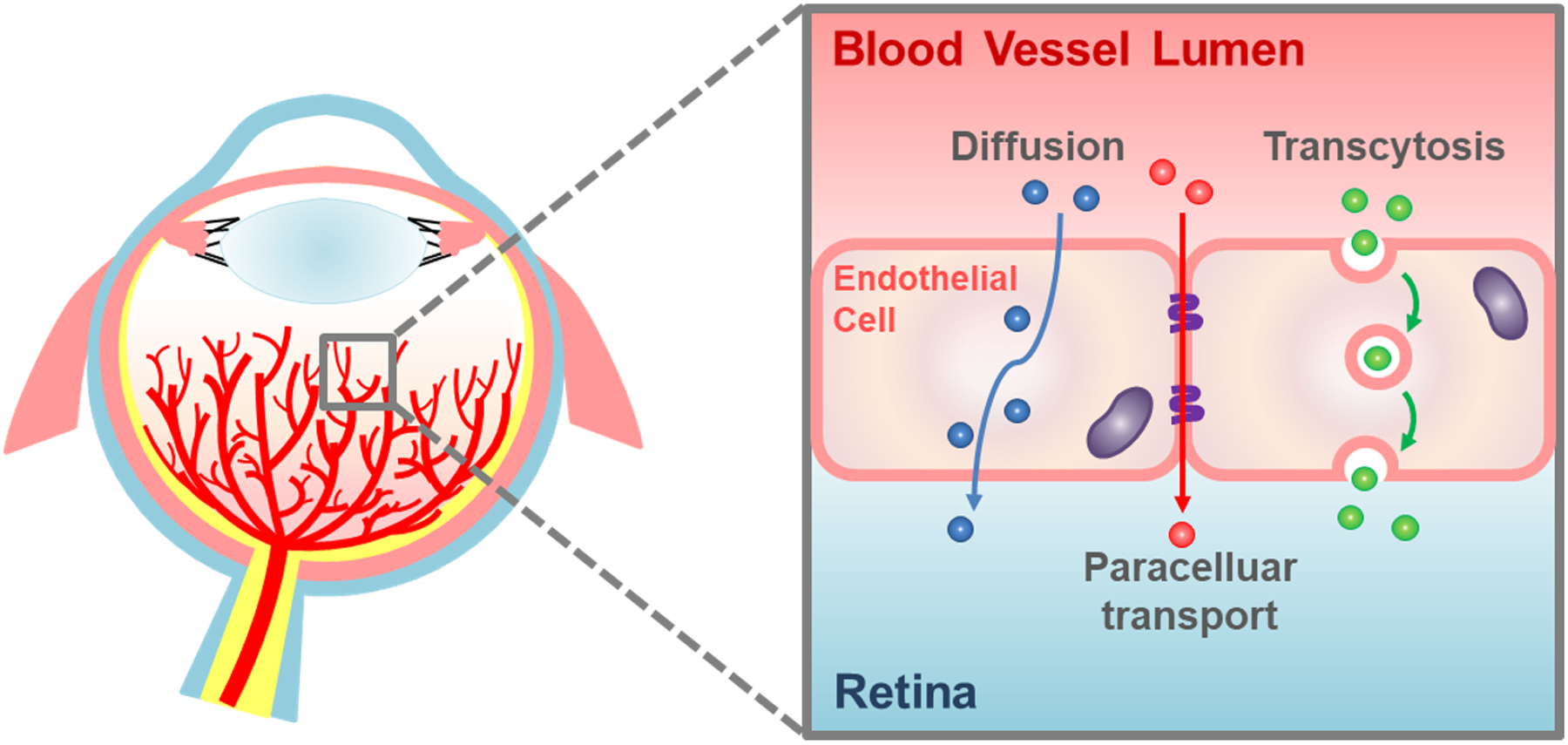
Schematic illustration showing different routes of molecular flux across retinal microvascular endothelial cells (RMECs). Transport across retinal vascular endothelial cells within the inner blood-retinal barrier takes place via two major routes: paracellular and transcellular pathways including transcytosis. This figure was adapted with permission from Yemanyi et al.5.
Vesicular transcytosis may be categorized as caveolin-mediated caveolar transcytosis, clathrin-dependent (and receptor-mediated) transcytosis, and clathrin-independent macropinocytosis (Figure 2). These vesicular transport processes involve different-sized vesicles, with macropinosomes being the largest (ranging from 200–500 nm) and caveolae being the smallest (averaging 50–100 nm), while clathrin-coated vesicles range from 70–150 nm10. Caveolae are flask-shaped lipid-rich plasma membrane invaginations with a protein coat, primarily composed of caveolin-1 that binds lipid membrane cholesterol and other structural and signaling proteins via their caveolin-scaffolding domain11. Caveolins work together with peripherally attached cavin to promote caveolae stabilization at the plasma membrane12. Caveolar membranes also may carry receptors for other molecules such as insulin, albumin, and circulating lipoproteins including high-density lipoprotein (HDL) and low-density lipoprotein (LDL) to assist their movement across endothelial cells13. During development, the formation of functional BRB depends on the suppression of EC transcytosis8. Mature retinal endothelium, hence, has relatively low levels of caveolae, caveolin-1, and albumin receptors with respect to other endothelial cells under physiological conditions, contributing to its barrier properties4,9.
Figure 2: Overview of transcytotic vesicular transport pathways across endothelial cells and their respective in vitro assessment.
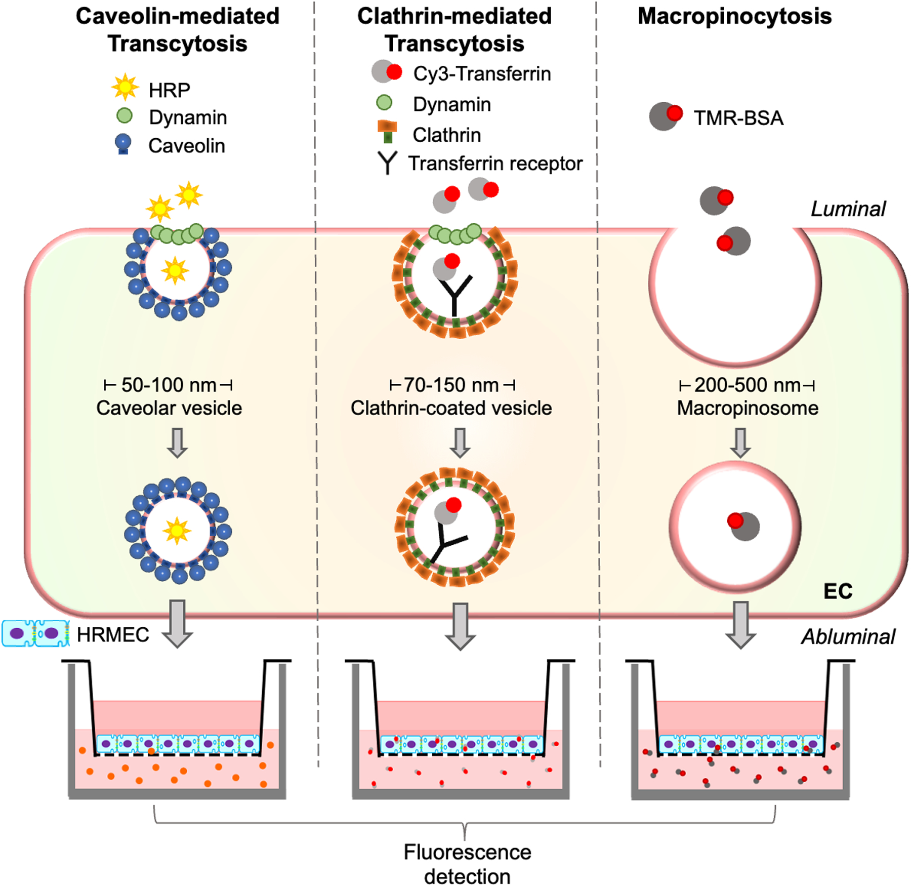
In endothelial cells (ECs), transcellular translocation of macromolecules occurs through three main types of vesicles: caveolar vesicles (50–100 nm), clathrin-coated vesicles (70–150 nm), or clathrin-independent macropinosomes (200–500 nm). Caveolae are flask-shaped, spherical, lipid-rich microdomains in the plasma membrane, composed of caveolin and cavins. Levels of transcytosis through these vesicles can be determined by fluorescence-based in vitro assays in EC culture using horseradish peroxidase (HRP) combined with fluorescent substrate, fluorescent-tagged transferrin (Cy3-Tf), or tetramethylrhodamine-tagged bovine serum albumin (TMR-BSA) for caveolae-mediated transcytosis, clathrin-mediated transcytosis, and macropinocytosis, respectively, to evaluate inner blood-retinal barrier (iBRB) permeability. While each pathway has distinct features and transport mechanisms, overlapping function and substance transport can occur, particularly between caveolar transport and macropinocytosis, both being clathrin-independent.
Because iBRB breakdown is a major hallmark of many pathological eye conditions, it is essential to develop methods to assess retinal vascular permeability in vivo and in vitro. These methods help provide probable insights into the mechanisms of compromised BRB integrity and assess the efficacy of potential therapeutic targets. Current in vivo imaging or quantitative vascular leakage assays typically employ fluorescent (sodium fluorescein and dextran), colorimetric (Evans Blue dye and horseradish peroxidase [HRP] substrate), or radioactive tracers14 to detect extravasation from the vasculature into surrounding retinal tissues with microscope imaging or in isolated tissue lysate. An ideal tracer for quantifying vascular integrity should be inert and large enough to freely permeate compromised vessels while confined within healthy and intact capillaries. Methods employing sodium fluorescein or fluorescein isothiocyanate-conjugated dextran (FITC-dextran) in live fundus fluorescein angiography (FFA) or isolated retinal flat mounts are widely used for quantitating retinal extravasation in vivo or ex vivo. FITC-dextran has the advantage of being available in different molecular weights ranging from 4–70 kDa for size-selective studies15,16,17. FITC-albumin (~68 kDa) is an alternative large-sized protein tracer of biological relevance for vascular leakage studies18. Evans Blue dye, injected intracardially19, retro-orbitally, or through the tail vein20, also relies on its binding with endogenous albumin to form a large molecule that can be quantified by mostly spectrophotometric detection or, less commonly, fluorescence microscopy in flat mounts20,21.
These quantitative or light imaging methodologies, however, often do not distinguish paracellular transport from trans-endothelial transport. For the specific analysis of transcytosis with ultrastructural visualization of transcytosed vesicles, tracer molecules such as HRP are typically used to locate transcytosed vesicles within endothelial cells that can be observed under an electron microscope22,23,24 (Figure 3A–C).
Figure 3: Visualization of EC transcytosis in the retina.
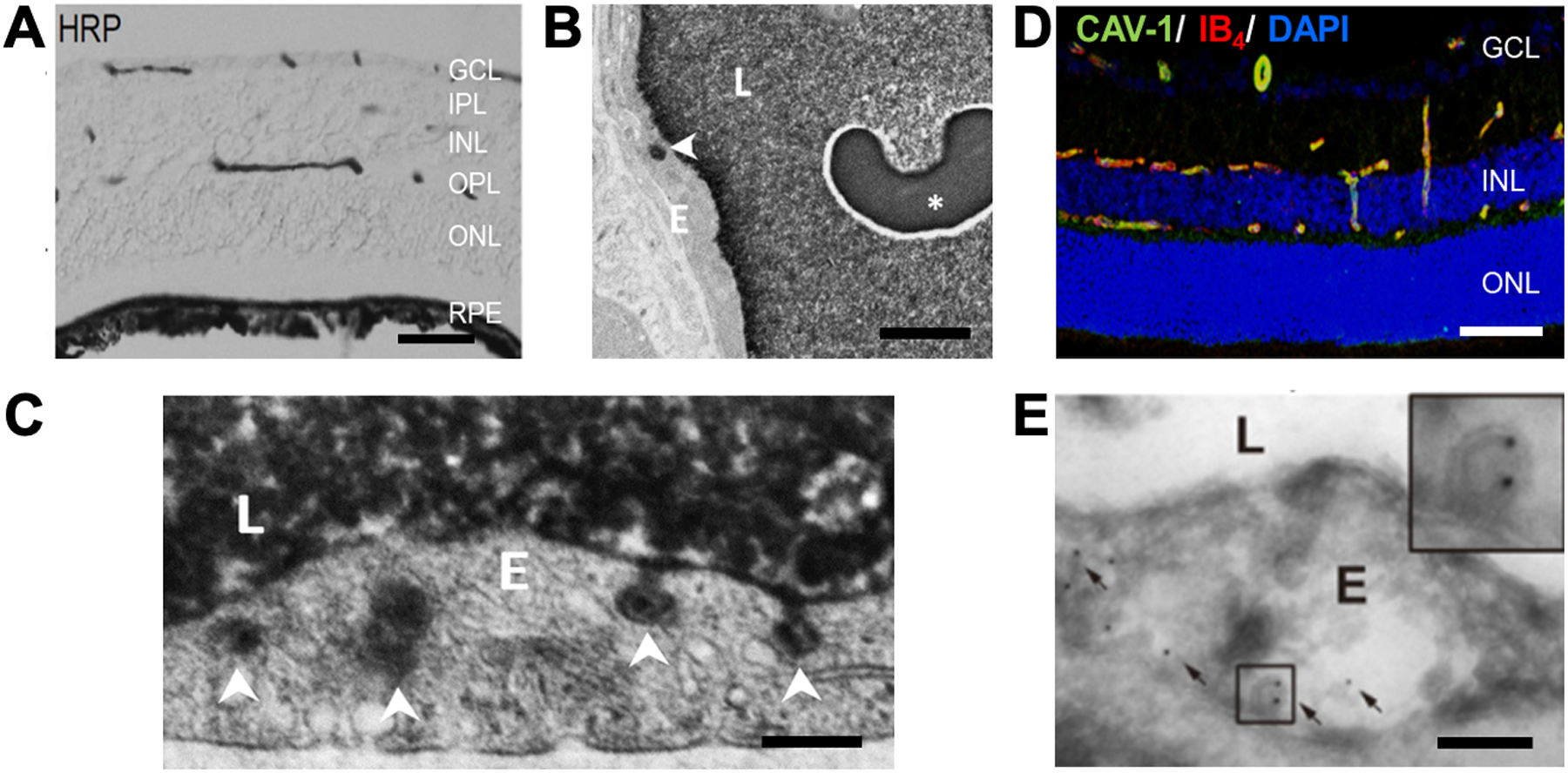
(A) Light microscope image shows an HRP-filled blood vessel lumen from a 3-month-old wild type (WT) mouse retinal section, stained with 3,3’-diamino benzidine (DAB) (black). HRP was retro-orbitally injected in WT mice, followed by eye isolation and tissue embedding. Thin sections were stained with electron-dense DAB substrate for the colorimetric detection of HRP as dark brown precipitate and imaged under a light microscope to reveal HRP within the retinal blood vessel lumen. (B,C) Samples were then further processed with transmission electron microscope (TEM) ultrathin sectioning to visualize HRP-containing transcytotic vesicles, depicting EC transcytosis across the iBRB. TEM images show a retinal section of a 3-month-old WT mouse with HRP-filled blood vessel lumen and HRP-containing vesicles within RMECs. (B) An occasional large vesicle potentially reflects macropinosome (arrowhead) within ECs, with the presence of red blood cells (asterisk) on the luminal side. (C) Small vesicles are likely caveolar vesicles (arrowheads). (D) Immunohistochemistry co-staining of CAV-1 antibody (green) and isolectin B4 (IB4, red, EC marker) demonstrates localization of CAV-1 in retinal blood vessels in 3-month-old WT mouse retina (DAPI, blue). (E) TEM image of immunogold-labeled CAV-1 (black dots, arrows) within the retinal ECs; inset shows a magnified image of a CAV-1 positive caveolar vesicle. Abbreviations: HRP = horseradish peroxidase; GCL = ganglion cell layer; IPL = inner plexiform layer; INL = inner nuclear layer; OPL = outer plexiform layer; ONL = outer nuclear layer; RPE = retinal pigment epithelium; E = endothelial cell; and L = blood vessel lumen. Magnification: (A, D) 20x. Scale bars: (A) 50 μm, (B) 2 μm, (D) 100 μm, and (C,E) 200 nm. Panel (E) was adapted with permission from Wang et al.24.
The development and use of in vitro iBRB models to evaluate the endothelial cell permeability could provide robust and high throughput assessment to complement in vivo experiments and aid the investigation of molecular regulators of vascular leakage. Commonly used assays to assess the paracellular transport and integrity of tight junctions include trans-endothelial electrical resistance (TEER), a measure of ionic conductance (Figure 4)2,25, and in vitro vascular leakage assay using small molecular weight fluorescent tracers26. In addition, transferrin-based transcytosis assays modeling BBB have been utilized to explore clathrin-dependent transcytosis27. Despite this, assays to evaluate BRB and, more specifically, retinal EC caveolar transcytosis in vitro are limited.
Figure 4: Validation of fully confluent HRMECs and increased vascular permeability in HRMECs after VEGF treatment.
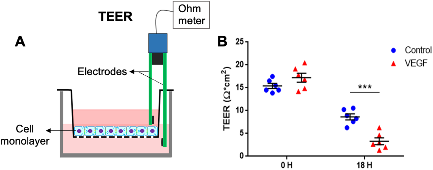
(A) Schematic diagram showing the positioning of electrodes in the apical and basolateral chambers of the wells for trans-endothelial electrical resistance (TEER) measurement as an assessment of vascular permeability and integrity of the cell monolayer. (B) Graph showing TEER recordings of fully confluent HRMECs with and without prior treatment with vascular endothelial growth factor (VEGF). Treatment with VEGF after 18 h (18 H) results in reduced TEER in fully confluent HRMECs31. *** p≤0.001. Panel (B) was adapted with permission from Tomita et al.31.
In this study, we describe an EC transcytosis assay using human retinal microvascular endothelial cells (HRMECs) as an in vitro model to determine iBRB permeability and EC transcytosis. This assay relies on the ability of HRMECs to transport transferrin or HRP via the receptor-mediated or caveolae-dependent transcytosis pathways, respectively (Figure 2). HRMECs cultured to full confluency in the apical chamber (i.e., filter insert) were incubated with fluorescent-conjugated transferrin (Cy3-Tf) or HRP to measure the fluorescence intensity corresponding to the levels of transferrin or HRP transferred to the bottom chamber through EC transcytosis solely. Confluency of the cell monolayer can be confirmed by measuring TEER, indicating the tight junction integrity25. To demonstrate the TEER and transcytosis assay technique, known molecular modulators of vascular permeability and EC transcytosis were used, including vascular endothelial growth factor (VEGF)28 and those in Wnt signaling (Wnt ligands: Wnt3a and Norrin)29.
Protocol
All animal experiments were approved by the Institutional Animal Care and Use Committee (IACUC) at Boston Children’s Hospital for the generation of light microscopy and EM images (Figure 3). Protocols for the in vivo studies can be obtained from Wang et al.24. All experiments involving human retinal microvascular endothelial cells (HRMECs) were approved by the Institutional Biosafety Committee (IBC) at Boston Children’s Hospital.
1. Preparation of Reagents
Solution for coating tissue culture dish: Prepare 0.1% gelatin solution by dissolving 1 mL of gelatin stock solution (40%−50%) in 500 mL of sterile tissue culture grade 1x PBS (pH = 7.4). Filter the solution through a 0.22 μm filter. Store 0.1% gelatin solution at 2–8°C. It can be stored at the said temperature for an indefinite time in a sterile condition.
Growth medium: Prepare 500 mL of endothelial cell growth complete medium (EGM) by dissolving EGM supplements into the endothelial cell growth basal medium (EBM) according to the manufacturer’s instructions (Table of Materials). Aliquot growth medium into 50 mL tubes and store the tubes at 4 °C. The shelf life of the growth medium is 12 months at 4 °C.
Trypsin solution: For 0.25% trypsin-EDTA solution, aliquot from the stock vial into 50 mL tubes and store at 4 °C (up to 2 weeks) or −20 °C (up to 24 months).
2. Culturing HRMECs
- Initial cell culture
-
Coat cell culture Petri dishes (100 mm diameter × 20 mm height) with 5 mL of 0.1% gelatin solution under a laminar flow hood and keep them undisturbed in the hood for 30 min at room temperature (RT) for uniform coating.NOTE: Gelatin coating of dishes enhances cell attachment. Alternatively, gelatin-coated T75 culture flasks can also be used.
-
Before seeding the cells, aspirate the gelatin solution using a vacuum pump. The vacuum pressure of the aspirator used was up to 724 mmHg.NOTE: Aspiration must be done immediately before seeding cells to prevent drying out of the coating solution.
-
Thaw a frozen vial of HRMECs (from storage in liquid nitrogen) either by using a water bath at 37 °C or by adding a warm growth medium into the vial. Once thawed, transfer the cell suspension to 9 mL of growth medium (assuming thawed vial of HRMECs is 1 mL).NOTE: The HRMECs were commercially obtained (Table of Materials). The growth medium added to the cells should always be prewarmed to 37 °C before use.
- Spin the cells at 200 × g for 5 min at RT and carefully aspirate the supernatant to avoid removing the cell pellet.
-
Resuspend the cell pellet in 10 mL of growth medium and transfer the resultant suspension onto a gelatin-coated Petri dish. Keep the cells in the incubator at 37 °C and 5% CO2. Typically, HRMECs are 70%−80% confluent after 72 h.NOTE: The growth medium is changed every other day.
-
- Subculture of HRMECs on permeable membrane inserts
-
Prepare cell culture filter inserts (6.5 mm inserts with 0.4 μm pore size polycarbonate membrane, placed in a 24-well plate) for seeding HRMECs by coating each insert (apical chamber) with 200 μL of 0.1% gelatin solution for 30 min under a laminar flow hood. Ensure the solution covers the entire bottom surface of the filter insert.NOTE: Each well has an apical and basolateral chamber separated by a porous membrane.
- Take out the Petri dish with cultured HRMECs (step 2.1.5.) from the incubator and aspirate the growth media. Gently rinse the cells 2x with 10 mL of 1x PBS under a laminar flow hood to get rid of potential floating/dead cells.
- Dissociate the cells with 0.5–1 mL of 0.25% trypsin-EDTA solution and place the Petri dish in the incubator (37 °C and 5% CO2) for 5 min.
- Quench the trypsin activity by adding 4.5–9 mL of growth media and transfer the cell suspension into a 15 mL tube using a 10 mL pipette.
- Spin the cells at 200 × g for 5 min at RT, carefully remove the supernatant, and resuspend the pellet in 3 mL of growth media.
- Count the number of cells using a manual hemocytometer or an automated cell counter and seed at a density of 4 × 10⁴ cells per filter insert (i.e., 1.25 × 10⁵ cells/cm2). The volume of cell suspension for each insert is 250 μL.
- Aspirate the coating solution from the wells containing permeable inserts and transfer 250 μL of the cell suspension per insert (apical chamber). Add only medium (i.e., without cells) to one of the inserts, which will be used as a “blank control” for TEER measurement. Simultaneously, also add 750 μL of medium per well, in the basolateral chambers.
- Keep the 24-well plate with permeable inserts in the incubator (37 °C and 5% CO2) for 7–12 days until the cultured cells become fully confluent and the desired TEER value of ~20 Ω·cm2 is achieved.
-
Change the growth medium every other day. During media change, carefully aspirate the media to minimize disruption of the cell monolayer and add 250 μL and 750 μL of fresh media per well to the apical and basolateral chambers, respectively.NOTE: Growth medium is changed for both the apical and basolateral chambers of the wells.
-
3. TEER measurements (Figure 4)
On day 14 post cell seeding (step 2.2.9.), measure the TEER for HRMECs using an epithelial volt-ohm meter (EVOM) electrical resistance (ER) system (Table of Materials) as follows.
Pre-charge the ER system and check the meter functionality using the STX04 test electrode. Calibrate, if required.
Connect the electrode to the meter and equilibrate the electrode by first soaking it in 70% ethanol for 15–20 min and then immersing it briefly in the cell culture EGM growth medium.
-
In the meantime, keep the cell-containing 24-well plate in the laminar flow hood at RT for 15–20 min for temperature equilibration.
NOTE: TEER measurements are affected by temperature, so the equilibration step is essential.
For TEER measurement, add fresh growth medium to both the apical (250 μL) and basolateral (750 μL) chambers of the wells.
-
Perform TEER measurement by carefully immersing the electrode such that the shorter tip is in the insert and the longer tip touches the bottom of the well. Measure the resistance across the blank control first. For each insert, measure TEER in triplicates.
NOTE: For rinsing the electrode in between the measurements, culture media is used. Ensure the electrode is held at a 90° angle to the bottom of the insert for stable readings.
Calculate electrical resistance (in Ω·cm2) across the monolayer using the formula, TEER = net resistance (Ω) × surface area of filter insert (cm2); here, net resistance is the difference between the resistance of each well (growth medium with cells) and the blank well (only growth medium).
-
Carry out further treatment of the cells only after the TEER value reaches ~20 Ω·cm2. If the desired TEER level is not reached, keep the plate in the incubator and measure the TEER the following day.
NOTE: HRMEC confluency can also be validated by morphological examination of cell shape (under a microscope) with typical cobblestone morphology and/or by the presence of cell junction proteins with immunohistochemistry staining separately.
4. Transcytosis assay
- Clathrin-mediated in vitro transcytosis assay using Cy3-tagged transferrin (Figure 5)
- Upon reaching confluency with TEER values around 20 Ω·cm2, serum deprive the cells for 24 h at 37 °C and 5% CO2 using 0.5% FBS in EBM (serum-reduced medium) in both chambers (apical chamber: 250 μL and basolateral chamber: 750 μL) prior to treatment with the ligand. Serum-reduced EBM was used throughout the assay.
-
Incubate the cells (using the serum-reduced medium in step 4.1.1.) in the apical chamber with fluorescent (cyanine 3)-tagged transferrin ligand (Cy3-Tf) (final concentration of 40 μg/mL) for 60 min at 37 °C.NOTE: Plates containing Cy3-Tf should be protected from light to avoid photobleaching of Cy3-Tf by wrapping in them aluminum foil and performing the experiment in a cell culture hood with the lights turned off.
-
After 1 h, place the plate on ice and wash the monolayer apically and basolaterally 4x (3–5 min per wash) with the serum-reduced medium at RT to remove the free unbound Cy3-Tf.NOTE: Washing is essential to remove free tracer molecules and allow accurate reading of transcytosis without potential leakage from the paracellular route.
- Add fresh serum-reduced medium (as in step 4.1.1.) to the thoroughly washed filter inserts containing cells and transfer the inserts to fresh wells of the 24-well plate containing pre-warmed serum-reduced medium.
- Incubate the cells for another 90 min in the incubator (37 °C and 5% CO2), and then collect the medium from the basolateral chamber.
- Record the fluorescence intensity of the solution from the basolateral chamber using a fluorescence detector. The levels of fluorescence intensity, measured as relative fluorescent units (RFU), indicate the amount of Cy3-Tf complex transcytosed across the HRMEC monolayer via clathrin-dependent transcytosis.
- Caveolae-mediated in vitro transcytosis assay using HRP (Figure 6)
- Upon reaching full confluency with TEER values around 20 Ω·cm2, serum deprive the cells for 24 h at 37 °C and 5% CO2 using 0.5% FBS-containing EBM medium (serum-reduced medium) before treatment (as in step 4.1.1.). Serum-reduced EBM medium was used throughout the entire assay.
-
Treat the cells in the apical chamber with the desired treatments and vehicle controls.NOTE: Here, we have used Wnt modulators as an example to demonstrate the regulation of caveolae-mediated transcytosis by the Wnt signaling pathway in HRMECs: human recombinant Norrin and Wnt inhibitor XAV939. In a typical experiment, cells were treated for 24 h in an incubator (37 °C and 5% CO2) with the following concentrations: Norrin (125 ng/mL), Norrin (125 ng/mL) + XAV939 (10 μM), and vehicle control solution. In addition, Wnt3a-conditioned medium (produced from L Wnt-3A cells) was also used.
- Incubate the cells in the apical chamber with HRP (5 mg/mL) for 15 min at 37 °C.
-
Afterward, place the 24-well plate on ice and wash the apical and basolateral chambers intensively 6x with P buffer (10 mM HEPES pH = 7.4, 1 mM sodium pyruvate, 10 mM glucose, 3 mM CaCl2, 145 mM NaCl) in order to remove the free extracellular HRP.NOTE: Washing is essential to remove free tracer molecules and allow accurate reading of transcytosis without potential leakage from the paracellular route.
- Add fresh serum-reduced medium (as in step 4.1.1.) to the apical chamber and transfer the inserts to a fresh well containing pre-warmed media.
- Incubate the monolayer for an additional 90 min in the incubator at 37 °C and 5% CO2 before collecting the medium from the basolateral chamber.
- To the collected medium, add 100 μL of HRP fluorogenic peroxidase substrate (Table of Materials) as per the manufacturer’s instructions, and incubate at RT for 10 min before stopping the reaction with 100 μL of Stop solution.
-
Detect the levels of HRP substrate reaction product in the media using a fluorescence plate reader. Measure the fluorescence intensity at 420 nm emission wavelength (with 325 nm excitation) and as relative fluorescent units (RFU). The values indicate the level of HRP transcytosed across the HRMEC layer through caveolae-mediated transcytosis.NOTE: The fluorescent product used here (Table of Materials) does not photobleach. Light protection against photobleaching is not needed.
Figure 5: A schematic illustration of clathrin-mediated EC transcytosis assay in HRMECs using transferrin (Tf).

(A) HRMECs were grown to full confluency on gelatin-coated inserts and serum starved overnight at 37 °C before assay. The cells were apically incubated with fluorescent Cy3-conjugated transferrin (Cy3-Tf) for 60 min at 37 °C. After incubation, the cells were washed intensively with a fresh medium to remove unbound extracellular Cy3-Tf. The inserts containing fresh medium were then transferred to a new plate containing warm medium in the basolateral chamber, and the cells were incubated at 37 °C for an additional 90 min to release endocytosed Cy3-Tf. The medium from the basolateral chamber can be collected, and the fluorescence intensity corresponding to clathrin-dependent (Cy3-Tf) EC transcytosis can be measured using a fluorescence plate reader. (B) Cy3-transferrin (red) was observed in HRMECs stained with DAPI (blue), confirming endocytosis.
Figure 6: Wnt signaling regulates caveolae-mediated HRMEC transcytosis in an in vitro HRP-based assay.
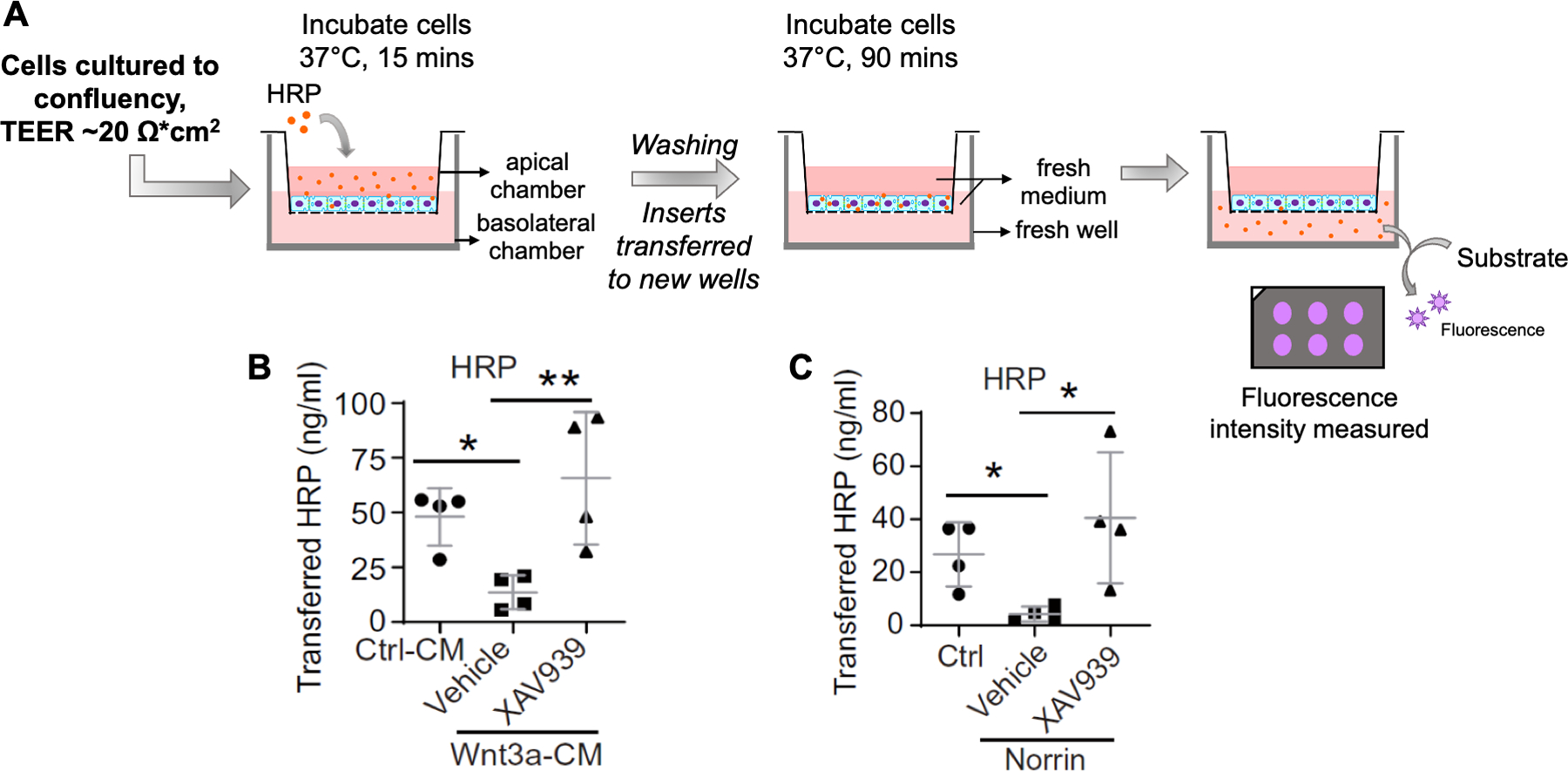
(A) Schematic illustration demonstrating the HRP-based assay to assess caveolae-mediated transcytosis. Fully confluent HRMECs cultured on gelatin-coated inserts were serum starved overnight in 0.5% fetal bovine serum (FBS) + EBM medium at 37 °C before treatment. The EC monolayer in the apical chamber was treated with the desired treatment for 24 h at 37 °C. Afterward, the cells were incubated with horseradish peroxidase (HRP) at 37 °C for 15 min and washed intensively to remove free extracellular HRP. Inserts containing fresh medium were then transferred to a new plate containing warm medium in basolateral chambers, and the cells were incubated for an additional 90 min at 37 °C. The medium from the basolateral chamber was collected, and the HRP levels were quantified by reaction with fluorogenic peroxidase substrate. Fluorescence corresponding to caveolae-mediated EC transcytosis was measured using a fluorescence plate reader. (B,C) In this example, cells were treated with Wnt pathway modulators: Wnt3a-conditioned medium (Wnt3a-CM) or its control-conditioned medium (Ctrl-CM), human recombinant Norrin or its control solution (Ctrl), and with or without Wnt/β-catenin signaling inhibitor (XAV939) or its vehicle control (vehicle). Their effects on HRMEC caveolar transcytosis were evaluated in the HRP-based assay. After treatment, the levels of HRP transferred to the basolateral chamber were measured, indicating caveolae-mediated EC transcytosis levels across HRMECs. Wnt activators reduced the levels of the HRP-based transcytosis, which were reversed by XAV939. * p≤0.05, ** p≤0.01. Panels (B) and (C) were adapted with permission from Wang et al.24.
Representative Results
EM images of retinal vascular endothelium show transcytotic vesicular transport and caveolar vesicles in endothelial cells in vivo.
EC transcytosis can be visualized in vivo within retinal cross-sections with dark brown precipitate reflecting HRP-containing blood vessels under a light microscope (Figure 3A) and as electron-dense precipitate indicative of HRP-containing transcytotic vesicles (Figure 3B,C) using a transmission electron microscope (TEM), thus demonstrating EC transcytosis across the iBRB. Large vesicles potentially reflect macropinocytosis (white arrowhead; Figure 3B), and small vesicles likely represent caveolar vesicles (white arrowheads; Figure 3C). Moreover, the localization of caveolae in retinal blood vessels can be seen as co-staining of caveolae marker CAV-1 antibody with isolectin (EC marker) in the blood vessel under a fluorescence microscope (Figure 3D). CAV-1 positive caveolar vesicles were identified under TEM by immunogold-labeled CAV-1 antibodies (black dots; Figure 3E) within the retinal vascular endothelial cells. Protocols for the in vivo studies can be obtained from Wang et al.24.
VEGF treatment increases vascular permeability in HRMECs as measured by trans-endothelial electrical resistance (TEER)
Before performing EC transcytosis assays, HRMECs should be cultured to full confluency, showing characteristic cobblestone morphology, which can be observed under a light microscope. Full cell confluency can be validated by trans-endothelial electrical resistance (TEER) measurement (Figure 4A), with readings reaching ~20 Ω·cm2 for confluent HRMECs30, suggestive of low levels of paracellular transport across the monolayer through tight or gap junctions. Vascular endothelial growth factor (VEGF) was used as an example to demonstrate TEER measurement. Treatment of HRMECs with VEGF significantly reduced TEER levels, reflective of increased HRMEC permeability (Figure 4B). A reduction in TEER values was also observed in control cells, potentially due to the duration of culture and multiple measurements taken at different time intervals that might be detrimental to cells31.
Transport of Cy3-transferrin can be utilized to assess clathrin-mediated EC transcytosis in HRMECs
For clathrin-mediated EC transcytosis, confluent HRMECs cultured on porous membrane were incubated with fluorescent (Cy3)-tagged transferrin to detect its transportation across ECs (Figure 5A). This assay exploits the fact that transportation of transferrin is a receptor-mediated transcytosis process. Cy3-transferrin endocytosis (red) was observed in HRMEC monolayer co-stained with nuclear stain, DAPI (blue) (Figure 5B), confirming its uptake into the cells. The fluorescence intensity of (Cy3)-tagged transferrin can be quantified from images to assess the endocytosis process and/or measured from the medium collected from the basolateral chamber of the wells to quantify the levels of clathrin-mediated transcytosis27.
Wnt signaling pathway regulates caveolae-mediated EC transcytosis across HRMECs in an HRP-based assay
Previously, it has been found that Wnt signaling regulates MFSD2A-dependent caveolae-mediated transcytosis across retinal vascular ECs to maintain iBRB24. The effects of Wnt modulators were evaluated in an HRP-based in vitro transcytosis assay (Figure 6A). Fully confluent HRMECs were treated with Wnt pathway activators: Wnt3a-conditioned medium (Wnt3a-CM) or human recombinant Norrin, with or without Wnt/β-catenin signaling inhibitor XAV939, and the levels of transcytosed HRP across HRMECs were detected. Treatment with Wnt3a-CM or Norrin revealed significantly decreased levels of transcytosed HRP, indicative of reduced caveolar transcytosis. In addition, combined treatment with the Wnt signaling inhibitor XAV939 demonstrated upregulated levels of transcytosed HRP in HRMECs, hence obliterating the effects of Wnt activators on HRMEC permeability (Figure 6B,C)24.
Discussion
BRB plays an essential role in retinal health and disease. In vitro techniques assessing vascular permeability have proven to be crucial tools in studies concerning barrier (BRB/BBB) development and function. The procedure described here could be utilized to study the molecular mechanisms underlying EC transcytosis or evaluate related molecular modulators affecting BRB permeability. In vitro EC transcytosis assays have multiple advantages over in vivo assays or techniques used for evaluating vascular permeability. They are fast to perform with high throughput and can be utilized to analyze the effects of many different variables or isolated molecules of specific signaling pathways with both genetic and pharmacological modulation. The pure EC culture system obliterates animal variation as is often observed in vivo and also limits the effects of possible modification or endocytosis of the target molecules by other cell types32. Handling of HRMEC cells is easy, requiring basic cell culture equipment available in most laboratories, and cell culture is often more cost-efficient compared with in vivo animal experiments. Although in vitro transcytosis assays do not fully replicate the in vivo physiological conditions33, they can be valuable tools to complement in vivo studies and advance our knowledge on BRB control.
Several important technical parameters require consideration, including the pore size of the filter insert, the type of substratum, and the cell seeding density. A larger pore size of the permeable filter inserts could result in undesirable migration and growth of cells on the underside of the insert, confounding the resulting measurement and its interpretation. While this propensity may vary with the different cell types, pore diameters ≤1 μm serve well for most cell types, including ECs34. The choice of a suitable substratum is a second crucial step because some cells demonstrate higher polarity and greater differentiation when grown on porous substratum and bathed in a culture medium on both sides35,36. Treated microporous polycarbonate filters are a better alternative for ECs because they are thinner and provide greater access of the reagents to the basolateral zone of the monolayer with minimal background fluorescence for immunoassays35. Finally, in order to achieve a fully confluent monolayer, the cell seeding density must be optimized based on the specific cell type. While too low seeding density could impact the differentiation state, too high seeding density, on the other hand, might favor rapidly attaching cells forming multiple cell layers instead of a monolayer, eventually altering the cellular morphology and resulting in inaccurate transcytosis measurement37.
The formation and integrity of the EC monolayer are crucial to this assay and can be validated by measuring TEER values to ensure the achievement of the desired TEER. A blank control, i.e., a filter insert without cells, should always be included to measure the basal levels of resistance across the permeable membrane to be subtracted from those from cell inserts. Various factors such as temperature, cell passage number, the composition of culture medium, culture duration, and junctional length changes may all cause minor variations in TEER values25,38,39. TEER values are also greatly affected by certain treatments, such as VEGF. Discovered initially as a potent inducer of vessel permeability40, VEGF affects vascular permeability by regulating paracellular transport, i.e., through phosphorylation of tight junction proteins ZO-1, occludin, and the disruption of tight junction protein claudin-541,42 as well as transcellular transport43. Additional validation of monolayer formation can be done with morphological examination and the presence of characteristic junction proteins with staining. Throughout the culture, changing media in both the apical and basolateral chambers at regular intervals is recommended to keep the cells in the optimal culture condition with the ideal pH range and nutrient availability.
Researchers using this assay should pay attention to a critical inherent feature of brain and retinal ECs: the apicobasal polarity, which is a cellular hallmark of the BBB44 and, similarly, the BRB. The direction of transcytosis is determined by the relative distribution of protein target receptors of interest and the associated transcytosis machinery in the apical/luminal vs. basolateral/abluminal domains of the EC membrane. Brain and retinal ECs are capable of releasing many factors from both apical/luminal and basolateral/abluminal membranes44. Clathrin-dependent transcytosis of transferrin, interestingly, appears to occur mostly unidirectionally from the blood side to the brain or the retina, since transferrin receptors are predominantly located on the apical/luminal membrane45. Caveolar transcytosis, on the other hand, occurs bi-directionally and can originate from either the apical/luminal or basolateral/abluminal membrane and across to the other side depending on the localization of vesicle cargo and their receptors46. MFSD2A, a transmembrane lipid receptor (for omega-3 fatty acid docosahexaenoic acid) and suppressor of caveolar transcytosis, is also located on the apical/luminal domains of ECs23,47. The expression of MFSD2A is transcriptionally regulated by Wnt signaling to mediate its impact on BRB control, as illustrated as an example in this protocol24. The technique described here, therefore, involves the detection of a tracer molecule placed on the upper/apical chamber following uptake by the EC monolayer and release into the bottom/basolateral chamber, mimicking transcytosis in the apical/luminal/blood to basolateral/abluminal/tissue direction. This protocol can be modified to detect transcytosis in the opposite direction by placing tracer molecules in the bottom chamber and monitoring uptake and release into the apical chamber to suit the need of investigators. Moreover, if needed, an additional step to determine intracellular accumulation/degradation of the endocytosed tracer-bound target molecule would yield further measurement of transcytotic vesicles remaining in the cytosol32.
The described assay has many advantages and serves as an essential tool in BRB/BBB-related studies, but there are a few limitations. It is an isolated system devoid of interactions from other cell types of the iBRB, such as pericytes and glial cells, and hence could not exactly mimic the physiological in-vivo environment. TEER values can vary between measurements owing to a change in temperature or positioning of the electrodes25,38. However, equilibrating the cells from 37 °C to room temperature before measurements, careful handling of the electrodes, and including blank controls can help minimize the fluctuation. Additionally, even with sufficient washing, leakage from the paracellular route, though in a trace amount, cannot be completely ruled out. Overlap of HRP transport among different transcytosis routes, for example, between caveolar transport and macropinocytosis, both being clathrin-independent, may also occur. As needed, the distinction can be further investigated using specific modulators and/or inhibitors of caveolae and micropinocytosis.
In summary, this paper describes an in vitro transcytosis assay permitting quantification of caveolae- or carrier-mediated transcytosis across the BRB/BBB. The in vitro assay could be of great aid in screening various pro-/anti-angiogenic molecules31, signaling pathway modulators24, or drug delivery systems27,48 pertaining to BRB/BBB control. Investigation of EC polarity and directional transcytosis can also benefit from this easy-to-use system. Considering the limited availability of in vitro models for iBRB and ocular research, the procedure outlined here could also be modified as a co-culture system together with pericytes, astrocytes, and 3-D organotypic cultures49, or using advanced cell-based models like microphysiological neurovascular systems50, in order to further investigate cellular interactions and better mimic physiological conditions in vivo.
Supplementary Material
Acknowledgments
This work was supported by NIH grants (R01 EY028100, EY024963, and EY031765) to JC. ZW was supported by a Knights Templar Eye Foundation Career Starter Grant.
Footnotes
A complete version of this article that includes the video component is available at http://dx.doi.org/10.3791/64076.
Disclosures
The authors have no conflicts of interest or financial interest to disclose.
References
- 1.Diaz-Coranguez M, Ramos C, Antonetti DA The inner blood-retinal barrier: Cellular basis and development. Vision Research. 139, 123–137 (2017). [DOI] [PMC free article] [PubMed] [Google Scholar]
- 2.Campbell M, Humphries P The blood-retina barrier: Tight junctions and barrier modulation. Advances in Experimental Medicine and Biology. 763, 70–84 (2012). [PubMed] [Google Scholar]
- 3.Chen J et al. Wnt signaling mediates pathological vascular growth in proliferative retinopathy. Circulation. 124 (17), 1871–1881 (2011). [DOI] [PMC free article] [PubMed] [Google Scholar]
- 4.Klaassen I, Van Noorden CJ, Schlingemann RO Molecular basis of the inner blood-retinal barrier and its breakdown in diabetic macular edema and other pathological conditions. Progress in Retinal and Eye Research. 34, 19–48 (2013). [DOI] [PubMed] [Google Scholar]
- 5.Yemanyi F, Bora K, Blomfield AK, Wang Z, Chen J Wnt signaling in inner blood-retinal barrier maintenance. International Journal of Molecular Sciences. 22 (21), 11877 (2021). [DOI] [PMC free article] [PubMed] [Google Scholar]
- 6.Naylor A, Hopkins A, Hudson N, Campbell M Tight junctions of the outer blood retina barrier. International Journal of Molecular Sciences. 21 (1), 211 (2019). [DOI] [PMC free article] [PubMed] [Google Scholar]
- 7.Erickson KK, Sundstrom JM, Antonetti DA Vascular permeability in ocular disease and the role of tight junctions. Angiogenesis. 10 (2), 103–117 (2007). [DOI] [PubMed] [Google Scholar]
- 8.Chow BW, Gu C Gradual suppression of transcytosis governs functional blood-retinal barrier formation. Neuron. 93 (6), 1325–1333 (2017). [DOI] [PMC free article] [PubMed] [Google Scholar]
- 9.Daruich A et al. Mechanisms of macular edema: Beyond the surface. Progress in Retinal and Eye Research. 63, 20–68 (2018). [DOI] [PubMed] [Google Scholar]
- 10.De Bock M et al. Into rather unexplored terrain-transcellular transport across the blood-brain barrier. Glia. 64 (7), 1097–1123 (2016). [DOI] [PubMed] [Google Scholar]
- 11.Rothberg KG et al. Caveolin, a protein component of caveolae membrane coats. Cell. 68 (4), 673–682 (1992). [DOI] [PubMed] [Google Scholar]
- 12.Kovtun O, Tillu VA, Ariotti N, Parton RG, Collins BM Cavin family proteins and the assembly of caveolae. Journal of Cell Science. 128 (7), 1269–1278 (2015). [DOI] [PMC free article] [PubMed] [Google Scholar]
- 13.Wolburg H, Dermietzel R, Spray DC, Nedergaard M The Endothelial Frontier. In Blood-Brain Interface: From Ontogeny to Artificial Barriers. 75–107. Wiley. Hoboken, NJ: (2006). [Google Scholar]
- 14.Saunders NR, Dziegielewska KM, Mollgard K, Habgood MD Markers for blood-brain barrier integrity: How appropriate is Evans blue in the twenty-first century and what are the alternatives? Frontiers in Neuroscience. 9, 385 (2015). [DOI] [PMC free article] [PubMed] [Google Scholar]
- 15.Atkinson EG, Jones S, Ellis BA, Dumonde DC, Graham E Molecular size of retinal vascular leakage determined by FITC-dextran angiography in patients with posterior uveitis. Eye. 5 (Pt 4), 440–446 (1991). [DOI] [PubMed] [Google Scholar]
- 16.Natarajan R, Northrop N, Yamamoto B Fluorescein isothiocyanate (FITC)-dextran extravasation as a measure of blood-brain barrier permeability. Current Protocols in Neuroscience. 79, 9.58.1–9.58.15 (2017). [DOI] [PMC free article] [PubMed] [Google Scholar]
- 17.Comin CH, Tsirukis DI, Sun Y, Xu X Quantification of retinal blood leakage in fundus fluorescein angiography in a retinal angiogenesis model. Scientific Reports. 11 (1), 19903 (2021). [DOI] [PMC free article] [PubMed] [Google Scholar]
- 18.Pietra GG, Johns LW Confocal- and electron-microscopic localization of FITC-albumin in H2O2-induced pulmonary edema. Journal of Applied Physiology. 80 (1), 182–190 (1996). [DOI] [PubMed] [Google Scholar]
- 19.Honeycutt SE, O’Brien LL Injection of Evans blue dye to fluorescently label and image intact vasculature. Biotechniques. 70 (3), 181–185 (2021). [DOI] [PMC free article] [PubMed] [Google Scholar]
- 20.Wu JH et al. Inhibition of Sema4D/PlexinB1 signaling alleviates vascular dysfunction in diabetic retinopathy. European Molecular Biology Organization (EMBO) Molecular Medicine. 12 (2), e10154 (2020). [DOI] [PMC free article] [PubMed] [Google Scholar]
- 21.Radu M, Chernoff J An in vivo assay to test blood vessel permeability. Journal of Visualized Experiments. (73), e50062 (2013). [DOI] [PMC free article] [PubMed] [Google Scholar]
- 22.Vinores SA Assessment of blood-retinal barrier integrity. Histology and Histopathology. 10 (1), 141–154 (1995). [PubMed] [Google Scholar]
- 23.Ben-Zvi A et al. Mfsd2a is critical for the formation and function of the blood-brain barrier. Nature. 509 (7501), 507–511 (2014). [DOI] [PMC free article] [PubMed] [Google Scholar]
- 24.Wang Z et al. Wnt signaling activates MFSD2A to suppress vascular endothelial transcytosis and maintain blood-retinal barrier. Science Advances. 6 (35), eaba7457 (2020). [DOI] [PMC free article] [PubMed] [Google Scholar]
- 25.Srinivasan B et al. TEER measurement techniques for in vitro barrier model systems. Journal of Laboratory Automation. 20 (2), 107–126 (2015). [DOI] [PMC free article] [PubMed] [Google Scholar]
- 26.Martins-Green M, Petreaca M, Yao M An assay system for in vitro detection of permeability in human “endothelium”. Methods in Enzymology. 443, 137–153 (2008). [DOI] [PubMed] [Google Scholar]
- 27.Sade H et al. A human blood-brain barrier transcytosis assay reveals antibody transcytosis influenced by pH-dependent receptor binding. PLoS One. 9 (4), e96340 (2014). [DOI] [PMC free article] [PubMed] [Google Scholar]
- 28.Feng Y et al. VEGF-induced permeability increase is mediated by caveolae. Investigative Ophthalmology and Visual Science. 40 (1), 157–167 (1999). [PubMed] [Google Scholar]
- 29.Wang Z, Liu CH, Huang S, Chen J Wnt signaling in vascular eye diseases. Progress in Retinal and Eye Research. 70, 110–133 (2019). [DOI] [PMC free article] [PubMed] [Google Scholar]
- 30.Suarez S et al. Modulation of VEGF-induced retinal vascular permeability by peroxisome proliferator-activated receptor-beta/delta. Investigative Ophthalmology and Visual Science. 55 (12), 8232–8240 (2014). [DOI] [PMC free article] [PubMed] [Google Scholar]
- 31.Tomita Y et al. Long-acting FGF21 inhibits retinal vascular leakage in in vivo and in vitro models. International Journal of Molecular Science. 21 (4), 1188 (2020). [DOI] [PMC free article] [PubMed] [Google Scholar]
- 32.Tuma P, Hubbard AL Transcytosis: Crossing cellular barriers. Physiological Reviews. 83 (3), 871–932 (2003). [DOI] [PubMed] [Google Scholar]
- 33.Moleiro AF, Conceicao G, Leite-Moreira AF, Rocha-Sousa A A critical analysis of the available in vitro and ex vivo methods to study retinal angiogenesis. Journal of Ophthalmology. 2017, 3034953 (2017). [DOI] [PMC free article] [PubMed] [Google Scholar]
- 34.Tucker SP, Melsen LR, Compans RW Migration of polarized epithelial cells through permeable membrane substrates of defined pore size. European Journal of Cell Biology. 58 (2), 280–290 (1992). [PubMed] [Google Scholar]
- 35.Butor C, Davoust J Apical to basolateral surface area ratio and polarity of MDCK cells grown on different supports. Experimental Cell Research. 203 (1), 115–127 (1992). [DOI] [PubMed] [Google Scholar]
- 36.Villars F et al. Ability of various inserts to promote endothelium cell culture for the establishment of coculture models. Cell Biology and Toxicology. 12 (4–6), 207–214 (1996). [DOI] [PubMed] [Google Scholar]
- 37.Liu F, Soares MJ, Audus KL Permeability properties of monolayers of the human trophoblast cell line BeWo. American Journal of Physiology. 273 (5), C1596–1604 (1997). [DOI] [PubMed] [Google Scholar]
- 38.Matter K, Balda MS Functional analysis of tight junctions. Methods. 30 (3), 228–234 (2003). [DOI] [PubMed] [Google Scholar]
- 39.Felix K, Tobias S, Jan H, Nicolas S, Michael M Measurements of transepithelial electrical resistance (TEER) are affected by junctional length in immature epithelial monolayers. Histochemistry and Cell Biology. 156 (6), 609–616 (2021). [DOI] [PMC free article] [PubMed] [Google Scholar]
- 40.Senger DR et al. Tumor cells secrete a vascular permeability factor that promotes accumulation of ascites fluid. Science. 219 (4587), 983–985 (1983). [DOI] [PubMed] [Google Scholar]
- 41.Antonetti DA, Barber AJ, Hollinger LA, Wolpert EB, Gardner TW Vascular endothelial growth factor induces rapid phosphorylation of tight junction proteins occludin and zonula occluden 1. A potential mechanism for vascular permeability in diabetic retinopathy and tumors. Journal of Biological Chemistry. 274 (33), 23463–23467 (1999). [DOI] [PubMed] [Google Scholar]
- 42.Argaw AT, Gurfein BT, Zhang Y, Zameer A, John GR VEGF-mediated disruption of endothelial CLN-5 promotes blood-brain barrier breakdown. Proceedings of the National Academy of Sciences of the United States of America. 106 (6), 1977–1982 (2009). [DOI] [PMC free article] [PubMed] [Google Scholar]
- 43.Penn JS et al. Vascular endothelial growth factor in eye disease. Progress in Retinal and Eye Research. 27 (4), 331–371 (2008). [DOI] [PMC free article] [PubMed] [Google Scholar]
- 44.Worzfeld T, Schwaninger M Apicobasal polarity of brain endothelial cells. Journal of Cerebral Blood Flow and Metabolism. 36 (2), 340–362 (2016). [DOI] [PMC free article] [PubMed] [Google Scholar]
- 45.Roberts RL, Fine RE, Sandra A Receptor-mediated endocytosis of transferrin at the blood-brain barrier. Journal of Cell Science. 104 (Pt 2), 521–532 (1993). [DOI] [PubMed] [Google Scholar]
- 46.Predescu SA, Predescu DN, Malik AB Molecular determinants of endothelial transcytosis and their role in endothelial permeability. American Journal of Physiology-Lung Cellular and Molecular Physiology. 293 (4), L823–842 (2007). [DOI] [PubMed] [Google Scholar]
- 47.Nguyen LN et al. Mfsd2a is a transporter for the essential omega-3 fatty acid docosahexaenoic acid. Nature. 509 (7501), 503–506 (2014). [DOI] [PubMed] [Google Scholar]
- 48.Pulgar VM Transcytosis to cross the blood brain barrier, new advancements and challenges. Frontiers in Neuroscience. 12, 1019 (2018). [DOI] [PMC free article] [PubMed] [Google Scholar]
- 49.Shamir ER, Ewald AJ Three-dimensional organotypic culture: Experimental models of mammalian biology and disease. Nature Reviews Molecular Cell Biology. 15 (10), 647–664 (2014). [DOI] [PMC free article] [PubMed] [Google Scholar]
- 50.Maurissen TL et al. Microphysiological neurovascular barriers to model the inner retinal microvasculature. Journal of Personalized Medicine. 12 (48), 148 (2022). [DOI] [PMC free article] [PubMed] [Google Scholar]
Associated Data
This section collects any data citations, data availability statements, or supplementary materials included in this article.


