Cystic lymphangiomas of the pancreas (CLP) were first described by Koch in 1913 1 . The origin of these benign lesions is not well defined; they could either be congenital malformations or secondary to a lymphatic vessel obstruction due to either radiotherapy, surgery, infection, or trauma 2 . The main challenge in pancreatic cystic lesions (PCLs) is the diagnostic certainty 3 . Needle-based confocal laser endomicroscopy (nCLE), described in pancreatic diseases in 2011 4 , is a very specific technique for the diagnosis of the main PCL 5 . The first description of the identifiable structures by nCLE in CLP is proposed below.
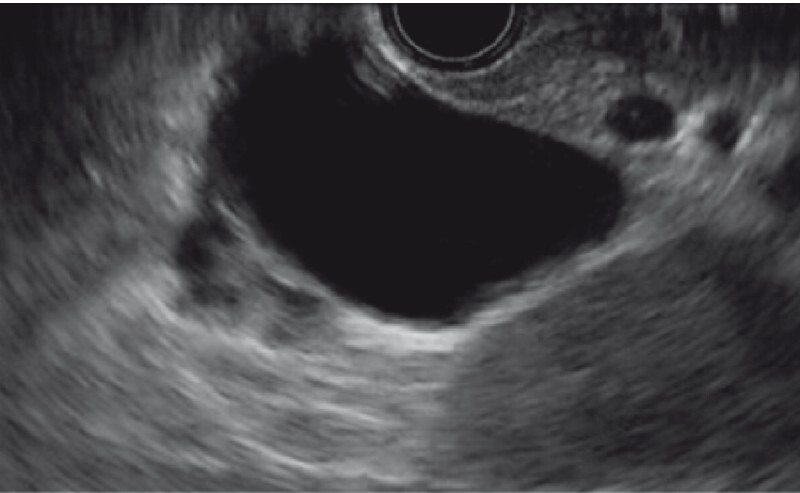
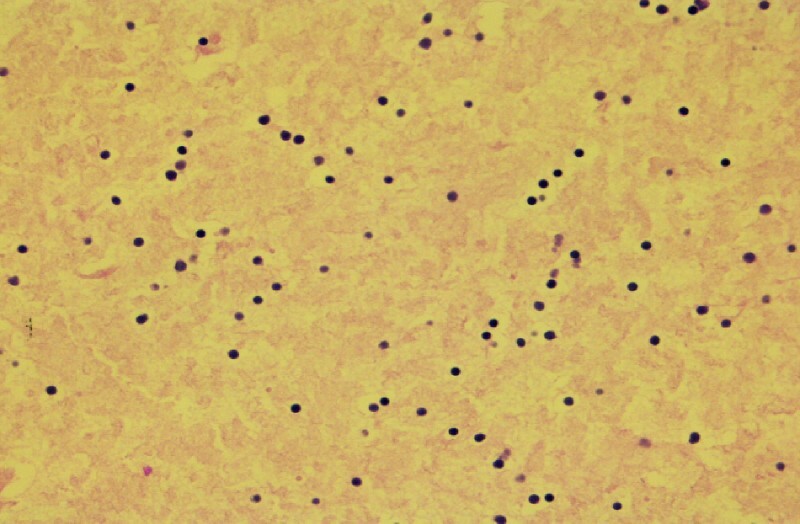
All cases of CLP seen between 2012 and 2020 in our center were reviewed. The study was approved by the institutional ethics committee (IRB00010835). Gold standard criterion for CLP diagnosis was cytology with or without histology. Herein, we report on six patients who all had endoscopic ultrasound imaging of the pancreatic cyst ( Fig. 1 ), cytology compatible with CLP showing small lymphocytes ( Fig. 2 ), with or without histologic confirmation on the surgical specimen (n = 3).
Fig. 1 .

Endoscopic ultrasound image of a cystic lymphangioma of the pancreas located in the body of the pancreas.
Fig. 2 .

Cytology obtained by endoscopic ultrasound-guided fine-needle aspiration.
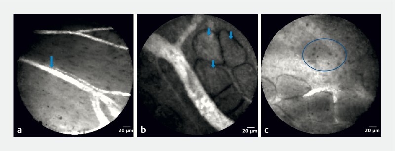
During nCLE, the main structure identified was vessels, qualified as “straight” in five patients (83.3 %) and “winding” in one (16.7 %), which were on a grey background in all patients ( Fig. 3 a ). The second structure was adipocytes, seen in four patients (66.7 %) ( Fig. 3 b ). Finally, all patients had small, disseminated cells on the grey background, suggesting small lymphocytes ( Fig. 3 c ).
Fig. 3.

Needle-based confocal laser endomicroscopy. a Straight vessels on grey background (arrow). b Adipocytes (arrows). c Small disseminated cells, suggesting small lymphocytes (circle).
To the best of our knowledge, this is the first case series describing nCLE characteristics in CLP; nCLE identified three main structures: rather large and straight vessels on a grey background, adipocytes, and small disseminated cells ( Video 1 ). When these signs are present, combined with the absence of the usual criteria for the most common PCLs, the diagnosis of CLP should be considered.
Video 1 Needle-based confocal laser endomicroscopy recording of a cystic pancreatic lymphangioma depicting adipocytes, straight vessels on grey background and small disseminated cells.
Endoscopy_UCTN_Code_CCL_1AF_2AZ_3AB
Footnotes
Competing interests B. Napoléon performs teaching sessions for Mauna Kea Technologies. C. Michoud, T. Khoury, A. Lisotti, R. Gincul, S. Leblanc, and A. I. Lemaistre declare that they have no conflict of interest.
Endoscopy E-Videos : https://eref.thieme.de/e-videos .
Endoscopy E-Videos is an open access online section, reporting on interesting cases and new techniques in gastroenterological endoscopy. All papers include a high quality video and all contributions are freely accessible online. Processing charges apply, discounts and wavers acc. to HINARI are available. This section has its own submission website at https://mc.manuscriptcentral.com/e-videos
References
- 1.Viscosi F, Fleres F, Mazzeo C et al. Cystic lymphangioma of the pancreas: a hard diagnostic challenge between pancreatic cystic lesions – review of recent literature. Gland Surg. 2018;7:487–492. doi: 10.21037/gs.2018.04.02. [DOI] [PMC free article] [PubMed] [Google Scholar]
- 2.Leung T K, Lee C M, Shen L K et al. Differential diagnosis of cystic lymphangioma of the pancreas based on imaging features. J Formos Med Assoc. 2006;105:512–517. doi: 10.1016/S0929-6646(09)60193-5. [DOI] [PubMed] [Google Scholar]
- 3.Krishna S G, Brugge W R, Dewitt J M et al. Needle-based confocal laser endomicroscopy for the diagnosis of pancreatic cystic lesions: an international external interobserver and intraobserver study (with videos) Gastrointest Endosc. 2017;86:644–654. doi: 10.1016/j.gie.2017.03.002. [DOI] [PubMed] [Google Scholar]
- 4.Konda V J, Aslanian H R, Wallace M B et al. First assessment of needle-based confocal laser endomicroscopy during EUS-FNA procedures of the pancreas (with videos) Gastrointest Endosc. 2011;74:1049–1060. doi: 10.1016/j.gie.2011.07.018. [DOI] [PubMed] [Google Scholar]
- 5.Napoleon B, Palazzo M, Lemaistre A I et al. Needle-based confocal laser endomicroscopy of pancreatic cystic lesions: a prospective multicenter validation study in patients with definite diagnosis. Endoscopy. 2019;51:825–835. doi: 10.1055/a-0732-5356. [DOI] [PubMed] [Google Scholar]


