

Abstract
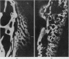
(1) The development of periarticular osteophytes in experimental osteoarthritis in the dog degins as early as 3 days after induction of the disease process. (2) Development of the osteophytes is still proceeding 48 weeks after induction. (3) The common site for development of the osteophyte is at the marginal zone where synovial membrane merges with fibrocartilage. (4) At this site the osteophyte begins as a deposition of outside the existing femoral bone cortex. (5) Further deposition of new bone and resorption lead to a remodelling which ultimately produces a mature osteophyte having a trabecular bone structure and free communication with the bone marrow spaces of the femur. (6) In some dogs there is also hyperplasia of bone with remodelling which takes place beneath the cartilage of the nonarticulating face of the trochlear ridge. This develops a mature trabecular structure later in the disease process and may become confluent with the osteophyte at the marginal zone. (7) The bone changes are not confined to development of the osteophyte. The whole distal end of the femur appears to have a marked increase in bone turnover, and there is also evidence of increased bone metabolism in the contralateral limb. (8) Dye injection techniques have shown that an increase in vascularity is associated with this development of new bone, and it is suggested that the results indicate the possible importance of a vascular component in the pathogenesis of osteoarthritis.
Full text
PDF


Images in this article


Selected References
These references are in PubMed. This may not be the complete list of references from this article.
- Arnoldi C. C., Linderholm H., Müssbichler H. Venous engorgement and intraosseous hypertension in osteoarthritis of the hip. J Bone Joint Surg Br. 1972 Aug;54(3):409–421. [PubMed] [Google Scholar]
- Brookes M., Helal B. Primary osteoarthritis, venous engortement and osteogenesis. J Bone Joint Surg Br. 1968 Aug;50(3):493–504. [PubMed] [Google Scholar]
- Brookes M. The vascular factor in osteoarthritis. Surg Gynecol Obstet. 1966 Dec;123(6):1255–1260. [PubMed] [Google Scholar]
- Marshall J. L., Olsson S. E. Instability of the knee. A long-term experimental study in dogs. J Bone Joint Surg Am. 1971 Dec;53(8):1561–1570. [PubMed] [Google Scholar]
- Marshall J. L. Periarticular osteophytes. Initiation and formation in the knee of the dog. Clin Orthop Relat Res. 1969 Jan-Feb;62:37–47. [PubMed] [Google Scholar]
- Rahn B. A., Perren S. M. Xylenol orange, a fluorochrome useful in polychrome sequential labeling of calcifying tissues. Stain Technol. 1971 May;46(3):125–129. doi: 10.3109/10520297109067836. [DOI] [PubMed] [Google Scholar]
- Rhinelander F. W. The normal microcirculation of diaphyseal cortex and its response to fracture. J Bone Joint Surg Am. 1968 Jun;50(4):784–800. doi: 10.2106/00004623-196850040-00016. [DOI] [PubMed] [Google Scholar]
- Suzuki H. K., Mathews A. Two-color fluorescent labeling of mineralizing tissues with tetracycline and 2,4-bis[N,N'-di-(carbomethyl)aminomethyl] fluorescein. Stain Technol. 1966 Jan;41(1):57–60. doi: 10.3109/10520296609116280. [DOI] [PubMed] [Google Scholar]