TO THE EDITOR: Lupus anticoagulant hypoprothrom-binemia syndrome (LAHPS) is a rare hematological entity, described in children only in a few case reports and case reports series. It was first reported in 1960, in a patient with systemic lupus erythematosus (SLE) who suffered from serious bleedings and had hypoprothrombinemia caused by an inhibitor impending prothrombin activity [1]. This acquired coagulopathy may be associated with an underlying autoimmune disease [2-6], or it may be a transient event following viral infection, particularly adenovirus [7-9]. Along with bleeding, which may be mild to life-threatening [2-4, 6, 10], typical laboratory findings are prolonged activated partial thromboplastin time (APTT), with or without prolonged prothrombin time (PT), along with positive lupus anticoagulant (LAC) and deficiency of prothrombin - factor II (FII) [1-12].
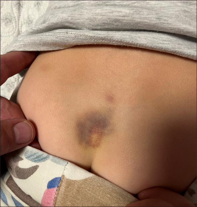
We herein present a case of a six-year-old, previously healthy girl, hospitalized at the Division of Pulmonology, Allergology, Immunology and Rheumatology, Department of Pediatrics, Children’s Hospital Zagreb, Croatia due to spontaneous bruising, without other manifest bleedings, accompanied with minor swelling and soreness of the right ankle. The patient was in a good general condition, afebrile, with multiple hematomas of various phases of resorption, mainly distributed on lower extremities and back, with the largest one in the sacral area (Fig. 1). The patient and her parents denied any severe trauma. A week before, she overcame an acute, presumably viral enterocolitis, treated symptomatically at home, while four months prior to the admission she had coronavirus disease 2019 (COVID-19) infection, confirmed by PCR testing, presenting as a common cold. The patient’s mother stated that the tendency to spontaneous bruising was present for a while and could chronologically be associated with the previous COVID-19 infection. At admission, rapid antigen test for COVID-19 was negative. Extensive laboratory work-up revealed a normal blood count, excluding thrombocytopenia as the cause of cutaneous bleeding, low inflammation parameters, unremarkable standard biochemistry findings (including liver synthesis markers and kidney function tests) and urine analysis. However, coagulation tests revealed prolongation of both PT and APTT, while C3 and C4 complement levels were reduced. This indicated the need for further immunological and hematological diagnostics to elucidate the underlying pathology, which confirmed the presence of LAC and a low F II activity level (Table 1). Since the patient was clinically stable during the whole hospital stay and did not develop any new bleeds, we decided to proceed with the watchful waiting approach, rather than introducing corticosteroid treatment, known to be the first line therapy for LAHPS. She was regularly evaluated; within a three-weeks period APTT (28.9 s) and PT (98% activity) normalized, as well as C3 (1.17 g/L) and C4 (0.18 g/L) complement levels. As the patient had an unremarkable family and personal medical history and no additional anamnestic data or clinical signs indicating autoimmune disease, such as SLE or antiphospholipid syndrome, the most probable diagnosis was a transient, viral-induced LAHPS. Finally, subsequent immunological testing ruled out an underlying autoimmune disease [negative antinuclear antibodies (ANA), extractable nuclear antigen (ENA) panel, antineutrophil cytoplasmic antibodies (ANCA), anticardiolipin and beta 2 glycoprotein antibodies]. The only question remaining unanswered was the exact microbial trigger to LAHPS. It might have been the acute viral enterocolitis that the patient recovered from a week before admission, but given the fact that bruises started appearing a few months before the admission, soon after the girl recovered from COVID-19 infection, COVID-19 was the more probable cause. At the time of writing this paper, our patient continues to be in a multidisciplinary follow-up, and so far, has not shown signs of disease relapse.
Fig. 1.
A large hematoma in the sacral area.
Table 1.
Laboratory findings upon admission.
| Parameter | Value | Reference range |
|---|---|---|
| RBC | 4.76×10¹²/L | 4.00–5.00×10¹²/L |
| Hb | 125 g/L | 109–138 g/L |
| Hct | 0.380 | 0.320–0.404 |
| Platelets | 455×109/L | 150–450×109/L |
| WBC | 8.32×10⁹/L | 5.0–13.0×10⁹/L |
| CRP | 2.1 mg/L | 0.1–2.8 mg/L |
| C3 complement component | 0.57 g/L | 0.9–1.8 g/L |
| C4 complement component | <0.08 g/L | 0.1–0.4 g/L |
| Anti-COVID-19 IgM and IgG antibodies | >250 U/mL | >0.8 U/mL–positive |
| PV | 59% activity | >70% activity |
| APTT | 64.0 s | 23–31.9 s |
| LA | 1.49 | <1.37 |
| F II | 26% activity | 70–120% activity |
Clinical and laboratory features in our patient are in accordance with case reports previously published on transient, post-infective LAHPS in pediatric age group. Children usually present with mild bleeds or may be asymptomatic but have a prolonged PT and APTT, along with the presence of LAC, following acute gastrointestinal or respiratory infection. The rarity of this syndrome leaves it without formulated evidence-based treatment guidelines. Therapeutical approach is mainly conservative, as children with transient LAHPS usually show spontaneous recovery within a few weeks, including normalization of coagulation tests [7, 8]. Immunosuppressants (corticosteroids, rituximab, cyclophosphamide, azathioprine) and intravenous immunoglobulins (IVIG), combined with life-saving supportive measures (blood transfusions, fresh frozen plasma, vitamin K, recombinant factor VII and/or antifibrinolytics), are used to treat children with major clinical hemorrhages [2-5, 9, 10]. LAHPS associated with autoimmune disease is usually more persistent and more commonly complicated by severe bleeding diathesis (fatal pulmonary hemorrhage [2], bilateral adrenal hemorrhage [6], deep tissue hematomas [5]), compared to the transient post-infectious LAHPS. However, serious episodes of hematemesis and hemarthrosis, have been reported in previously healthy patients with non-SLE associated LAHPS [12].
The pathophysiological mechanism in LAHPS was first explained by Bajaj et al. [11] Although LAC is usually associated with thrombotic events, the authors postulated that in the case of LAHPS the presence of non-neutralizing anti-prothrombin antibodies induces rapid clearance of prothrombin antigen-antibody complexes in the reticuloendothelial system, eventually resulting in hypoprothrombinemia and bleeding diathesis. A report of one case of LAHPS in a familial infectious context speculated that there may exist a genetic predisposition to anti-prothrombin antibodies [8]. More recently, LAC-positive coagulopathy was observed in adult patients with COVID-19 infection. It presents mainly as isolated APTT prolongation, but without bleeding tendency, so the laboratory finding is not a contraindication for antithrombotic therapy [13]. However, to our knowledge, a case of a possible post-COVID-19 pediatric LAHPS has not yet been reported in the literature.
We believe that the prevalence of post-infectious LAHPS is underestimated, as there are described asymptomatic cases of transient coagulopathy, detected only due to extensive laboratory work-up in acute illness. However, in the setting of an acute respiratory or intestinal infection (especially if caused by adenovirus), followed by mucocutaneous bleeding, one must always think of LAHPS. We therefore suggest performing coagulation indices in these patients as a screening method for LAHPS to avoid rare, but possibly life-threatening hemorrhagic episodes. Depending on the severity of symptoms, watchful waiting or therapeutical approach should commence, corticosteroids being the first and usually most easily available choice. We also emphasize the importance of further regular follow-up for patients with positive LAC, particularly if combined with positive antinuclear antibodies (ANA), as LAHPS in these cases may precede or be the first manifestation of an autoimmune disease.
Footnotes
Authors’ Disclosures of Potential Conflicts of Interest
No potential conflicts of interest relevant to this article were reported.
REFERENCES
- 1.Rapaport SI, Ames SB, Duvall BJ. A plasma coagulation defect in systemic lupus erythematosus arising from hypoprothrombinemia combined with antiprothrombinase activity. Blood. 1960;15:212–27. doi: 10.1182/blood.V15.2.212.212. [DOI] [PubMed] [Google Scholar]
- 2.Kim JS, Kim MJ, Bae EY, Jeong DC. Pulmonary hemorrhage in pediatric lupus anticoagulant hypoprothrombinemia syndrome. Korean J Pediatr. 2014;57:202–5. doi: 10.3345/kjp.2014.57.4.202.364f9c68e02543d4afac7ebfda6975b1 [DOI] [PMC free article] [PubMed] [Google Scholar]
- 3.Mazodier K, Arnaud L, Mathian A, et al. Lupus anticoagulant- hypoprothrombinemia syndrome: report of 8 cases and review of the literature. Medicine (Baltimore) 2012;91:251–60. doi: 10.1097/MD.0b013e31826b971f. [DOI] [PubMed] [Google Scholar]
- 4.Komvilaisak P, Wisanuyotin S, Jettrisuparb A, Wiangnon S. Lupus anticoagulant-hypoprothrombinemia syndrome (LAC-HPS) in children with systemic lupus erythematosus: report of 3 cases. J Pediatr Hematol Oncol. 2017;39:e521–4. doi: 10.1097/MPH.0000000000000891. [DOI] [PubMed] [Google Scholar]
- 5.Yacobovich JR, Uziel Y, Friedman Z, Radnay J, Wolach B. Diffuse muscular haemorrhage as presenting sign of juvenile systemic lupus erythematosus and lupus anticoagulant hypoprothrom-binaemia syndrome. Rheumatology (Oxford) 2001;40:585–7. doi: 10.1093/rheumatology/40.5.585. [DOI] [PubMed] [Google Scholar]
- 6.Sakamoto A, Ogura M, Hattori A, et al. Lupus anticoagulant hypoprothrombinemia syndrome associated with bilateral adrenal haemorrhage in a child: early diagnosis and intervention. Thromb J. 2021;19:19. doi: 10.1186/s12959-021-00271-0.49fdf6c8e3a445758245d4ec2c434d59 [DOI] [PMC free article] [PubMed] [Google Scholar]
- 7.Jaeger U, Kapiotis S, Pabinger I, Puchhammer E, Kyrle PA, Lechner K. Transient lupus anticoagulant associated with hypoprothrombinemia and factor XII deficiency following adenovirus infection. Ann Hematol. 1993;67:95–9. doi: 10.1007/BF01788133. [DOI] [PubMed] [Google Scholar]
- 8.Appert-Flory A, Fischer F, Amiral J, Monpoux F. Lupus anticoagulant-hypoprothrombinemia syndrome (HLAS): report of one case in a familial infectious context. Thromb Res. 2010;126:e139–40. doi: 10.1016/j.thromres.2009.11.025. [DOI] [PubMed] [Google Scholar]
- 9.Schmugge M, Tölle S, Marbet GA, Laroche P, Meili EO. Gingival bleeding, epistaxis and haematoma three days after gastroenteritis: the haemorrhagic lupus anticoagulant syndrome. Eur J Pediatr. 2001;160:43–6. doi: 10.1007/PL00008415. [DOI] [PubMed] [Google Scholar]
- 10.Baca V, Montiel G, Meillón L, et al. Diagnosis of lupus anticoagulant in the lupus anticoagulant-hypoprothrombinemia syndrome: report of two cases and review of the literature. Am J Hematol. 2002;71:200–7. doi: 10.1002/ajh.10194. [DOI] [PubMed] [Google Scholar]
- 11.Bajaj SP, Rapaport SI, Fierer DS, Herbst KD, Schwartz DB. A mechanism for the hypoprothrombinemia of the acquired hypoprothrombinemia-lupus anticoagulant syndrome. Blood. 1983;61:684–92. doi: 10.1182/blood.V61.4.684.684. [DOI] [PubMed] [Google Scholar]
- 12.Becton DL, Stine KC. Transient lupus anticoagulants associated with hemorrhage rather than thrombosis: the hemorrhagic lupus anticoagulant syndrome. J Pediatr. 1997;130:998–1000. doi: 10.1016/S0022-3476(97)70291-9. [DOI] [PubMed] [Google Scholar]
- 13.Bowles L, Platton S, Yartey N, et al. Lupus anticoagulant and abnormal coagulation tests in patients with Covid-19. N Engl J Med. 2020;383:288–90. doi: 10.1056/NEJMc2013656. [DOI] [PMC free article] [PubMed] [Google Scholar]


