

Abstract
Chitin nanofibers have recently received increased attention and are considered to be a promising material for a wide range of applications because of their excellent characteristics. In this study, 2,2,6,6-tetramethylpiperidine-1-oxyl (TEMPO)-oxidized chitin nanofibers (CNFs) with various oxidation times were prepared and characterized. CNFs with different oxidation times were then utilized for enzyme immobilization, using chymotrypsin as a model enzyme. The effects of oxidation time on enzyme immobilization were explored. Results showed characteristics of chitin nanofibers can be controlled by adjusting oxidation time. CNFs treated with TEMPO for 360 min showed the lowest crystallinity (79.13 ± 1.43%), the shortest length (241.70 ± 74.61 nm), the largest width (12.67 ± 3.43 nm), and the highest transmittance (73.01% at 800 nm). The activity of immobilized enzymes and enzyme loading showed good correlation to the carboxylate content of CNFs. The enzyme efficiency based on CNFs and the content of carboxylate groups peaked at the oxidization time of 60 min. When the additional amount of chymotrypsins (CTs) was 500 or 2000 mg/g carrier, the highest loading amount of CTs was 307.17 ± 4.08 or 726.82 ± 12.05 mg/g carrier, respectively.
Electronic supplementary material
The online version of this article (10.1007/s42995-020-00054-y) contains supplementary material, which is available to authorized users.
Keywords: Chitin, Nanofiber, Chymotrypsin, Immobilization
Introduction
Chitin is a natural mucopolysaccharide that consists of 2-acetamino-2-deoxygen-d-glucose and 2-amino-2-deoxygen-d-glucose linked by a β (1 → 4) glycosidic bond (Kumar 2000; Shahidi et al. 1999). The amount of chitin in nature is large, as it is found in crabs and shrimps, fungus, and the cell walls of lower plants (Rinaudo 2006). Chitin also has distinctive biological characteristics; it is non-toxic, antibacterial, biocompatible, and biodegradable (Hirano 1999; Jayakumar et al. 2010; Kurita 2006). Moreover, chitin can show low immunogenicity as well as it has film forming properties and can form fibers (Zheng et al. 2002).
Functional groups, such as acetyl groups, hydroxyl groups, and carbonyl groups, in the main chain structure of the chitin molecule make it easy to combine with itself or other hydroxyl compounds by hydrogen bonds (Rinaudo 2006; Tamura et al. 2006, 2011). As a result, chitin is difficult to dissolve in most acidic or alkaline solutions and organic solvents, limiting its usage (Tamura et al. 2011). To be usable, chitin needs to be chemically modified, such as through deacetylation and esterification, before application in the medical, food, agricultural, biotechnical, and environmental fields, resulting in an increased interest in chitin nanofibers (Ifuku et al. 2009; Ifuku and Saimoto 2012; Jayakumar et al. 2010; Wang et al. 2018a, b). Chitin nanofibers are generally referred to as a linear fiber or whisker with a nanometer diameter and a high aspect ratio (Ifuku and Saimoto 2012). They have been prepared by a variety of methods, including mechanical treatment, dissolution and regeneration, TEMPO oxidation, and electrostatic spinning (Jia et al. 2002; Kato et al. 2004; Muzzarelli et al. 1999; Zhong et al. 2010). The nanofibers prepared by chitin not only have the features of chitin itself as mentioned above but also are characterized by a high specific surface area and stability in an aqueous solution (Ifuku and Saimoto 2012). The nanofibers are easier to process further and utilize in the food and biomedical industries, e.g., chitin/chitosan packaging film and cell attachment (Jayakumar et al. 2010; Mushi et al. 2014).
Enzyme immobilization, the restriction of enzymes on solid materials, is a requisite for the application of enzymes as biocatalysts (Han et al. 2016; Sheldon 2007; Wang and Caruso 2005). Enzyme immobilization can prevent instability and unrepeatability of free enzymes because the support of chitin nanofibers facilitates recovery from reaction mixtures (Wang et al. 2009). Nanofibers have been applied to immobilization, with immobilization methods including covalent binding, physical adsorption, and polymer embedding (Iyer and Ananthanarayan 2008; Sheldon 2007; Zhang et al. 2009). In enzyme immobilization, chitin nanofibers are a kind of inert immobilized carrier, making the enzyme reusable, easy to separate, and more stable (Iyer and Ananthanarayan 2008; Khoshnevisan et al. 2011; Mateo et al. 2007). Magnetic nanoparticles, used in enzyme immobilization, can be rapidly separated and immobilized enzymes can be recovered from the reaction system using an external magnet (Deng et al. 2009; Xiao et al. 2017). The use of magnetic supports can also reduce operation costs and prevent problems that can limit recovery (Khoshnevisan et al. 2011; Pan et al. 2009).
We previously developed a chitin-nanofiber-based support for enzyme immobilization (Huang et al. 2018). Chitin-based supports for enzyme immobilization with high biocompatibility, stability, and recyclability have been previously studied (Huang et al. 2018). However, the relationship and optimization between oxidized time of immobilization support and enzyme efficiency have not been studied. In the present study, chitin nanofibers were prepared using the TEMPO/NaBr/NaClO system with various oxidation times and then used for chymotrypsin immobilization. The characteristics of the TEMPO-oxidized chitin nanofibers were measured by X-ray diffraction (XRD), transmission electron microscopy (TEM), dynamic light scattering (DLS), Fourier transform infrared spectrometry (FT-IR), ultraviolet–visible (UV/VIS) spectrophotometry, and optical microscopy. The relationship between different nanofibers and their carboxylate content, crystallinity, and light transmittance was analyzed. Finally, the effects of oxidation time on the chymotrypsin immobilization were evaluated.
Results
Characterization of chitin nanofibers
X-ray diffraction patterns of oxidized chitin
The diffraction pattern of the original chitin was the crystalline form of the α-chitin. The XRD patterns are shown in Supplementary Fig. S1 and the crystallinity in Table 1. The peaks at 9.3°, 12.8°, 19.2°, 21.3°, 23.2°, and 26.3° correspond to the chitin crystal planes of (020), (021), (110), (120), (130), and (013), respectively (Zhang et al. 2005). The crystallinity of the starting chitin was more than 85%. When the oxidation time was less than 60 min, the small peaks at 12.8° or 23.2° disappeared; however, the (020) and (110) peaks of the oxidized chitin were stable. Furthermore, the crystallinity increased slightly with a short oxidation time (less than 60 min), indicating the disordered regions of chitin may have been destroyed (Jiang et al. 2018). The XRD curves were obviously altered when the oxidation time was between 60 and 480 min. The crystallinity decreased significantly by 10.57% and the intensities of the (020) and (110) peaks were also reduced. These results indicated chitin chains underwent depolymerization via TEMPO oxidation (Kato et al. 2004; Muzzarelli et al. 1999). The water-insoluble CNFs obtained were below 1% when the oxidation time of chitin was 480 min, making it impossible to analyze the characteristics of CNFs.
Table 1.
Crystallinity of TEMPO-oxidation chitin with various oxidation times
| Oxidization time/min | ICR ± RSD/% |
|---|---|
| 0 | 89.70 ± 0.64 |
| 20 | 89.91 ± 0.77 |
| 40 | 90.16 ± 0.94 |
| 60 | 87.02 ± 1.12 |
| 120 | 84.37 ± 2.15 |
| 240 | 83.30 ± 1.20 |
| 360 | 80.89 ± 1.29 |
| 480 | 79.13 ± 1.43 |
Transmission electron microscopy images of chitin nanofibers
The water-insoluble CNFs were obtained by centrifugation and the depolymerized part was removed. The TEM images are shown in Fig. 1 and size distribution of CNFs is shown in Fig. 2. The prepared nanofibers were needle-shaped or whisker-shaped (Fig. 1). The width of the CNFs increased by about 1.4 nm with an increased oxidization time, as shown in Fig. 2. The increased width was related to the inter-whisker linkages formed by electrostatic interactions between carboxyl and amino groups (Fan et al. 2007). However, the average length of the CNFs decreased from 450.38 ± 122.11 nm (at an oxidation time of 20 min) to 241.70 ± 74.61 nm (at an oxidation time of 360 min). These results were consistent with the patterns of dynamic light scattering (Supplementary Table S1 and Fig. S2). The proportion of CNFs with lengths in the range of 200–500 nm was about 68% when the TEMPO-mediated time was 360 min, compared with 32% at 20 min and 49% at 60 min, indicating TEMPO-oxidation could shorten the length of nanofibers with ultrasonic treatment. Meanwhile, the CNFs with lengths of more than 5000 nm with a short oxidation time were about 5% because blocks of chitin fibers existed in the form of aggregation or bundles in the CNFs dispersion.
Fig. 1.

Transmission electron microscopy (TEM) images of chitin nanofibers with various oxidization times (magnification: a–c, × 200,000; d–f × 100,000)
Fig. 2.
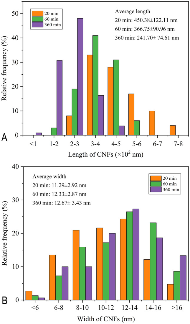
Size distribution of chitin nanofibers calculated by TEM images
Infrared spectroscopy characterization of chitin nanofibers
The FT-IR spectra of the TEMPO-oxidized chitins are shown in Fig. 3. The absorption peaks at 3431 and 3262 cm−1 are attributed to the O–H and N–H stretching vibrations, respectively. The absorption peaks at 1654 and 1622 cm−1 correspond to the amide I band. These bands are associated with the typical features of α-chitin (Wang et al. 2018a, b). The sodium carboxylate groups of CNFs were converted to protonated carboxyl groups under acidic condition. An absorption band appeared at approximately 1740 cm−1, indicating the hydroxyl groups had been successfully oxidized. The C–O stretching bands at approximately 1030 cm−1 were the internal standard band. As shown in Supplementary Fig. S3, the absorption ratios A1740/A1030 corresponded to the carboxylate contents of the CNFs. The degree of acetylation was plotted as the ratio of A1560/A1030, where the absorbance of the amide II bands appeared at 1560 cm−1 (Shigemasa et al. 1996). The carboxylate contents increased when the oxidation time was shorter than 60 min; the maximum appeared at 60 min. The contents decreased slightly with longer oxidation times. However, the degree of acetylation of CNFs treated with various times was mostly constant, indicating that almost no deacetylation occurred during the TEMPO/NaClO/NaBr oxidation.
Fig. 3.
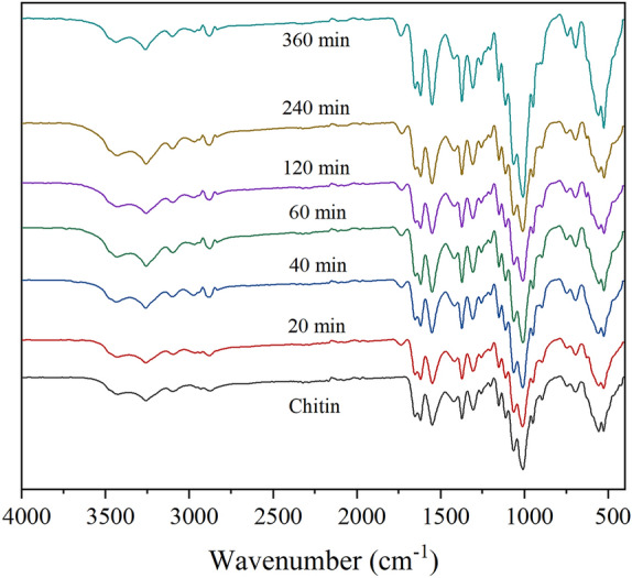
FT-IR spectra of the chitin nanofibers prepared with various oxidized times
The transmittance of chitin nanofibers
The transmittance of chitin nanofibers was evaluated by full wavelength scanning using an ultraviolet–visible spectrophotometer (Fig. 4). While the oxidation time was less than 40 min, the dispersive properties of CNFs were poor and the light transmittance was under 10%. When the oxidation time was 20 min, the transmittance was less than 2% (i.e., the highest transmittance was 1.15% at a wavelength of 800 nm), and the chitin crystal bundles made the dispersion muddy (Fig. 5). As seen in the optical microscopy images (Fig. 6), the CNFs oxidized for 20 min were flake shaped. Results showed there was undissolved chitin or chitin derivatives in the dispersion, making the dispersion non-transparent. The light transmittance was also continuously increased until the oxidation time was 360 min. Meanwhile, the dispersion tended to be transparent. There was no macroscopic flake chitin derivative in the dispersion treated for more than 120 min, suggesting CNFs subjected to ultrasonic treatment tended to be individual rather than aggregated.
Fig. 4.
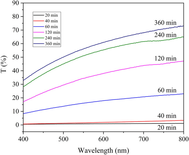
Light transmittance of chitin nanofibers’ dispersions treated with various oxidized times
Fig. 5.

Photographs of CNFs dispersions prepared with various oxidized times
Fig. 6.

Optical micrographs of chitin nanofibers treated with various oxidation times (magnification: a–c × 100; d–f × 400)
Content of carboxylate groups
The carboxylate contents of CNFs are shown in Fig. 7. The carboxylate content increased during TEMPO-treatment of up to 60 min. The content of carboxylate groups was 0.52 mmol/g CNFs, corresponding well to the tendency with the A1740/A1030 ratio (Supplementary Fig. S3). The carboxylate groups slightly decreased with oxidation times beyond 60 min. The results showed depolymerization and excessive oxidation can lead to an increase in water-soluble chitin with large consumption of NaOH and NaClO, with the content of carboxyl groups declining in the water-insoluble part.
Fig. 7.
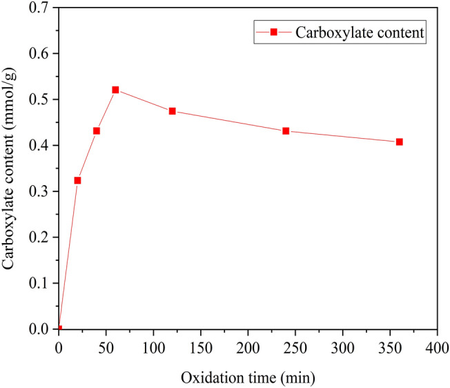
The content of carboxylate group about different chitin nanofibers’ dispersions
Immobilization enzymes
CNFs can be applied to enzyme immobilization because carboxyl groups of CNFs have covalent bindings with amino groups. The effects of oxidation time on immobilization are presented in Fig. 8. The enzyme loading increased with increased addition of enzymes when the CNFs were a type of immobilization carrier. While the additional amount of CTs was 500 or 2000 mg/g carrier, the highest loading immobilization and relative enzyme activity were at the oxidation time of 60 min. The highest enzyme loading was 307.17 ± 4.08 mg/g carrier with the addition of CTs at 500 mg/g carrier. When the additional amount was 2000 mg/g carrier, the maximum was 726.82 ± 12.05 mg/g carrier. When the oxidation time was below 60 min, the enzyme loading and relative activity increased with the increase of carboxyl group content. When the oxidation time was beyond 60 min, the CTs immobilization capacity decreased. While the oxidation time was 360 min, the catalytic activity of immobilized CTs only retained about 90% of the maximum relative enzyme because of the decreased carboxyl group content.
Fig. 8.
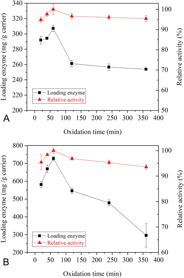
The immobilization of NH2-Fe3O4 modified chitin nanofibers (CNFs) and chymotrypsins (a 500 mg/g carrier; b 2000 mg/g carrier)
Discussion
Chitin has high crystallinity because of the hydrogen bond, and it is insoluble in water or organic solvents, leading to limited usage of chitin. Improving its solubility is a key factor for the industrial application of chitin. TEMPO is a type of organic radical that is widely used as a catalyst for oxidation. The C6 hydroxyl groups on the carbon chain can be converted into carboxylate groups using the TEMPO/NaBr/NaClO system (Muzzarelli et al. 1999). Hence, TEMPO oxidation can effectively improve the solubility and dispersion of chitin. When the oxidation time was less than 60 min, TEMPO could not completely oxidize chitin. Therefore, TEMPO-oxidized chitin was present only on the surface of the chitin bundles, with most chitin residues remaining unreacted in the product (Fan et al. 2007). As a result, the dispersity of the TEMPO-meditated chitin nanofibers with short oxidation time was low and bundles of the fibers and chitin flakes remained. Conversely, long-time TEMPO-oxidation can cause depolymerization of chitin, with the degrees of polymerization of products generally lower than 50 (Muzzarelli et al. 1999). The resulting crystal form of chitin was deformed and the crystallinity decreased. Additionally, solubility and the dispersity of the CNFs increased because of the TEMPO-causing depolymerization.
CNFs have great potential as a support for enzyme immobilization in various biotechnological applications (Wang et al. 2018a, b). Through the activation of the carboxylate groups using EDC/NHS, the CNFs can immobilize the NH2-Fe3O4 and CTs by covalent bonding. The degree of oxidation of chitin has a significant effect on the immobilization efficiency. As shown in Fig. 8, the enzyme activity and loading capacity of the immobilized CTs increased with the carboxylate group content of the chitin nanofibers. These results occurred because the enzymes were immobilized onto the chitin nanofibers via interaction with the carboxylate groups. Moreover, more carboxylate groups could conjugate more enzymes, resulting in increased enzyme activity and loading capacity of the immobilized enzymes.
Conclusion
In this study, TEMPO-oxidized chitin nanofibers treated with various oxidation time were prepared and characteristics analyzed. Chitin nanofibers were applied to enzyme immobilization using chymotrypsin as a model enzyme. It was demonstrated that enzyme efficiency could be improved by controlling TEMPO-oxidation time by using chitin nanofibers as support. The CNFs treated with TEMPO for 60 min retained the highest activity of immobilized enzymes and enzyme loading, corresponding to the maximum carboxylate content (0.52 mmol/g CNFs). While the additional amount of CTs was 500 or 2000 mg/g carrier, the highest loading amount of CTs was 307.17 ± 4.08 or 726.82 ± 12.05 mg/g carrier, respectively. The immobilized enzymes showed good correlation to the TEMPO oxidation time. Therefore, the optimal enzyme immobilization achieved with various oxidation times can reduce production costs when used in industrial reactions.
Materials and methods
Materials
2,2,6,6-Tetramethylpiperidine-1-oxyl (TEMPO) was purchased from Adamas Reagent Ltd. Chitin was purchased from Yuanye Bio-Technology Co. (Shanghai, China). Sodium hypochlorite (NaClO), N-hydroxysuccinimide (NHS) and chymotrypsins (CTs) were purchased from Macklin Biochemical Technology Co. (Shanghai, China). 2-morpholinoethanesulfonic acid (MES) and 1-ethyl-3-(3-dimethylaminopropyl) carbodiimide hydrochloride (EDC) were purchased from Aladdin Co. (Shanghai, China). Other materials were purchased from Sinopharm Group Co. Ltd. (Shanghai, China). All chemical reagents were of analytical grade.
Preparation of chitin nanofibers
The method to prepare chitin nanofibers (CNFs) was similar to that used by Fan et al. (2007) and Huang et al. (2018). Chitin powder (5.00 g, degree of acetylation was 96%) was suspended in water (417.5 ml) containing 1 mmol/L TEMPO (0.08 g) and 10 mmol/L sodium bromide (0.51 g). The oxidation of chitin was started by adding NaClO solution (4%, 82.50 ml, equal to 10 mmol/g chitin) into the slurry. The pH of the chitin slurry was maintained at 10 through 1 mol/L NaOH with magnetic stirring at room temperature. After different reaction times (20–480 min), the oxidization reaction ended by adding ethanol until the pH did not change. The pH of the mixture was adjusted to 7.0 with 0.5 mol/L HCl or 0.5 mol/L NaOH. The slurry was centrifuged at 9500 rpm for 15 min. The precipitate was washed with deionized water through repeated centrifugation and reduplicative suspension. After the precipitate was mixed with deionized water and a consistent concentration (0.1%) remained, the CNFs were treated with ultrasonic disruption (360 W, 30 min, Scientz IID). The CNFs were freeze-dried for further use.
Characterization of chitin nanofibers
The original chitin and the CNFs of different TEMPO-oxidized times were converted to powder and measured by XRD. The range of diffraction angle (2θ) was from 5° to 60° in an X-ray diffraction (Bruker D2 PHASE, German) that used Co Kα at 30 kV and 10 mA. The crystallinity (ICR, %) was calculated as ICR = (I020 − Iam)/I020 × 100, where I020 is the maximum intensity at about 9.6° and Iam is the intensity of amorphous diffraction at 16° (Zhang et al. 2005). The average size and size distribution of the chitin nanofibers were obtained by dynamic light scattering (DLS, Malvern Zetasizer Nano ZS 90) before 1‰ (m/v) CNF suspensions were filtered through 5 μm mixed cellulose ester (MCE) filtration. The images of CNFs were detected through transmission electron microscopy (MIC-JEM 1200EX) at an accelerating voltage of 120 kV after the dried CNFs were dispersed into 0.5% (m/v) dispersion. CNFs with free carboxylate groups were prepared by immersing them in 0.01 mol/L HCl, repeatedly washing them with deionized water, then drying them at 50 °C using a vacuum dryer for FT-IR measurement (Thermo Fisher-Nicolet 6700).
The dispersion properties were characterized by optical transmittance. The CNFs were dispersed in water suspensions of 1% (m/v). The transmittance of each sample was measured through a UV–Vis spectrophotometer (Shimadzu UV-2550) with the wavelength ranging from 400 to 800 nm (Saito and Isogai 2004). The CNFs were then observed through optical microscopy (BM-1000).
Determination of carboxylate groups content
The carboxylate content of the CNFs was determined by the electrical conductivity titration method (Fan et al. 2007). Water (40 ml) was added to the dried CNFs (0.20 g), the mixture stirred for 2 h to prepare a well-dispersed slurry, then 50 mmol/l HCl was added to the mixture to set the pH at 2.5. Next, 50 mmol/L NaOH solution was added at a rate of 0.1 ml/min up to pH 11 using a pH–Stat titration system. The carboxylate content and consumption of NaOH curves were obtained to reflect the content of both carboxylate groups.
Enzyme immobilization
The synthesis of NH2-Fe3O4
Preparation of the amine-functionalized magnetic nanoparticles (NH2-Fe3O4) was done using the same procedure as Mohapatra et al. (2007). FeCl2·4H2O (1.25 g) and FeCl3·6H2O (3.40 g) were dissolved in 100 ml deionized water under nitrogen protection. Ammonium hydroxide (6.00 ml) was added and stirred for 30 min. The system was then magnetically separated and precipitate was washed with deionized water and ethyl alcohol. The magnetic materials (Fe3O4) were vacuum dried for further use. Fe3O4 (1.00 g) was dispersed through a 50% (v/v) ethyl alcohol solution (100 ml) and the pH was adjusted to 4.0 by 0.1 mol/L HCl at 70 °C. Aminopropyltriethoxysilane (APTES, 3.00 ml) was added and stirred for 12 h under nitrogen atmosphere; then NH2-Fe3O4 was washed with deionized water and ethyl alcohol.
Immobilization of chymotrypsins and chitin nanofibers
Enzyme immobilization was performed using the method of Huang et al. (2018). CTs were used for immobilization. CNFs were activated through the EDC/NHS system to covalently immobilize the enzymes onto the surface of the supports. CNFs [1% (m/v)] treated with different oxidation times were dispersed in an MES buffer (100 mmol/L pH 6) containing EDC (10 mg/ml) and NHS (50 mg/ml). The mixture was then shaken for 3 h. The activated CNFs were separated via centrifugation at 13,000 r/min for 10 min. The water-insoluble part was washed with deionized water and centrifuged repeatedly.
In previous work, the CTs were immobilized onto CNFs without magnetic nanoparticles; however, the immobilized CTs were aggregated and enzyme activities not detected. Therefore, the CNFs (1 mg) were covalently bonded with NH2-Fe3O4 (0.5 mg) for quick separation. The suspension was shaken for 1 h at 20 °C. The magnetic chitin nanofibers were separated by external magnets. The citric acid–sodium citrate buffer solution (50 mmol/L pH 5) containing the CTs (500 or 2000 mg/g CNFs) was added to the CNFs. The resulting mixture was incubated for 2 h at 20 °C under shaking. The immobilized CTs were separated by external magnets and washed with a citric acid–sodium citrate buffer solution (50 mmol/L, pH 5). The concentration of CTs without immobilization was determined by the Bradford method. The activities of the immobilized enzymes were analyzed using the Lowry–Folin method (Bradford 1976; Kawamura et al. 1981).
Electronic supplementary material
Below is the link to the electronic supplementary material.
Acknowledgements
This work was supported by the National Key Research and Development Program of China (no. 2019YFD0901902), National Natural Science Foundation of China (31922072), China Agriculture Research System (CARS-48), Taishan Scholar Project of Shandong Province (tsqn201812020).
Author contributions
W-CH and XM conceived and designed the study. RC and WW performed the experiments. RC and W-CH wrote the paper. W-CH and XM reviewed and edited the manuscript. All authors read and approved the manuscript.
Compliance with ethical standards
Conflict of interest
The authors have no conflicts of interest to declare.
Animal and human rights statement
This article does not contain any studies with human participants or animals performed by any of the authors.
Footnotes
Edited by Xin Yu.
Rui Chen and Wen-Can Huang contributed equally.
References
- Bradford MM. A rapid and sensitive method for the quantitation of microgram quantities of protein utilizing the principle of protein-dye binding. Anal Biochem. 1976;72:248–254. doi: 10.1016/0003-2697(76)90527-3. [DOI] [PubMed] [Google Scholar]
- Deng Y, Deng C, Qi D, Liu C, Liu J, Zhang X, Zhao D. Synthesis of core/shell colloidal magnetic zeolite microspheres for the immobilization of trypsin. Adv Mater. 2009;21:1377–1382. doi: 10.1002/adma.200801766. [DOI] [Google Scholar]
- Fan Y, Saito T, Isogai A. Chitin nanocrystals prepared by TEMPO-mediated oxidation of α-chitin. Biomacromolecules. 2007;9:192–198. doi: 10.1021/bm700966g. [DOI] [PubMed] [Google Scholar]
- Han H, Zhou Y, Li S, Wang Y, Kong XZ. Immobilization of lipase from Pseudomonas fluorescens on porous polyurea and its application in kinetic resolution of racemic 1-phenylethanol. ACS Appl Mater Interfaces. 2016;8:25714–25724. doi: 10.1021/acsami.6b07979. [DOI] [PubMed] [Google Scholar]
- Hirano S. Chitin and chitosan as novel biotechnological materials. Polym Int. 1999;48:732–734. doi: 10.1002/(SICI)1097-0126(199908)48:8<732::AID-PI211>3.0.CO;2-U. [DOI] [Google Scholar]
- Huang WC, Wang W, Xue C, Mao X. Effective enzyme immobilization onto a magnetic chitin nanofiber composite. ACS Sustain Chem Eng. 2018;6:8118–8124. doi: 10.1021/acssuschemeng.8b01150. [DOI] [Google Scholar]
- Ifuku S, Nogi M, Abe K, Yoshioka M, Morimoto M, Saimoto H, Yano H. Preparation of chitin nanofibers with a uniform width as α-chitin from crab shells. Biomacromol. 2009;10:1584–1588. doi: 10.1021/bm900163d. [DOI] [PubMed] [Google Scholar]
- Ifuku S, Saimoto H. Chitin nanofibers: preparations, modifications, and applications. Nanoscale. 2012;4:3308–3318. doi: 10.1039/C2NR30383C. [DOI] [PubMed] [Google Scholar]
- Iyer PV, Ananthanarayan L. Enzyme stability and stabilization—aqueous and non-aqueous environment. Process Biochem. 2008;43:1019–1032. doi: 10.1016/j.procbio.2008.06.004. [DOI] [Google Scholar]
- Jayakumar R, Prabaharan M, Nair SV, Tamura H. Novel chitin and chitosan nanofibers in biomedical applications. Biotechnol Adv. 2010;28:142–150. doi: 10.1016/j.biotechadv.2009.11.001. [DOI] [PubMed] [Google Scholar]
- Jia H, Zhu G, Vugrinovich B, Kataphinan W, Reneker DH, Wang P. Enzyme-carrying polymeric nanofibers prepared via electrospinning for use as unique biocatalysts. Biotechnol Prog. 2002;18:1027–1032. doi: 10.1021/bp020042m. [DOI] [PubMed] [Google Scholar]
- Jiang J, Ye W, Yu J, Fan Y, Ono Y, Saito T, Isogai A. Chitin nanocrystals prepared by oxidation of α-chitin using the O2/laccase/TEMPO system. Carbohydr Polym. 2018;189:178–183. doi: 10.1016/j.carbpol.2018.01.096. [DOI] [PubMed] [Google Scholar]
- Kato Y, Kaminaga J, Matsuo R, Isogai A. TEMPO-mediated oxidation of chitin, regenerated chitin and N-acetylated chitosan. Carbohydr Polym. 2004;58:421–426. doi: 10.1016/j.carbpol.2004.08.011. [DOI] [Google Scholar]
- Kawamura Y, Nakanishi K, Matsuno R, Kamikubo T. Stability of immobilized α-chymotrypsin. Biotechnol Bioeng. 1981;23:1219–1236. doi: 10.1002/bit.260230605. [DOI] [Google Scholar]
- Khoshnevisan K, Bordbar AK, Zare D, Davoodi D, Noruzi M, Barkhi M, Tabatabaei M. Immobilization of cellulase enzyme on superparamagnetic nanoparticles and determination of its activity and stability. Chem Eng J. 2011;171:669–673. doi: 10.1016/j.cej.2011.04.039. [DOI] [Google Scholar]
- Kumar MNR. A review of chitin and chitosan applications. React Funct Polym. 2000;46:1–27. doi: 10.1016/S1381-5148(00)00038-9. [DOI] [Google Scholar]
- Kurita K. Chitin and chitosan: functional biopolymers from marine crustaceans. Mar Biotechnol. 2006;8:203. doi: 10.1007/s10126-005-0097-5. [DOI] [PubMed] [Google Scholar]
- Mateo C, Palomo JM, Fernandez-Lorente G, Guisan JM, Fernandez-Lafuente R. Improvement of enzyme activity, stability and selectivity via immobilization techniques. Enzyme Microb Technol. 2007;40:1451–1463. doi: 10.1016/j.enzmictec.2007.01.018. [DOI] [Google Scholar]
- Mohapatra S, Pramanik N, Mukherjee S, Ghosh SK, Pramanik P. A simple synthesis of amine-derivatised superparamagnetic iron oxide nanoparticles for bioapplications. J Mater Sci. 2007;42:7566–7574. doi: 10.1007/s10853-007-1597-7. [DOI] [Google Scholar]
- Mushi NE, Utsel S, Berglund LA. Nanostructured biocomposite films of high toughness based on native chitin nanofibers and chitosan. Front Chem. 2014;2:99. doi: 10.3389/fchem.2014.00099. [DOI] [PMC free article] [PubMed] [Google Scholar]
- Muzzarelli RAA, Muzzarelli C, Cosani A, Terbojevich M. 6-Oxychitins, novel hyaluronan-like regiospecifically carboxylated chitins. Carbohydr Polym. 1999;39:361–367. doi: 10.1016/S0144-8617(99)00027-2. [DOI] [Google Scholar]
- Pan C, Hu B, Li W, Sun Y, Ye H, Zeng X. Novel and efficient method for immobilization and stabilization of β-d-galactosidase by covalent attachment onto magnetic Fe3O4-chitosan nanoparticles. J Mol Catal B Enzym. 2009;61:208–215. doi: 10.1016/j.molcatb.2009.07.003. [DOI] [Google Scholar]
- Rinaudo M. Chitin and chitosan: properties and applications. Prog Polym Sci. 2006;31:603–632. doi: 10.1016/j.progpolymsci.2006.06.001. [DOI] [Google Scholar]
- Saito T, Isogai A. TEMPO-mediated oxidation of native cellulose. The effect of oxidation conditions on chemical and crystal structures of the water-insoluble fractions. Biomacromol. 2004;5:1983–1989. doi: 10.1021/bm0497769. [DOI] [PubMed] [Google Scholar]
- Shahidi F, Arachchi JKV, Jeon YJ. Food applications of chitin and chitosans. Trends Food Sci Technol. 1999;10:37–51. doi: 10.1016/S0924-2244(99)00017-5. [DOI] [Google Scholar]
- Sheldon RA. Enzyme immobilization: the quest for optimum performance. Adv Synth Catal. 2007;349:1289–1307. doi: 10.1002/adsc.200700082. [DOI] [Google Scholar]
- Shigemasa Y, Matsuura H, Sashiwa H, Saimoto H. Evaluation of different absorbance ratios from infrared spectroscopy for analyzing the degree of deacetylation in chitin. Int J Biol Macromol. 1996;18:237–242. doi: 10.1016/0141-8130(95)01079-3. [DOI] [PubMed] [Google Scholar]
- Tamura H, Furuike T, Nair SV, Jayakumar R. Biomedical applications of chitin hydrogel membranes and scaffolds. Carbohydr Polym. 2011;84:820–824. doi: 10.1016/j.carbpol.2010.06.001. [DOI] [Google Scholar]
- Tamura H, Nagahama H, Tokura S. Preparation of chitin hydrogel under mild conditions. Cellulose. 2006;13:357–364. doi: 10.1007/s10570-006-9058-z. [DOI] [Google Scholar]
- Wang Q, Yan X, Chang Y, Ren L, Zhou J. Fabrication and characterization of chitin nanofibers through esterification and ultrasound treatment. Carbohydr Polym. 2018;180:81–87. doi: 10.1016/j.carbpol.2017.09.010. [DOI] [PubMed] [Google Scholar]
- Wang W, Guo N, Huang WC, Zhang Z, Mao X. Immobilization of chitosanases onto magnetic nanoparticles to enhance enzyme performance. Catalysts. 2018;8:401. doi: 10.3390/catal8090401. [DOI] [Google Scholar]
- Wang Y, Caruso F. Mesoporous silica spheres as supports for enzyme immobilization and encapsulation. Chem Mater. 2005;17:953–961. doi: 10.1021/cm0483137. [DOI] [Google Scholar]
- Wang ZG, Wan LS, Liu ZM, Huang XJ, Xu ZK. Enzyme immobilization on electrospun polymer nanofibers: an overview. J Mol Catal B Enzym. 2009;56:189–195. doi: 10.1016/j.molcatb.2008.05.005. [DOI] [Google Scholar]
- Xiao A, Xiao Q, Lin Y, Ni H, Zhu Y, Cai H. Efficient immobilization of agarase using carboxyl-functionalized magnetic nanoparticles as support. Electron J Biotechnol. 2017;25:13–20. doi: 10.1016/j.ejbt.2016.10.007. [DOI] [Google Scholar]
- Zhang YT, Zhi TT, Zhang L, Huang H, Chen HL. Immobilization of carbonic anhydrase by embedding and covalent coupling into nanocomposite hydrogel containing hydrotalcite. Polymer. 2009;50:5693–5700. doi: 10.1016/j.polymer.2009.09.067. [DOI] [Google Scholar]
- Zhang Y, Xue C, Xue Y, Gao R, Zhang X. Determination of the degree of deacetylation of chitin and chitosan by X-ray powder diffraction. Carbohydr Res. 2005;340:1914–1917. doi: 10.1016/j.carres.2005.05.005. [DOI] [PubMed] [Google Scholar]
- Zheng H, Zhou J, Du Y, Zhang L. Cellulose/chitin films blended in NaOH/urea aqueous solution. J Appl Polym Sci. 2002;86:1679–1683. doi: 10.1002/app.11043. [DOI] [Google Scholar]
- Zhong C, Cooper A, Kapetanovic A, Fang Z, Zhang M, Rolandi M. A facile bottom-up route to self-assembled biogenic chitin nanofibers. Soft Matter. 2010;6:5298–5301. doi: 10.1039/c0sm00450b. [DOI] [Google Scholar]
Associated Data
This section collects any data citations, data availability statements, or supplementary materials included in this article.