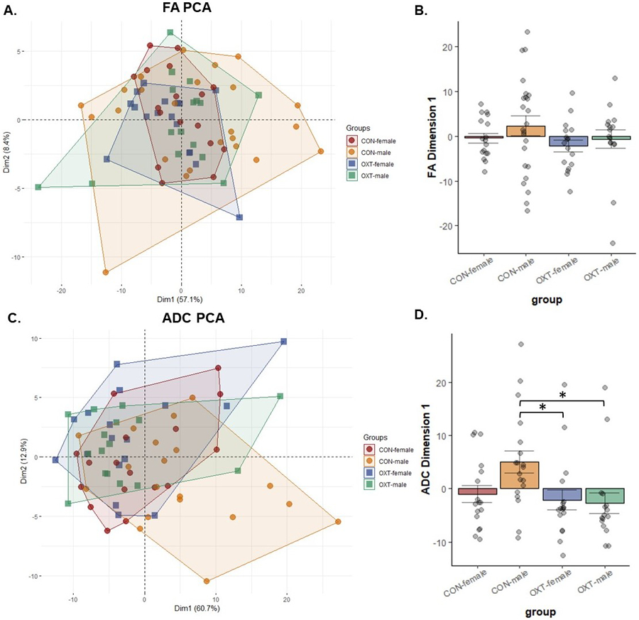
Figure 3.

Diffusion-weighted imaging (DWI) measures for fractional anisotropy (FA, panels A and B) and apparent diffusion coefficient (ADC, panels C and D) from 111 brain regions were loaded into a principal component analysis. (B) There were no significant differences in FA. (D) Control males had greater dimension 1 scores than OXT-exposed males (* p = 0.026) and OXT-exposed females (* p = 0.042). in terms of ADC.
