Abstract
Black queen cell virus (BQCV) is a ubiquitous honeybee virus and a significant pathogen to queen bee (Apis mellifera) larvae. However, many aspects of the virus remain poorly understood, including the transmission dynamics. In this study, we used next-generation sequencing to identify BQCV in Aedes vexans (n = 4,000) collected in 2019 and 2020 from Manitoba, Canada. We assembled de novo the nearly complete (>96%) genome sequence of the virus, which is the first available from North America and the first report of BQCV being harbored by mosquitoes. Phylogenetic tree reconstructions indicated that the genome had 95.5% sequence similarity to a BQCV isolate from Sweden. Sequences of a potential vector (Varroa destructor) and a microsporidian associated with BQCV (Nosema apis) were not identified in the mosquito samples, however, we did detect sequences of plant origin. We, therefore, hypothesize that the virus was indirectly acquired by mosquitoes foraging at the same nectar sources as honeybees.
Keywords: apiary, Aedes, nectar, Picornavirales, transmission
Highly valued as pollinators, honeybees (Apis mellifera L.) can be infected by a myriad of potentially detrimental viruses (Allen and Ball 1996, McMenamin and Genersch 2015; Grozinger and Flenniken 2019). Since it was first reported in 1955, Black queen cell virus (BQCV) is one of the most common and widespread honeybee viruses (Tentcheva et al. 2004, Ellis and Munn 2005, Mondet et al. 2014). Adult bees infected with the virus remain largely asymptomatic, however, BQCV can induce considerable mortality in the developing queen bee larvae, causing their necrotic carcasses to blacken pupal cells (Spurny et al. 2017). Despite its importance and prevalence, BQCV remains among the least understood honeybee viruses.
BQCV is classified as a Triatovirus, within the Dicistroviridae family and the order Picornavirales. The viral genome is composed of linear single-stranded, positive sense RNA of ~8,550 nucleotides in length (Kubaa et al. 2020). This includes two open reading frames (ORFs) encoding polyproteins containing non-structural (ORF1) and structural (ORF2) subunits (Spurny et al. 2017). The viral genome sequence is currently available from several geographical locations, including Europe (Tapaszti et al. 2009, Spurny et al. 2017, Kubaa et al. 2020), Asia (Li et al. 2023), Africa (Leat et al. 2000), and Australia (unpublished). However, to the best of our knowledge, no BQCV genome sequence has been reported from the Americas.
The precise mode of BQCV transmission has not yet been fully determined, but it likely involves multiple routes. Indeed, there is evidence that the virus is both venereally (Prodělalová et al. 2019) and vertically (Chen et al. 2006b, Naggar and Paxton 2020) transmitted. The presence of the microsporidian Nosema apis has also been linked to BQCV (Leat et al. 2000), but its role (if any) in virus transmission is unclear. Further, Varroa infestations have been associated with BQCV and the virus has been isolated from these mites (Ribière et al. 2008). However, the capacity of mites to serve as vectors of BQCV remains unresolved.
Another potential mode of BQCV transmission is through the foraging excursions of adult bees (Spurny et al. 2017). Honeybees collect nectar from flowers and orally pass it between workers, gradually converting the nectar into honey through biochemical processes and moisture loss (Leach and Drummond 2018). Although there is no direct evidence for nectar transmission of BQCV, honeybees have been shown to deposit the virus on flowers (Alger et al. 2019) and BQCV-positive pollen and honey have been identified (Chen et al. 2006a). Further, BQCV has been identified in both bumblebees (Peng et al. 2011) and solitary bees (Murray et al. 2018), which may be attributed to interspecies transfer of the virus through contaminated nectar sources.
Several methods have been employed to detect honeybee viruses, including enzyme-linked immunosorbent assay (ELISA), immunodiffusion, reverse transcriptase PCR (RT-PCR), and enhanced chemiluminescent (ECL) immunoblotting (Stoltz et al. 1995, Allen and Ball 1996, Benjeddou et al. 2001, Milićević et al. 2018). Next-generation sequencing (NGS) technologies are a relatively recent development and provide superior resolution and sensitivity to the aforementioned approaches. It has become increasingly used to detect viruses in insects, including honeybees (Marzoli et al. 2021, Li et al. 2022). In this investigation, we provide molecular evidence via NGS of Aedes vexans mosquitoes from Manitoba, Canada, harboring BQCV. We further speculate that the virus originated in flowers foraged by nectar-feeding mosquitoes.
Materials and Methods
To conduct this research, we collected mosquitoes in 2019 and 2020 in conjunction with provincial (Manitoba) surveillance programs. CDC miniature light traps (Model 1012, John W. Hock, Gainesville, FL) were hung on trees ~1.5 m off the ground, which released carbon dioxide (CO2; a female mosquito attractant) at 15 pounds per square inch (PSI) from dusk until dawn. In 2019, we placed traps in ten locations across the city of Brandon (49°50ʹ54″N 099°57ʹ00″W) in coordination with the City of Brandon and Manitoba Public Health. Trapping took place two nights per week for ten weeks, from July to September. In 2020, one-time (July) satellite traps from nine additional locations throughout the central and eastern regions of the province were provided to us by the City of Winnipeg Insect Control Branch. The collected mosquitoes were sorted and Ae. vexans were identified using relevant mosquito identification keys (Wood et al. 1979, Thielman and Hunter 2007). It should be noted that only females were captured and sequenced, as CO2 does not elicit host-seeking behaviors in males. Mosquitoes were stored at −80°C in location- and date-specific Petri dishes.
A maximum of fifty mosquitoes were pooled and RNA was isolated using the RNeasy Mini Kit (Qiagen, Hilden, Germany) according to the manufacturer’s recommendations. We then combined the RNA into two pools (by year) and ~2 µg per sample was sent to the Génome Québec Innovation Centre (McGill University, Montreal, QC, Canada) for mRNA library preparation (New England Biolabs, Ipswich, MA, USA) and paired-read sequencing (100 bp) using the NovaSeq 6000 System (Illumina, San Diego, CA). A total of 1,783 and 2,208 pooled Ae. vexans individuals were sequenced from collections in 2019 and 2020, respectively. Raw RNA sequencing reads can be retrieved from the NCBI short sequence read archive under the SRA accession number PRJNA866544. We processed the raw reads and performed de novo contig assembly using the CLC Genomics Workbench version 20 and optimized parameters: mismatch cost = 2, insertion cost = 3, deletion cost = 3, length fraction = 0.7, and similarity fraction = 0.95. To identify BQCV, each contig was mapped against the NCBI non-redundant (nr) database using BLASTn (E-value < 1 × 10100). We confirmed the presence of BQCV and the viral genome assembly using default settings with the Chan Zuckerberg ID Metagenomic Pipeline v6.8 (Chan Zuckerberg Biohub; CZID), an open-sourced cloud-based bioinformatics platform (Kalantar et al. 2020).
To explore the evolutionary relationship among BQCV isolates, we constructed a maximum likelihood tree based on a 7,961 bp region of the genome (>93% of the complete sequence). In addition to our Canadian strain, BQCV genomic sequences were retrieved from the NCBI database to represent a breadth of geographical viral isolates. We retained only genomic sequences that were >99% complete, and when more than one isolate sequence was available we selected the two most divergent isolates for further analysis. The sequences were aligned using default parameters (pairwise gap opening penalty = 15, gap extension penalty = 6.66) and tested for the best evolutionary model in MEGA X (Kumar et al. 2018). The tree with the highest likelihood was selected (-35842.56) and the phylogeny was then inferred using a maximum likelihood approach and a General Time Reversible (GTR) model with discrete Gamma distribution (5 categories; +G, parameter = 0.1639), with 1,000 bootstrap iterations (Nei and Kumar 2000). All codon positions were included in the analysis, while gaps and missing data were discarded from the final analysis.
Results
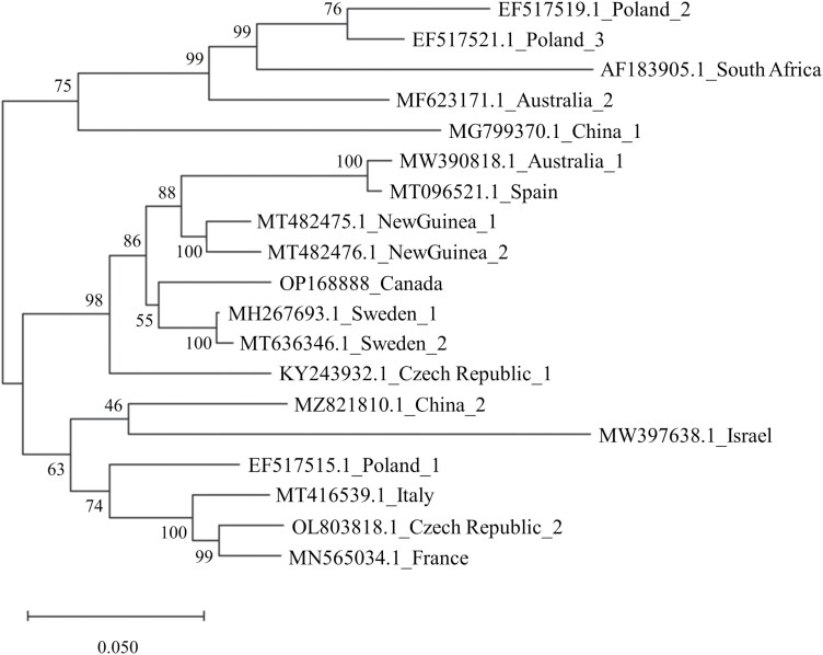
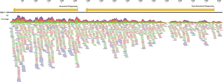
RNA sequencing of two pooled Ae. vexans RNA samples (2019 and 2020) generated 91,331,949 raw sequence reads. The reads were assembled into 28,566 (2019) and 33,563 (2020) contigs which, as expected, were primarily of Aedes origin. In 2019, one contig matched to BQCV, which had 95.5% sequence similarity to a BQCV isolate from Sweden in NCBI (MH267693.1; Fig. 1). The sequence represented ~95% (8,122 bp) of the viral genome with ~329 bp of the 5ʹ region not present, presumably due to low coverage of that region. For 2020, seven smaller BQCV contigs (between 267 and 826 bp) were assembled representing ~38% (3,233 bp) of the genome. We constructed a coverage map of the BQCV isolate from Canada using sequencing reads from both 2019 and 2020, which are displayed in Fig. 2. Genomic features of the BQCV sequence (e.g., ORFs, predicted proteins) were consistent with those previously reported (Leat et al. 2000, Spurny et al. 2017, Kubaa et al. 2020). The genomic sequence of the BQCV isolates reported in this study has been deposited in the GenBank database under the accession number OP168888.
Fig. 1.
Maximum likelihood inference of evolutionary relationship amongst BQCV isolates worldwide. BQCV sequences were retrieved and aligned to the Canadian isolate (OP168888) genomic sequences in MEGA X (see methods). The tree is drawn to scale with branch lengths representing the average number of substitutions per site analyzed. Numbers near branches represent the percentage of trees supporting the proposed topology between isolates.
Fig. 2.
Coverage map of the BQCV genome of the Canadian isolate. The map was generated by mapping sequencing reads from both positive Aedes vexans samples to the most similar BQCV genome in NCBI (MH267693.1). The mapping was carried out using CLC and default settings.
To provide insights into the possible origins of BQCV in Ae. vexans, we searched each contig sequence for orthologue matches that may be derived from potential vectors (mites), nectar (plants), and a microsporidian associated with the presence of BQCV in infected honey bee larvae (Nosema apis). For both samples, no contigs were identified that mapped to the most recent genome assemblies of Varroa destructor (Vdes_3.0) or Nosema apis (NapisBRLv01). However, a subset of contigs from the 2020 collections (n = 3) was of chloroplast origin (Table 1). All three are contigs mapped to multiple species with identical coverage/sequence similarity, including flowering plants, trees, and shrubs.
Table 1.
Contig sequences of plant origin found within Aedes vexans samples that were positive for BQCV
| Contig size (bp) | Year collected | Top match | Query coverage | Sequence similarity | Sequence |
|---|---|---|---|---|---|
| 399 | 2020 | Chloroplast | 100% | 100% | CGAATAGGTCAACCTTTCGAACTGCTGCTGAATCCATGGGCAGGCAAGAGACAACCTGGCGAACTGAAACATCTTAGTAGCCAGAGGAAAAGAAAGCAAAAGCGATTCCCGTAGTAGCGGCGAGCGAAATGGGAGCAGCCTAAACCGCGAAAACGGGGTTGTGGGAGAGCAATACAAGCGTCGTGCTGCTAGGCGAAGCGGTGGAGTGCTGCACCCTAGATGGCGAGAGTCCAGTAGCCGAAAGTATCACTAGCTTACGCTCTGACCCGAGTAGCATGGGACACGTGGAATCCCGTGTGAATCAGCAAGGACCACCTTGCAAGGCTAAATACTCCTGGGTGACCGATAGTGAAGTAGTACCGTGAGGGAAGGGTGAAAAGAACCCCCATCGGGGAGTGA |
| 473 | 2020 | Chloroplast | 100% | 99.15% | CTACCTTAGGACCGTTATTGTTACGGCCGCCGTTCACCGGGGCTTCGGTCGCCGGCTCCCCAGTCATCAGGTCACCAACATCCTTGACCTTCCGGCACTGGGCAGGCGTCAGCCCCCATACATGGTCTTACGACTTTGCGGAGACCTGTGTTTTTGGTAAACAGTCGCCCGGGCCTGGTCACTGCGACCCCCTTTGTGAGGAGGCACCCCTTCTCCCGAAGTTACGGGGCTATTTTGCCGAGTTCCTTAGAGAGAGTTGTCTCGCGCCCCTAGGTATTCTCTACCTACCCACCTGTGTCGGTTTCGGGTACAGGTACCCTTTTGTTGAAGGTCGTTCGAGCTTTTCCTGGGAGTATGGCATCGGTTACTTCAGCGCCGTAGCGCCTTGGTACTCGAACATTGGCTCGAGGCATTTTCTCGACCCCTTCTTACCCTGAAAAAGCAGGGGCACCTTGCGTCCTTGAACCGATAAC |
| 226 | 2020 | Chloroplast | 100% | 100% | GCTAAGCGATCTGCCGAAGCTGTGGGATGTAAAAATGCATCGGTAGGGGAGCGTTCCGCCTAGAGGGAAGCACCCGCGCGAGCAGTGGTAGACGAAGCGGAAGCGAGAATGTCGGCTTGAGTAACGCAAACATTGGTGAGAATCCAATGCCCCGAAAACCTAAGGGTTCCTCTGCAAGGTTCGTCCACGGAGGGTGAGTCAGGGCCTAAGATCAGGCCGAAAGGCG |
All three sequences could not be resolved to the species level, with each having top matches to flowering plants, shrubs, and trees.
Discussion
The results of our study strongly suggest the presence of BQCV in Ae. vexans is due to nectar foraging behaviors. This may seem counterintuitive as females of this mosquito species are hematophagous, primarily feeding on the blood of large mammals (e.g., deer, horses, and cows) (Nasci 1984). The nutrients in vertebrate blood are required for egg production by the vast majority of mosquito species, including iron and amino acids (Goldstrohm et al. 2003, Zhou et al. 2007). However, nectar represents a key source of nutrition for adult mosquitoes of both sexes (Barredo and DeGennaro 2020). For females, sugar deprivation has been associated with both reduced survival and fecundity (Foster 1995, Fernandes and Briegel 2005, Chadee et al. 2014). Since all of the plant sequences identified in our Ae. vexans samples were derived from chloroplasts, which are highly conserved (Cheng et al. 2020), we were unable to resolve the nectar sources to the species level. Plant preferences differ by mosquito species and availability, with a variety of semiochemicals elicited by plants that serve as attractants (Barredo and DeGennaro 2020). There is currently no evidence that BQCV can replicate in mosquitoes or be transmitted by mosquitoes, indicating Ae. vexans is likely a dead-end host for the virus. However, our findings of the virus in the same mosquito species across multiple years suggest that BQCV may be commonly associated with nectar foraging Ae. vexans.
To our knowledge, this is the first report of BQCV detected in mosquitoes or any other dipteran. In addition to mites, the virus has been identified in several Hymenopterans including ants (Payne et al. 2020), bumblebees (Peng et al. 2011), solitary bees (Murray et al. 2018), and wasps (Singh et al. 2010). Interspecies transmission of BQCV has been hypothesized to be due to direct (e.g., parasitism, predation, and scavenging) and/or indirect (foraging at the same nectar sources) interactions between honeybees and these arthropods (Grozinger and Flenniken 2019, Payne et al. 2020). Future studies aimed at directly sampling nectar-feeding Ae. vexans and validating the presence of BQCV in both the nectar and mosquito would better implicate foraging behaviors as a direct source of this virus in mosquitoes.
Our study also highlights the capabilities of massive parallel NGS technologies to characterize aspects of the host microbiome. Although it requires considerable integration of bioinformatics (Conesa et al. 2016), many limitations of traditional approaches for pathogen identification (e.g., PCR-based methods and serological testing) can be overcome using NGS. In addition to its greater resolution and sensitivity, NGS does not require a priori knowledge of the nucleic acid to be sequenced or specific antibodies (Díaz Cruz et al. 2019). Indeed, we had no expectation of identifying BQCV in Ae. vexans or isolating the near complete viral genome sequence. It is conceivable that NGS could be harnessed to determine the prevalence of key pollinator pathogens (e.g., Deformed wing virus, Acute bee paralysis virus, Kashmir bee virus, and Sacbrood virus) in a given region, either by sequencing the animal or the nectar. Moreover, if the virus can be readily obtained from nectar, it could serve as a biomarker to determine pollinator plant preferences and/or potential outbreaks.
In conclusion, we present the first report of BQCV detected in mosquitoes as well as the first comprehensive genome sequence of the virus from North America. While other studies have detected the virus in arthropods using traditional approaches (e.g., PCR), we demonstrate that BQCV can also be readily detected using NGS technologies. We hypothesize that Ae. vexans acquired BQCV by foraging at the same nectar sources as honeybees harboring the virus. Future research is needed to investigate whether mosquitoes are capable of transmitting BQCV to honeybees, either directly or indirectly. Further, studies aimed at determining the pervasiveness of BQCV in mosquito species and the contaminated nectar sources would provide added insights into the transmission dynamics of this virus.
Acknowledgments
We thank Ben Pilling, Milah Mikkelsen, Jessica Sparrow, and Carlyn Duncan for assistance with mosquito collections. The authors also thank the City of Winnipeg Insect Control Branch for assistance with mosquito collections and Manitoba Public Health for use of trapping equipment. We are grateful to Sheena Pharand and Kerri Weir for project support.
Contributor Information
Cole Baril, Department of Biology, Brandon University, Brandon, Manitoba R7A 6A9, Canada.
Christophe M R LeMoine, Department of Biology, Brandon University, Brandon, Manitoba R7A 6A9, Canada.
Bryan J Cassone, Department of Biology, Brandon University, Brandon, Manitoba R7A 6A9, Canada.
Funding
This project was funded through a grant from the Public Health Agency of Canada (PHAC) Infectious Disease and Climate Change Fund (IDCCF), awarded to Bryan Cassone.
Author Contributions
Cole Baril (Data curation-Lead, Formal analysis-Lead, Methodology-Lead, Writing – review & editing-Equal), Christophe LeMoine (Formal analysis-Supporting, Investigation-Supporting, Methodology-Equal, Software-Supporting, Writing – review & editing-Equal), Bryan Cassone (Conceptualization-Lead, Data curation-Supporting, Formal analysis-Equal, Funding acquisition-Lead, Investigation-Supporting, Methodology-Equal, Project administration-Lead, Resources-Lead, Software-Lead, Supervision-Lead, Writing – original draft-Lead)
References
- Alger SA, Burnham PA, Brody AK.. Flowers as viral hot spots: honeybees (Apis mellifera) unevenly deposit viruses across plant species. PLoS One. 2019:14:e0221800e0225295. 10.1371/journal.pone.0221800 [DOI] [PMC free article] [PubMed] [Google Scholar]
- Allen M, Ball B.. The incidence and world distribution of the honey bee viruses. Bee World. 1996:77(3):141–162. 10.1080/0005772x.1996.11099306 [DOI] [Google Scholar]
- Barredo E, DeGennaro M.. Not just from blood: mosquito nutrient acquisition from nectar sources. Trends Parasitol. 2020:36(5):473–484. 10.1016/j.pt.2020.02.003 [DOI] [PubMed] [Google Scholar]
- Benjeddou M, Leat N, Allsopp M, Davison S.. Detection of Acute bee paralysis virus and Black queen cell virus from honeybees by reverse transcriptase PCR. Appl Environ Microbiol. 2001:67(5):2384–2387. 10.1128/AEM.67.5.2384-2387.2001 [DOI] [PMC free article] [PubMed] [Google Scholar]
- Chadee DD, Sutherland JM, Gilles JRL.. Diel sugar feeding and reproductive behaviours of Aedes aegypti mosquitoes in Trinidad: with implications for mass release of sterile mosquitoes. Acta Trop. 2014:132:S86–S90. [DOI] [PubMed] [Google Scholar]
- Chen YP, Evans J, Feldlaufer MF.. Horizontal and vertical transmission of viruses in the honey bee, Apis mellifera. J Invert Pathol. 2006a:92:152–159. [DOI] [PubMed] [Google Scholar]
- Chen YP, Pettis JS, Collins A, Feldlaufer MF.. Prevalence and transmission of honeybee viruses. Appl Environ Microbiol. 2006b:72(1):606–611. 10.1128/AEM.72.1.606-611.2006 [DOI] [PMC free article] [PubMed] [Google Scholar]
- Cheng Y, Zhang L, Qi J, Zhang L.. Complete chloroplast genome sequence of Hibiscus cannabinus and comparative analysis of the Malvaceae family. Front Genet. 2020:11:227. 10.3389/fgene.2020.00227 [DOI] [PMC free article] [PubMed] [Google Scholar]
- Conesa A, Madrigal P, Tarazona S, Gomez-Cabrero D, Cervera A, McPherson A, Szcześniak MW, Gaffney DJ, Elo LL, Zhang X, et al. A survey of best practices for RNA-seq data analysis. Genome Biol. 2016:17:13. 10.1186/s13059-016-0881-8 [DOI] [PMC free article] [PubMed] [Google Scholar]
- Díaz Cruz G, Smith CM, Wiebe KF, Villanueva SM, Klonowski AR, Cassone BJ.. Applications of next generation sequencing for large scale pathogen diagnoses in soybean. Plant Dis. 2019:103:1075–1083. [DOI] [PubMed] [Google Scholar]
- Ellis JD, Munn PA.. The worldwide health status of honeybees. Bee World. 2005:86(4):88–101. 10.1080/0005772x.2005.11417323 [DOI] [Google Scholar]
- Fernandes L, Briegel H.. Reproductive physiology of Anopheles gambiae and Anopheles atroparvus. J Vector Ecol. 2005:30:11–26. [PubMed] [Google Scholar]
- Foster WA. Mosquito sugar feeding and reproductive energetics. Annu Rev Entomol. 1995:40:443–474. 10.1146/annurev.en.40.010195.002303 [DOI] [PubMed] [Google Scholar]
- Goldstrohm DA, Pennington JE, Wells MA.. The role of hemolymph proline as a nitrogen sink during blood meal digestion by the mosquito Aedes aegypti. J Insect Physiol. 2003:49(2):115–121. 10.1016/s0022-1910(02)00267-6 [DOI] [PubMed] [Google Scholar]
- Grozinger CM, Flenniken ML.. Bee viruses: ecology, pathogenicity, and impacts. Annu Rev Entomol. 2019:64:205–226. 10.1146/annurev-ento-011118-111942 [DOI] [PubMed] [Google Scholar]
- Kalantar KL, Carvalho T, A de Bourcy CF, et al. IDseq-An open source cloud-based pipeline and analysis service for metagenomic pathogen detection and monitoring. GigaScience. 2020:9:giaa111. [DOI] [PMC free article] [PubMed] [Google Scholar]
- Kubaa RA, Giampetruzzi A, Addante R, Saponari M.. Coding-complete genome sequence of a Black queen cell virus isolate from honeybees (Apis mellifera) in Italy. Microbiol Resour Announc. 2020:9:e00552–e00520. [DOI] [PMC free article] [PubMed] [Google Scholar]
- Kumar S, Stecher G, Li M, Knyaz C, Tamura K.. MEGA X: molecular evolutionary genetics analysis across computing platforms. Mol Biol Evol. 2018:35(6):1547–1549. 10.1093/molbev/msy096 [DOI] [PMC free article] [PubMed] [Google Scholar]
- Leach ME, Drummond F.. A review of native wild bee nutritional health. Int J Ecol. 2018:208:9607246. [Google Scholar]
- Leat N, Ball B, Govan V, Davison S.. Analysis of the complete genome sequence of black queen-cell virus, a picorna-like virus of honey bees. J Gen Virol. 2000:81(Pt 88):2111–2119. 10.1099/0022-1317-81-8-2111 [DOI] [PubMed] [Google Scholar]
- Li N, Huang Y, Li W, Xu S.. Virome analysis reveals diverse and divergent RNA viruses in wild insect pollinators in Beijing, China. Viruses. 2022:14(2):227. 10.3390/v14020227 [DOI] [PMC free article] [PubMed] [Google Scholar]
- Li N, Li C, Hu T, Li J, Zhou H, Ji J, Wu J, Kang W, Holmes EC, Shi W, et al. Nationwide genomic surveillance reveals the prevalence and evolution of honeybee viruses in China. Microbiome. 2023:11(1):6. 10.1186/s40168-022-01446-1 [DOI] [PMC free article] [PubMed] [Google Scholar]
- Marzoli F, Forzan M, Bortolotti L, Pacini MI, Rodríguez-Flores MS, Felicioli A, Mazzei M.. Next generation sequencing study on RNA viruses of Vespa velutina and Apis mellifera sharing the same foraging area. Transbound Emerg Dis. 2021:68(4):2261–2273. 10.1111/tbed.13878 [DOI] [PubMed] [Google Scholar]
- McMenamin AJ, Genersch E.. Honey bee colony losses and associated viruses. Curr Opin Insect Sci. 2015:8:121–129. 10.1016/j.cois.2015.01.015 [DOI] [PubMed] [Google Scholar]
- Milićević V, Radojičić S, Kureljušić J, Šekler M, Nešić K, Veljović L, Maksimović Zorić J, Radosavljević V.. Molecular detection of black queen cell virus and Kashmir bee virus in honey. AMB Expr. 2018:8:128. [DOI] [PMC free article] [PubMed] [Google Scholar]
- Mondet F, de Miranda JR, Kretzschmar A, Le Conte Y, Mercer AR.. On the front line: quantitative virus dynamics in honeybee (Apis mellifera L.) colonies along a new expansion front of the parasite Varroa destructor. PLoS Pathog. 2014:10(8):e1004323. 10.1371/journal.ppat.1004323 [DOI] [PMC free article] [PubMed] [Google Scholar]
- Murray EA, Burand J, Trikoz N, Schnabel J, Grab H, Danforth BN.. Viral transmission in honeybees and native bees, supported by a global black queen cell virus phylogeny. Environ Microbiol. 2018:21:972–983. [DOI] [PubMed] [Google Scholar]
- Naggar YA, Paxton R.. Mode of transmission determines the virulence of black queen cell virus in adult honeybees, posing a future threat to bees and apiculture. Viruses. 2020:12:535. [DOI] [PMC free article] [PubMed] [Google Scholar]
- Nasci RS. Variations in the blood-feeding patterns of Aedes vexans and Aedes trivittatus (Diptera: Culicidae). J Med Entomol. 1984:21(1):95–99. 10.1093/jmedent/21.1.95 [DOI] [PubMed] [Google Scholar]
- Nei M, Kumar S.. Molecular evolution and phylogenetics. New York: Oxford University Press; 2000. [Google Scholar]
- Payne AN, Shepherd TF, Rangel J.. The detection of honey bee (Apis mellifera)-associated viruses in ants. Sci Rep. 2020:10:2923. [DOI] [PMC free article] [PubMed] [Google Scholar]
- Peng W, Li J, Boncristiani H, Strange JP, Hamilton M, Chen Y.. Host range expansion of honey bee Black Queen Cell Virus in the bumble bee, Bombus huntii. Apidologie. 2011:42(5):650–658. 10.1007/s13592-011-0061-5 [DOI] [Google Scholar]
- Prodělalová J, Moutelíková R, Titěra D.. Multiple virus infections in western honeybee (Apis mellifera L.) ejaculate used for instrumental insemination. Viruses. 2019:11(4):306. 10.3390/v11040306 [DOI] [PMC free article] [PubMed] [Google Scholar]
- Ribière M, Ball B, Aubert M.. Natural history and geographical distribution of honey bee viruses. In Aubert M, editor. Virology and the honey bee. Luxembourg: European Communities; 2008. p. 15–84. [Google Scholar]
- Singh R, Levitt AL, Rajotte EG, Holmes EC, Ostiguy N, Vanengelsdorp D, Lipkin WI, Depamphilis CW, Toth AL, Cox-Foster DL.. RNA viruses in Hymenopteran pollinators: Evidence of inter-taxa virus transmission via pollen and potential impact on non-apis Hymenopteran species. PLoS One. 2010:5(12):e14357. 10.1371/journal.pone.0014357 [DOI] [PMC free article] [PubMed] [Google Scholar]
- Spurny R, Přidal A, Pálková L, Khanh H, Kiem T, de Miranda JR, Plevka P.. Virion structure of black queen cell virus, a common honeybee pathogen. J Virol. 2017:91:e02100–e02116. [DOI] [PMC free article] [PubMed] [Google Scholar]
- Stoltz D, Shen XR, Boggis C, Sisson G.. Molecular diagnosis of Kashmir bee virus infection. J Apic Res. 1995:34:153–160. [Google Scholar]
- Tapaszti Z, Forgach P, Kovago C, Topolska G, Nowotny N, Rusvai M, Bakonyi T.. Genetic analysis and phylogenetic comparison of Black queen cell virus genotypes. Vet Microbiol. 2009:139(3-4):227–234. 10.1016/j.vetmic.2009.06.002 [DOI] [PubMed] [Google Scholar]
- Tentcheva D, Gauthier L, Zappulla N, Dainat B, Cousserans F, Colin ME, Bergoin M.. Prevalence and seasonal variations of six bee viruses in Apis mellifera L. and Varroa destructor mite populations in France. Appl Environ Microbiol. 2004:70(12):7185–7191. 10.1128/AEM.70.12.7185-7191.2004 [DOI] [PMC free article] [PubMed] [Google Scholar]
- Thielman AC, Hunter FF.. Photographic key to the adult female mosquitoes (Diptera: Culicidae) of Canada. Can J Arthropod Identif. 2007:4. 10.3752/cjai.2007.04. Available online at: http://www.ualberta.ca/bsc/ejournal/th_04/th_04.html. [DOI] [Google Scholar]
- Wood DM, Dang PT, Ellis RA.. The mosquitoes of Canada (Diptera: Culicidae). In: The insects and arachnids of Canada, part 6: agriculture Canada. Publication 1686, 390; 1979. [Google Scholar]
- Zhou G, Kohlhepp P, Geiser D, Carmen Frasquillo M, Vazquez-Moreno L, Winzerling JJ.. Fate of blood meal iron in mosquitos. J Insect Physiol. 2007:53:1169–1178. [DOI] [PMC free article] [PubMed] [Google Scholar]


