Abstract
Background
The frequency of proteinase 3 gene (PRTN3) polymorphisms in patients with antineutrophil cytoplasmic antibody (ANCA)-associated vasculitis (AAV) is not fully characterised. We hypothesise that the presence of a PRTN3 gene polymorphism (single nucleotide polymorphism (SNP) rs351111) is relevant for clinical outcomes.
Methods
DNA variant calling for SNP rs351111 (chr.19:844020, c.355G>A) in PRTN3 gene assessed the allelic frequency in patients with PR3-AAV included in the Rituximab in ANCA-Associated Vasculitis trial. This was followed by RNA-seq variant calling to characterise the mRNA expression. We compared clinical outcomes between patients homozygous for PRTN3-Ile119 or PRTN3-Val119.
Results
Whole blood samples for DNA calling were available in 188 patients. 75 patients with PR3-AAV had the allelic variant: 62 heterozygous PRTN3-Val119Ile and 13 homozygous for PRTN3-Ile119. RNA-seq was available for 89 patients and mRNA corresponding to the allelic variant was found in 32 patients with PR3-AAV: 25 heterozygous PRTN3-Val119Ile and 7 homozygous for PRTN3-Ile119. The agreement between the DNA calling results and mRNA expression of the 86 patients analysed by both methods was 100%. We compared the clinical outcomes of 64 patients with PR3-AAV: 51 homozygous for PRTN3-Val119 and 13 homozygous for PRTN3-Ile119. The frequency of severe flares at 18 months in homozygous PRTN3-Ile119 was significantly higher when compared with homozygous PRTN3-Val119 (46.2% vs 19.6%, p=0.048). Multivariate analysis identified homozygous PR3-Ile119 as main predictor of severe relapse (HR 4.67, 95% CI 1.16 to 18.86, p=0.030).
Conclusion
In patients with PR3-AAV, homozygosity for PRTN3-Val119Ile polymorphism appears associated with higher frequency of severe relapse. Further studies are necessary to better understand the association of this observation with the risk of severe relapse.
Keywords: Autoantibodies; Granulomatosis with polyangiitis; Polymorphism, Genetic; Outcome Assessment, Health Care; Systemic vasculitis
WHAT IS ALREADY KNOWN ON THIS TOPIC
The implications of proteinase 3 gene (PRTN3) polymorphisms in exonic sequences of the gene have not been studied.
WHAT THIS STUDY ADDS
The PRTN3-Val119Ile polymorphism homozygosity was associated with higher frequency and risk of severe relapse in patients with PR3-antineutrophil cytoplasmic antibody (ANCA)-associated vasculitis (AAV) but not in myeloperoxidase-AAV.
Our data suggest that conformational changes of the autoantigen may modulate its interactions with pathogenic PR3-ANCA promoting disease activity.
HOW THIS STUDY MIGHT AFFECT RESEARCH, PRACTICE OR POLICY
Homozygosity for PRTN3-Val119Ile polymorphism may represent an additional identifiable risk factor for relapse.
Therefore, relapse risk might be stratified allowing the tailoring of treatment and adequate monitoring of patients at higher risk of relapse.
Introduction
The genetic background of antineutrophil cytoplasmic antibody (ANCA) associated vasculitis (AAV) is recognised as relevant for the disease pathogenesis.1 Evidence from genome-wide association studies (GWAS) has contributed decisively to question the original concept of clinical phenotypes in AAV.2–4 Patients are customarily classified according to shared clinical and histopathological features into granulomatosis with polyangiitis (GPA), microscopic polyangiitis (MPA) and eosinophilic granulomatosis with polyangiitis.5 However, over the last decade, studies have shown that clinical outcomes separate more clearly when patients are classified according with ANCA specificity into antiproteinase 3 (PR3)-AAV and antimyeloperoxidase (MPO)-AAV.5–7
GWAS in AAV demonstrates that PR3-AAV and MPO-AAV have different genetic risk associations.2–4 PR3-AAV is associated with the HLA-DPB1*04:01 which is related with the presentation of PR3 antigen to the immune system.2 3 8 Furthermore, the balance between α1-antitrypsin (SERPINA1) and PR3 (PRTN3) gene expression influences PR3 levels.2 3 In PR3-AAV, the role of PRTN3 gene polymorphisms has been previously studied.9 Eight single nucleotide polymorphisms (SNP) have been identified.9 The SNP in the promoter region might affect a putative transcription factor binding site, whereas the role of SNP in exonic sequences has not been defined.9 10 However, the actual frequency of PRTN3 polymorphisms in patients with ANCA-associated vasculitis (AAV) is not well characterised. The SNP rs351111 (chr.19:844020) has a minor allelic frequency between 0.497 and 0.322 and results in a nucleotide change from G to A in the position 355 (c.355G>A) in the PRTN3 gene.10 This change leads to a missense protein with an amino-acid change from Valine to Isoleucine in the position 119 of the PR3 protein (Val119Ile) and amino-acid numbering based on National Center for Biotechnology Information (NCBI) nomenclature.10 This variant has been previously described in PR3-AAV, and the allelic frequency of the variant was 35.3% vs 64.7% when compared with the wild type, and this was not different when compared with controls.10 The homozygous variants of the PRTN3 gene polymorphism (SNP rs351111) result in uniquely different proteins in affected patients. These different PR3 proteins could interact differently with potentially pathogenic PR3-ANCA. Therefore, we hypothesise that the PRTN3 gene polymorphism (SNP rs351111) might impact clinical outcomes.
This study aimed to (i) characterise the frequency of the SNP rs351111 and (ii) determine the association of the polymorphic conformation with disease presentation, clinical phenotypes and clinical outcomes such as relapse rate and severity of the disease in a very well-characterised clinical trial cohort.
Materials and methods
Study population and design
We studied the presence of PRTN3 Val119Ile polymorphism in samples obtained from patients enrolled in the RAVE (Rituximab in ANCA-Associated Vasculitis) trial.11 In brief, the RAVE trial was a multicentre, randomised, double-blind, non-inferiority controlled trial conducted between 2004 and 2008 that compared rituximab (RTX) to cyclophosphamide (CYC) for remission-induction in patients with AAV. The study enrolled 197 newly diagnosed or relapsing patients with AAV (GPA and MPA) in which 99 patients were assigned to treatment with intravenous RTX at a dose of 375 mg/m2/week for 4 weeks plus daily placebo of CYC and 98 patients were assigned to the control group and received treatment with placebo RTX infusions plus daily CYC (2 mg/kg). The mean (±SD) Birmingham Vasculitis Activity Score/Wegener Granulomatosis (BVAS/WG) at entry were 8.5±3.2 in the RTX group and 8.2±3.2 in the control group. The main clinical diagnoses were GPA in 148 patients (75%) and MPA in 48 patients (24%). ANCA specificity was PR3 in 133 patients (67.5%) and MPO in 66 patients (33.5%).
We initially performed a DNA variant calling analysis to characterise the presence of SNP rs351111 in our sample as this is the only SNP reported to result in an amino-acid change in PR3 (Val to Ile in position 119) with high allelic frequency.10 To determine the protein expressed and correct assignment of status to each patient through the DNA variant calling analysis, we performed RNA variant calling and determined the agreement. Finally, we correlated the zygosity status with clinical presentation and clinical outcomes.
DNA extraction, sequencing and variant calling analysis
Chromosome 19 and the PRTN3 gene were sequenced from whole blood samples and SNP and multinucleotide polymorphisms, insertions and deletions were characterised. These data were generated in collaboration with Immune Tolerance Network (ITN) and Genentech.
For primer design, ‘NCBI primer blast for design’ and manual search were used to encompass the flanking regions of 300 bp both upstream and downstream of the desired targets for optimal coverage, which resulted in the generation of 6 amplicons per sample to analyse the regions of interest. Amplicon generation for each of the six targets was performed using Q5 Hot Start High-Fidelity 2X Master Mix (NEB) (instrument: Tetrad Thermal Cycler, Bio-Rad) with the same DNA input amount across samples. The resulting amplicons were QC’ed with respect to yield and fragment length distribution using the Fragment Analyzer (Advanced Analytical). Based on this quantitation of the amplicons, one pool per 96-well plate was quantified again on the Agilent 2100 Bioanalyzer (Agilent Technologies) for final pooling of four plate-pools into one pool, respectively. The resulting three library pools underwent a final quantitation for loading on the MiSeq. The library-pools were sequenced on MiSeq instruments (Illumina) using the 2×300 bp (PE) run mode with double indexing.
Quality of the read sequences were performed using Trimmomatic (V.0.36, http://www.usadellab.org/cms/index.php?page=trimmomatic).12 Sequences with the following criteria were removed to improve on the subsequent analyses: possible adapter sequences, low-quality bases (Phred score<20) or shorter than a length threshold of 40 base pairs.
Mapping of reads to reference sequences was performed using BWA-MEM (V.0.7.12-r1039, http://bio-bwa.sourceforge.net/). The reads and their associated alignment positions are provided as binary alignment map (BAM) formatted files. Variant analysis was performed using VarScan (V.2.4.3, http://varscan.sourceforge.net). Only reads with a minimum alignment quality of 1 were used for variant analysis. This excludes all reads with ambiguous alignments. VarScan takes the deduplicated reads as input and searches for sequence variants that meet the following criteria: minimum read depth 8, minimum base quality of 15 (Phred Quality Score), alternative allele is supported by at least 2 reads, alternative allele has a frequency of at least 5%, the p value for variant is better than 0.98, the variant was observed on both strands. The called SNPs and InDels were further processed using snpEff (V.4.3p).13 The snpEff tool compares the called variants to a preprocessed database (snpEff DB-name: hg38). Based on the comparison, snpEff adds various annotations and predicts possible effects of each variant, for example, amino acid changes, frame shifts.
RNA extraction, sequencing and variant calling analysis
The methodology for RNA extraction from whole blood, deep sequencing, alignment, quantification of large RNA and quantitative reverse transcription-polymerase chain reaction (qRT-PCR) analysis were described elsewhere.14
FASTQ reads from RNA sequencing were aligned to human genome hg19 using STAR V.2.7.5b.12 The aligned reads in BAM files were then processed using GATK V.3.7.0 according to the best practice Variants were called from the processed BAM files using GATK HaplotypeCaller (V.3.7.0) or bcftools (V.1.9) (RNAseq short variant discovery [SNPs+Indels] – GATK (broadinstitute.org)).
Clinical presentation and outcomes
The clinical presentation and the clinical outcomes were retrieved from the RAVE trial.11 In brief, the main outcomes addressed were: complete remission, defined as BVAS/WG of 0 and prednisone dose of 0 mg/day; relapse, defined as an increase in the BVAS/WG of 1 point or more and severe relapse, defined as the presence of BVAS/WG ≥3 or the recording of a major BVAS/WG item after complete remission was achieved.
Statistical analysis
Categorical variables were presented as count (per cent) while continuous variables were presented as mean (SD) if they were normally distributed as determined by Shapiro-Wilk test or as median (IQR) if not normally distributed. For comparisons of categorical variables between groups, Pearson’s χ2 test was used. For comparison of continuous variables between two groups, an unpaired Student’s t-test for independent samples was used for distributions consistent with normality, and the Mann-Whitney U test was used otherwise. Bonferroni correction for multiple testing was applied.
The Kaplan Meier method was used to assess cumulative incidence of severe relapse after randomization and Cox proportional hazards regression model were used to determine predictive factors for severe relapse at 12 months.
IBM SPSS Statistics for MacOS, V.27 (IBM, Armonk, New York, USA) was used for all data analysis.
Results
DNA variant calling
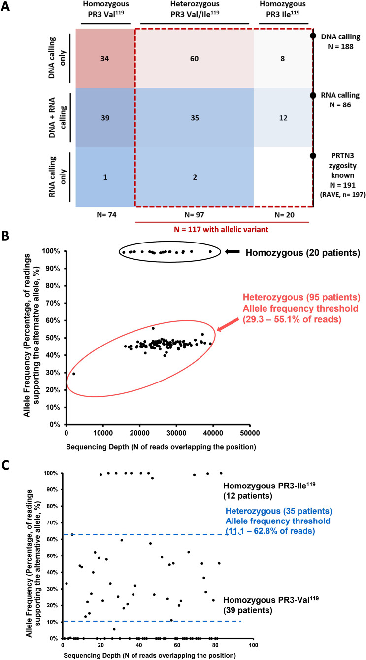
Available samples from 188 patients (95.4% of RAVE population) were analysed by next generation sequencing (targeted sequencing) (figure 1A and table 1). The allelic variant of the SNP rs351111 was detected in 115 patients with AAV (61.1%) and 75 patients with PR3-ANCA (39.9%) (figure 1A). From the 125 patients with PR3-ANCA evaluated, 50 (40.0%) were homozygous for PRTN3-Val119, 62 (49.6%) were heterozygous for PRTN3-Val119Ile and 13 (10.4%) were homozygous for PRTN3Ile119. The allele frequency threshold of readings for heterozygous varied from 29.3% to 55.1% of the reads (figure 1B).
Figure 1.
RAVE trial population distribution according to the zygosity status for PRTN3 gene (patients with PR3 and MPO-ANCA) and DNA or RNA variant calling (A) and allelic frequency for rs351111, chr.19:844 020 (c.355G>A) in PRTN3 among patients with AAV (PR3 and MPO-ANCA) in the DNA (B) and RNA (C) variant calling. AAV, ANCA-associated vasculitis; ANCA, antineutrophil cytoplasmic antibody; MPO, myeloperoxidase; PR3, proteinase 3; RAVE, Rituximab in ANCA-Associated Vasculitis.
Table 1.
PRTN3 gene polymorphism (SNP rs351111) DNA zygosity of the 188 patients included in the RAVE trial
| DNA zygosity, n (%) | Total (n=188) | PR3 (n=125) | MPO (n=63) |
| Homozygous PRTN3 Val119 | 73 (38.8) | 50 (40.0) | 23 (36.5) |
| Homozygous PRTN3 Ile119 | 20 (10.6) | 13 (10.4) | 7 (11.1) |
| Heterozygous PRTN3 Val119/Ile119 | 95 (50.5) | 62 (49.6) | 33 (52.4) |
ANCA, antineutrophil cytoplasmic antibody; MPO, myeloperoxidase; n, number; PR3, proteinase 3; RAVE, Rituximab in ANCA-Associated Vasculitis; SNP, single nucleotide polymorphism.
mRNA-sequencing
RNA zygosity was determined in 89 patients from the RAVE trial (figure 1A and table 2).11 The mRNA corresponding to the allelic variant was present in 47 patients (52.8%) and in 32 patients with PR3-ANCA (37.2%) (figure 1A). From the 60 patients with PR3-ANCA (67.4%) evaluated, 25 (28.1%) were homozygous for PRTN3-Val119, 28 (31.5%) were heterozygous for PRTN3-Val119Ile and 7 (7.9%) were homozygous for PRTN3Ile119. The allele frequency threshold of readings for heterozygous varied from 11.1% to 62.8% of the reads (figure 1C). Combining both analyses, we were able to determine the zygosity regarding the PRTN3-Val119Ile polymorphism in 191 patients (figure 1A). The agreement between the DNA calling results and the mRNA expression of the 86 patients that were analysed by both methods was 100%. When needed, we used the mRNA to confirm DNA zygosity (ie, patients with lower allelic frequency threshold in the DNA variant calling). In one additional patient in which a sample was not available for the DNA calling, it was possible to attribute the status of homozygosity for PRTN3-Val119 through the mRNA-sequencing.
Table 2.
PRTN3 gene polymorphism (SNP rs351111) mRNA zygosity of the 89 patients included in the RAVE trial with results available
| RNA zygosity, n (%) | Total (n=89) | PR3 (n=60) | MPO (n=23) |
| Homozygous PRTN3 Val119 | 39 (43.8) | 28 (46.7) | 11 (47.8) |
| Homozygous PRTN3 Ile119 | 12 (13.5) | 7 (11.7) | 5 (21.7) |
| Heterozygous PRTN3 Val119/Ile119 | 35 (39.3) | 25 (41.7) | 10 (43.5) |
| Missing in the DNA calling | 3 (3.4) (1 Homozygous PRTN3 Val119, 2 Heterozygous PRTN3 Val119/Ile119) | 3 (5.0) | – |
ANCA, antineutrophil cytoplasmic antibody; MPO, myeloperoxidase; n, number; PR3, proteinase 3; RAVE, Rituximab in ANCA-Associated Vasculitis.
Outcomes
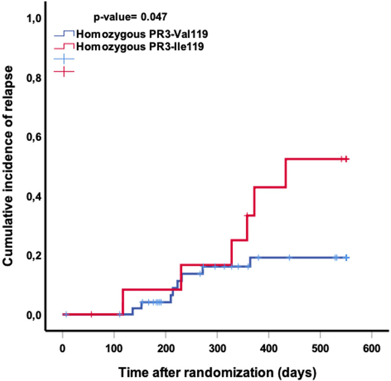
We analysed the clinical features and outcomes of 64 PR3-ANCA-positive patients: 51 patients homozygous for PRTN3-Val119 and 13 patients with PRTN3Ile119 (tables 3 and 4). There were no differences in the clinical presentation or treatment (table 3). We found that the proportion of severe flares experienced by 18 months in homozygous PRTN3-Ile119 carriers was more than two times higher when compared with homozygous PRTN3-Val119 patients (46.2% vs 19.6%, p=0.048) (table 4). Time to severe relapse after randomisation was also shorter in patients homozygous for PRTN3-Ile119 when compared with homozygous for PRTN3-Val119 (424 vs 489 days, p=0.047) (figure 2). We also reviewed the clinical presentation (individual BVAS/WG components) of each of the severe relapses of the patients homozygous for either PRTN3-Val119 or PRTN3-Ile119 and found that all had at least one disease manifestation typically attributed to inflammation of capillaries (capillaritis), that is, glomerulonephritis, alveolar haemorrhage, scleritis and so on, whereas the non-severe flares did not (data not shown).
Table 3.
Clinical characteristics and treatment of patients to PR3-ANCA according with PRTN3 zygosity
| PR3-ANCA (n=64) | Homozygous PRTN3-Val119 (n=51) |
Homozygous PRTN3-Ile119 (n=13) |
P value |
| Age at informed consent, median (IQR) years | 50 (38.0–57.0) | 55 (49.5–60.0) | 0.070 |
| Male, n (%) | 30 (58.8) | 6 (46.2) | 0.411 |
| Disease presentation, n (%) | 0.574 | ||
| AAV new diagnosis | 31 (62.0) | 9 (69.2) | |
| AAV relapse | 20 (39.2) | 4 (30.8) | |
| AAV, n (%) | 0.607 | ||
| MPA | 1 (2.0) | 0 (0.0) | |
| GPA | 50 (98.0) | 13 (100.0) | |
| BVAS/WG, median (IQR) | 8 (5–10) | 8 (7–9.5) | 0.485 |
| Any renal involvement, n (%) | 33 (64.7) | 9 (69.2) | 0.759 |
| Alveolar haemorrhage, n (%) | 15 (29.4) | 2 (15.4) | 0.307 |
| Capillaritis, n (%) | 42 (82.4) | 10 (76.9) | 0.654 |
| Randomised treatment group, n (%) | 0.662 | ||
| CYC | 24 (47.1) | 7 (53.8) | |
| RTX | 27 (52.9) | 6 (46.2) |
AAV, antineutrophil cytoplasmic antibody-associated vasculitis; ANCA, antineutrophil cytoplasmic antibody; BVAS/WG, Birmingham Vasculitis Activity Score for Wegener Granulomatosis; CYC, cyclophosphamide; GPA, granulomatosis with polyangiitis; MPA, microscopic polyangiitis; n, number; PR3, proteinase 3; PRTN3, proteinase 3 gene; RTX, rituximab.
Table 4.
Outcomes of patients with PR3-ANCA according with PRTN3 zygosity
| PR3-AAV (n=64) | Homozygous PRTN3-Val119 (n=51) |
Homozygous PRTN3-Ile119 (n=13) |
P value* |
| Remission, n (%) | 45 (88.2) | 13 (100) | 0.194 |
| Complete remission, n (%) | 36 (70.6) | 10 (76.9) | 0.650 |
| Any flare by 18 months, n (%) | 30 (58.8) | 7 (53.8) | 0.746 |
| Severe* flare by 18 months, n (%) | 10 (19.6) | 6 (46.2) | 0.048 |
*Relapse was considered severe according with the RAVE trial definition: BVAS/WG >3 or new major item.
AAV, antineutrophil cytoplasmic antibody associated vasculitis; ANCA, antineutrophil cytoplasmic antibody; BVAS/WG, Birmingham Vasculitis Activity Score for Wegener Granulomatosis; n, number; PR3, proteinase 3; PRTN3, proteinase 3 gene; RAVE, Rituximab in ANCA-Associated Vasculitis.
Figure 2.
Kaplan Meier curve of the severe relapse cumulative incidence after 550 days of randomisation according with the status of PRTN3 polymorphism: time to relapse is shorter in patients with homozygous for PRTN3-Ile119 when compared with homozygous for PRTN3-Val119 (424 vs 489 days, log-rank test p=0.047). PRTN3, proteinase 3 gene.
We used multivariable Cox regression to adjust our analysis to demographics, clinical characteristics and remission-induction treatment: homozygosity to PRTN3-Ile119 (HR 4.67, 95% CI 1.16 to 18.86, p=0.030) was the main predictor of severe relapse at 18 months, whereas any degree of kidney involvement was protective (HR 0.149, 95% CI 0.034 to 0.659, p=0.012), and this holds true when adjusted to the presence of any capillaritis (HR 2.823, 95% CI 0.426 to 19.156, p=0.288), alveolar haemorrhage (HR 0.919, 95% CI 0.211 to 3.994, p=0.910), baseline BVAS/WG (HR 0.971, 95% CI 0.756 to 1.246, p=0.817), age at informed consent (HR 0.971, 95% CI 0.929 to 1.016, p=0.205), gender (HR 0.392, 95% CI 0.133 to 1.152, p=0.089), enrolment as a relapser (HR 2.879, 95% CI 0.574 to 14.437, p=0.199) and remission-induction treatment with RTX (HR 0.767, 95% CI 0.226 to 2.610, p=0.672).
Finally, we also analysed the proportion of severe flares at 18 months in the patients with MPO-ANCA. There were no differences in the proportion of severe flares at 18 months in homozygous PRTN3-Ile119 when compared with the homozygous PRTN3-Val119 patients (14.3% vs 17.4%, p=0.847).
Discussion
Our study shows that in patients with PR3-AAV, the homozygosity for the allelic variant on the PRTN3-Val119Ile polymorphism was associated with more than double the risk for severe relapse in patients who achieve remission. In addition, we found that the agreement between the DNA variant calling results and mRNA-sequencing is 100%, suggesting that the DNA is sufficient to determine zygosity. We identified a genetic association that has potential prognostic relevance for the relapse risk stratification and management of PR3-AAV.
Reducing the occurrence of relapses is one of the most relevant unmet needs in the treatment of AAV.15 The accrual of damage is cumulative and relapse results in significant morbidity.1 Patients with PR3-AAV have an increased risk for relapse when compared with patients with MPO-AAV.15 In addition, granulomatous disease is more commonly associated with relapse.1 Relapse is also dependent on the treatment received for remission-induction.15 Moreover, patients who experienced previous relapses have a higher risk of subsequent relapse.1 Nevertheless, stratifying the pretreatment risk of relapse based on the presence of a biomarker or other differentiating feature has been challenging. Identifying patients at higher risk of relapse could be useful to personalise their management.
We hypothesise that, in vivo, the presence of polymorphisms in proteins might modulate their antigenicity particularly during states of severe inflammation, similarly to what was found in vitro. We previously showed that remote mutations and engagement of distal epitopes by an anti-PR3 monoclonal antibody can increase the local mobility and expose latent PR3 epitopes, leading to conformational adaptation facilitating or necessary for antibody binding.16 17 This type of epitope activation—achieved in vitro by remote mutations, or in vivo either by polymorphisms or by binding of another autoantibody or protein ligand—may be an important contributor of PR3-AAV pathogenesis.16 17 Our observation that homozygosity for the PRTN3-Val119Ile polymorphism is associated with a different risk for severe relapses is consistent with the hypothesis that the two different PR3 antigen variants may interact differently with pathogenic PR3-ANCA. The pathogenic potential of ANCA is best documented for disease manifestations caused by capillaritis, such as glomerulonephritis, alveolar haemorrhage, scleritis and so on, that is, those captured as ‘major’ disease activity items on the BVAS/WG instrument, which is in contrast to ‘minor’ BVAS/WG items. This may explain why we found a significant difference for severe relapses, but not when all non-severe relapses a lumped into the analysis.
In addition, we found that in patients with MPO-AAV, the presence of homozygosity to PRTN3-Ile119 is not associated with an increased risk of severe relapses at 18 months. This finding further supports the strength of our main results that for the first time identify a genetic association with an increased risk of severe relapse in PR3-AAV, but not MPO-AAV.
Our current study is unique because we analysed whether there is a correlation between a polymorphism and the risk of relapse in patients with PR3-AAV. Specifically, we linked the presence of the PRTN3-Val119Ile polymorphism to the risk of relapse in a well-characterised population that was treated similarly, and in which the outcomes were determined in a standardised and protocolised fashion. Because the agreement between DNA sequence and mRNA sequence was 100%, it is safe to assign a zygosity state based only on DNA sequencing particularly in patients that are homozygous for the presence or absence of variant. In patients that are heterozygous for the variant, it is not possible to determine what proportion of protein with each polymorphism is produced and if this is modified by an inflammatory state.
Our study also has limitations. First, more studies are needed to validate these findings. However, in this case, the validation will only be attainable with the analysis of a similar or larger population of patients in which clinical outcomes are comparable and evaluated in a similar standardised fashion. Second, as a result of effective remission induction regimens and longitudinal monitoring of patients, relapse is now a rare outcome, and severe relapse even more so. Hence, the small number of events in this rare disease is not unexpected and represents a challenge for the design of similar studies.
In conclusion, the findings in this cohort of patients with PR3-AAV suggest an association of homozygosity for the PRTN3-Ile119 polymorphism with higher frequency of severe relapse. Consequently, homozygosity for this polymorphism, which is the only one resulting in a change of the amino acid sequence of PR3, may represent an additional identifiable risk factor for severe relapse. Moreover, our data suggest that the quantity of PR3 expressed in patients with AAV compared with healthy controls and conformational changes of the autoantigen may modulate its interactions with pathogenic PR3-ANCA promoting disease activity.17 18 Further studies are necessary to validate and better understand the association of this observation with the risk of severe relapse and its possible molecular mechanisms.
Footnotes
Twitter: @martacasalmoura
Presented at: This work was presented at EULAR 2022, and it is published in abstract format (10.1136/annrheumdis-2022-eular.4977) at Annals of Rheumatic Diseases.
Contributors: MCM and US designed this project. ZD, SRB and PG obtained the RNA sequencing results. WT and MP obtained the DNA sequencing results. MCM, FF, PAM, PG and US analysed the data. MCM and US drafted the initial manuscript. All the authors revised and provided input to the final version of the manuscript. MCM and US were responsible for the overall content as the guarantor.
Funding: The Rituximab in ANCA-Associated Vasculitis (RAVE) trial was performed with the support of the Immune Tolerance Network (NIH Contract #N01 AI15416), an international clinical research consortium supported by the National Institute of Allergy and Infectious Diseases and the Juvenile Diabetes Research Foundation. Genentech and Biogen Idec provided the study medications and partial funding. At the Mayo Clinic and Foundation, the trial was supported by a Clinical and Translational Science Award from the National Center for Research Resources (NCRR) (RR024150-01); at Johns Hopkins University, by grants from the NCRR (RR025005) and career development awards (K24 AR049185 to JHS and K23 AR052820 to PS) and at Boston University, by a Clinical and Translational Science Award (RR 025771), grants from the National Institutes of Health (M01 RR00533) and a career development award (K24 AR02224 to PAM). PM was also supported by an Arthritis Investigator Award from the Arthritis Foundation. The present study was supported funds from the Connor Group Foundation and the Mayo Foundation to US as well as Roche Inc (DNA variant calling) and NIAMS (m-RNA sequencing). MSM is partially supported through a Vasculitis Clinical Research Consortium-Vasculitis Foundation Fellowship.
Competing interests: None declared.
Provenance and peer review: Not commissioned; externally peer reviewed.
© Author(s) (or their employer(s)) 2023. Re-use permitted under CC BY-NC. No commercial re-use. See rights and permissions. Published by BMJ.
Data availability statement
Data are available upon reasonable request. The data are available for consult per request sent to Dr. Ulrich Specks. The data are not part of public genetic database.
Ethics statements
Patient consent for publication
Not applicable.
Ethics approval
The RAVE trial was approved by the institutional review board at each participating site. Participants gave informed consent to participate in the study before taking part.
References
- 1.Kitching AR, Anders H-J, Basu N, et al. ANCA-associated vasculitis. Nat Rev Dis Primers 2020;6:71. 10.1038/s41572-020-0204-y [DOI] [PubMed] [Google Scholar]
- 2.Lyons PA, Rayner TF, Trivedi S, et al. Genetically distinct subsets within ANCA-associated vasculitis. N Engl J Med 2012;367:214–23. 10.1056/NEJMoa1108735 [DOI] [PMC free article] [PubMed] [Google Scholar]
- 3.Merkel PA, Xie G, Monach PA, et al. Identification of functional and expression polymorphisms associated with risk for antineutrophil cytoplasmic autoantibody-associated vasculitis. Arthritis Rheumatol 2017;69:1054–66. 10.1002/art.40034 [DOI] [PMC free article] [PubMed] [Google Scholar]
- 4.Lyons PA, Peters JE, Alberici F, et al. Genome-wide association study of eosinophilic granulomatosis with polyangiitis reveals genomic loci stratified by ANCA status. Nat Commun 2019;10:5120. 10.1038/s41467-019-12515-9 [DOI] [PMC free article] [PubMed] [Google Scholar]
- 5.Mahr A, Specks U, Jayne D. Subclassifying ANCA-associated vasculitis: a unifying view of disease spectrum. Rheumatology (Oxford) 2019;58:1707–9. 10.1093/rheumatology/kez148 [DOI] [PubMed] [Google Scholar]
- 6.Lionaki S, Blyth ER, Hogan SL, et al. Classification of antineutrophil cytoplasmic autoantibody vasculitides: the role of antineutrophil cytoplasmic autoantibody specificity for myeloperoxidase or proteinase 3 in disease recognition and prognosis. Arthritis Rheum 2012;64:3452–62. 10.1002/art.34562 [DOI] [PMC free article] [PubMed] [Google Scholar]
- 7.Schirmer JH, Wright MN, Herrmann K, et al. Myeloperoxidase-antineutrophil cytoplasmic antibody (ANCA) -positive granulomatosis with polyangiitis (Wegene’'s) is a clinically distinct subset of ANCA-associated vasculitis: a retrospective analysis of 315 patients from a German vasculitis referral center. Arthritis Rheumatol 2016;68:2953–63. 10.1002/art.39786 [DOI] [PubMed] [Google Scholar]
- 8.Chen DP, McInnis EA, Wu EY, et al. Immunological interaction of HLA-DPB1 and proteinase 3 in ANCA vasculitis is associated with clinical disease activity. J Am Soc Nephrol 2022;33:1517–27. 10.1681/ASN.2021081142 [DOI] [PMC free article] [PubMed] [Google Scholar]
- 9.Willcocks LC, Lyons PA, Rees AJ, et al. The contribution of genetic variation and infection to the pathogenesis of ANCA-associated systemic vasculitis. Arthritis Res Ther 2010;12:202. 10.1186/ar2928 [DOI] [PMC free article] [PubMed] [Google Scholar]
- 10.Gencik M, Meller S, Borgmann S, et al. Proteinase 3 gene polymorphisms and Wegener’s granulomatosis. Kidney Int 2000;58:2473–7. 10.1046/j.1523-1755.2000.00430.x [DOI] [PubMed] [Google Scholar]
- 11.Stone JH, Merkel PA, Spiera R, et al. Rituximab versus cyclophosphamide for ANCA-associated vasculitis. N Engl J Med 2010;363:221–32. 10.1056/NEJMoa0909905 [DOI] [PMC free article] [PubMed] [Google Scholar]
- 12.Bolger AM, Lohse M, Usadel B. Trimmomatic: a flexible trimmer for illumina sequence data. Bioinformatics 2014;30:2114–20. 10.1093/bioinformatics/btu170 [DOI] [PMC free article] [PubMed] [Google Scholar]
- 13.Cingolani P, Platts A, Wang LL, et al. A program for annotating and predicting the effects of single nucleotide polymorphisms, snpeff. Fly 2012;6:80–92. 10.4161/fly.19695 [DOI] [PMC free article] [PubMed] [Google Scholar]
- 14.Grayson PC, Carmona-Rivera C, Xu L, et al. Neutrophil-related gene expression and low-density granulocytes associated with disease activity and response to treatment in antineutrophil cytoplasmic antibody-associated vasculitis. Arthritis Rheumatol 2015;67:1922–32. 10.1002/art.39153 [DOI] [PMC free article] [PubMed] [Google Scholar]
- 15.Specks U, Merkel PA, Seo P, et al. Efficacy of remission-induction regimens for ANCA-associated vasculitis. N Engl J Med 2013;369:417–27. 10.1056/NEJMoa1213277 [DOI] [PMC free article] [PubMed] [Google Scholar]
- 16.Pang Y-P, Casal Moura M, Thompson GE, et al. Remote activation of a latent epitope in an autoantigen decoded with simulated B-factors. Front Immunol 2019;10:2467. 10.3389/fimmu.2019.02467 [DOI] [PMC free article] [PubMed] [Google Scholar]
- 17.Casal Moura M, Thompson GE, Nelson DR, et al. Activation of a latent epitope causing differential binding of antineutrophil cytoplasmic antibodies to proteinase 3. Arthritis Rheumatol 2022. 10.1002/art.42418 [DOI] [PMC free article] [PubMed] [Google Scholar]
- 18.Chen DP, Aiello CP, McCoy D, et al. PRTN3 variant correlates with increased autoantigen levels and relapse risk in PR3-ANCA versus MPO-ANCA disease. JCI Insight 2023;8:e166107. 10.1172/jci.insight.166107 [DOI] [PMC free article] [PubMed] [Google Scholar]
Associated Data
This section collects any data citations, data availability statements, or supplementary materials included in this article.
Data Availability Statement
Data are available upon reasonable request. The data are available for consult per request sent to Dr. Ulrich Specks. The data are not part of public genetic database.


