

Abstract
A central nervous system disorder is usually referred to as epilepsy. In epilepsy brain activity becomes abnormal, leading to times of abnormal behavior or seizures, and at times loss of awareness. Consequently, epilepsy patients face problems in daily life due to precautions they must take to adapt to this condition, particularly when they use heavy equipment, e.g., vehicle derivation. Epilepsy studies rely primarily on electroencephalography (EEG) signals to evaluate brain activity during seizures. It is troublesome and time-consuming to manually decide the location of seizures in EEG signals. The automatic detection framework is one of the principal tools to help doctors and patients take appropriate precautions. This paper reviews the epilepsy mentality disorder and the types of seizure, preprocessing operations that are performed on EEG data, a generally extracted feature from the signal, and a detailed view on classification procedures used in this problem and provide insights on the difficulties and future research directions in this innovative theme. Therefore, this paper presents a review of work on recent methods for the epileptic seizure process along with providing perspectives and concepts to researchers to present an automated EEG-based epileptic seizure detection system using IoT and machine learning classifiers for remote patient monitoring in the context of smart healthcare systems. Finally, challenges and open research points in EEG seizure detection are investigated.
Keywords: Epilepsy, Electroencephalography (EEG), Features extraction, Classification, Artificial intelligence, IoT
Introduction
Epilepsy, a neurological issue, has been considered a worldwide problem and is one of the principal dangers to human lives. As demonstrated by the reports of the World Health Organization (WHO), around 50 million individuals around the world are suffering from epilepsy, which makes it quite possibly the most broadly recognized global neurological disease [13, 27]. Epilepsy influences both females, males, and even children. The seizure’s symptoms can vary broadly [20, 41].
A few groups of individuals with epilepsy just stare blankly for a couple of moments during a seizure, while others repeatedly jerk their arms or legs. Having one seizure attack doesn’t mean you have epilepsy. At least two seizure attacks without stimulus are generally required for an epilepsy diagnosis. To detect brain abnormalities, EEG is one of the widely common techniques utilized to measure electrical disturbances in the human brain for the diagnosis of epileptic seizures [88, 142].
The normal shape of EEG signals gets modified during an epileptic seizure. Thus, based on the variety of EEG signal characteristics, epileptic patients’ states can be grouped into three phases, normal, preictal, and ictal. Many electrical disturbances start happening in the cerebrum of epileptic patients before a seizure’s actual onset, which is termed a preictal stage. For recognition of seizures at this stage, such electrical disturbances in the patient’s brain need to be recorded during the transition from normal to the ictal stage [109]. Consequently, this process of early recognition of epileptic seizures at the preictal stage could save the existence of patients by enabling them to take precautionary measures to prevent injurious and life-threatening accidents. In EEG tests, electrodes are associated with your scalp using a paste-like substance or cap. The electrodes register the electrical activity of your brain [47, 67, 107].
As previously stated, there is an urgent need for an automatic efficient method for early recognition of epileptic seizures to save the lives of thousands of epileptic patients every year, capable of alerting the patients, their families, and nearby hospitals before the actual occurrence of epileptic seizures. so, this system could help epileptic patients in the case of an emergency to save their lives and for improving their quality of life [3, 39].
Recently, research is being attempted for the detection of epileptic seizures to support for automatic diagnosis system to help clinicians from burdensome work. In this respect, an enormous number of research papers is published for the identification of epileptic seizures. Automatic seizure detection can prove efficient by making the process reliable and faster. Therefore, this domain attracts the researchers to investigate several types of techniques and domains such as the frequency domain, time domain, time-frequency domain, Empirical mode decomposition, and nonlinear methods. Nevertheless, experiments showed significant performance improvements when two or more conventional methods are combined. U. Rajendra discusses various entropies utilized for an automated diagnosis of epilepsy using EEG signals, and also the applications of entropies with their advantages and disadvantages [4]. In [124] automated epileptic seizure detection techniques based on multi-domain approaches were reviewed. The authors in [102] review pattern recognition techniques to detect seizures from EEG data, and also study the performance of DWT features with different classifiers. Focal and non-focal characterization are identified to determine areas affected by seizures [5]. Md Shafiqul Islam suggested a dynamic approach utilizing a deep learning model (Epileptic-Net) to detect an epileptic seizure. This approach included dense convolutional blocks, feature attention modules, residual blocks, and the hypercolumn technique [49]. Gaetano Zazzaro and Luigi Pavone evaluate the performance of a seizure detection system by studying its performance in correctly identifying seizures and in minimizing false alarms and to decide if it is generalizable to several patients [146]. In [74] explore the possibilities of wearable multi-modal monitoring in epilepsy and identify effective strategies for seizure-detection.
Nowadays, the Internet of Things (IoT) is playing a dynamic vital role in medical care by providing significant solutions for many medical and healthcare applications. The IoT technologies provide continuous and real-time observation of patients’ health using wearable devices. These technologies are also applied for the acquisition and transmission of EEG signals of epileptic patients. Along with such technologies, machine learning algorithms provide promising solutions for the effective detection of seizure stages from received EEG signals. Also, IoT utilized in combination with AI procedures and cloud computing services has arisen as a powerful technology to resolve many problems in the medical care area. The need to present an automatic epileptic seizure recognition framework for early identification of epileptic seizures utilizing existing communication technologies in collaboration with machine learning, IoT, and cloud computing [3, 93].
This work will bring researchers up to date on the significant feature extraction techniques, statistical and machine learning classifiers, and recent deep learning algorithms. Another contribution of this review is to help researchers to identify publicly available databases of recorded epileptic seizure signals. Finally, based on this current review, suggestions on future research directions are provided. In conclusion, the primary contributions of this work can condense as follows:
Provide an overview of EEG signals and explore the seizure detection process along with providing information on available EEG datasets.
Reviewing works done using various deep learning models for automated detection of epileptic seizures by various modalities.
Explore challenges in the detection of epileptic seizures as well as analyze the best performing model for various modalities of data.
Explore and present AI-based seizure detection and IoT-based automated seizure detection.
Provide opportunities and future research directions for this cutting-edge research subject.
This paper is organized as follows, Section 2 defines preliminaries for EEG data, available datasets, and the seizure detection process, which is separated into three steps: preprocessing step, feature extraction techniques, and classification algorithms. Section 3 presents the AI-based Seizure detection. The IoT-based automated seizure detection is provided in Section 4. Section 5 shows the challenges and the future research direction. Finally, the paper ended with the conclusion and references.
EEG signal acquisition
This section explores the basic knowledge of EEG signals and the available datasets with understanding the methodology of EEG signal acquisition.
EEG data
Epilepsy is a neurological issue described by the erratic intrusion of typical cerebrum exercises. In epilepsy, certain zones or all zones of the cerebrum are overactive, this prompts seizures. Epileptic seizures fluctuate from one person to another one, they may last for a few seconds and unnoticed, they might influence just a single arm or one leg or the entire body, and sometimes individuals end up oblivious. Thusly, there are two primary classes of epileptic seizure as shown in Fig. 1:
Generalized seizures
Partial seizures
Fig. 1.

Types of seizures and sub-types
Generalized seizures affect the whole brain. It results in a lack of consciousness and makes the whole frame convulse. Focal or partial epileptic seizures include just a specific piece of the cerebrum. As in Fig. 1, generalized epileptic seizures can be subdivided into absence (petit mal), tonic-clonic (grand mal), atonic, myoclonic, clonic, and tonic seizures [92]. To diagnose epileptic seizures, Various screening techniques have been developed. This is accomplished by using various methods like Electro Encephalography (EEG), Magneto Encephalography (MEG), functional Magnetic Resonance Imaging (fMRI), and Near Infra-Red Spectroscopy (NIRS). Table 1 presents a comparison between the various kinds of brain signal acquisition.
Table 1.
Brain signal acquisition methods

EEG is the most widely preferred signal acquisition method. This is due to EEG signals being economical, portable, and obtaining clear rhythms in the frequency domain. The EEG provides the voltage variations produced by the ionic current of neurons in the brain, which indicate the brain’s bioelectric activity. Diagnosing epilepsy with EEG signals is time-consuming and strenuous, as the epileptologist or neurologist needs to screen the EEG signals minutely. Also, there is a possibility of human error, and hence, developing a computer-based diagnosis may alleviate these problems.
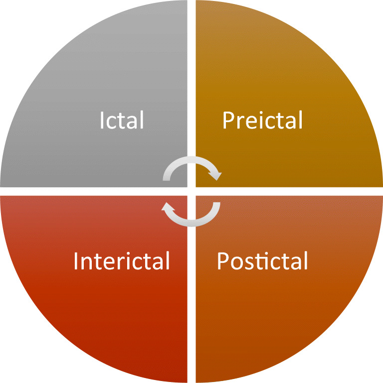
Electroencephalography (EEG) is a simple test that estimates electrical activity in the cerebrum. It is generally utilized for the recognition and examination of epileptic seizures. EEG signals can be partitioned into four stages, which are marked by epilepsy patients. These stages are an ictal, preictal, postictal, and interictal state as in Fig. 2 [17, 61].
Fig. 2.
Different stages of EEG signals
EEG signals divided into five main rhythms are perceived from an EEG recording: Alpha 0.5 − 4 Hz, Beta 4 − 8 Hz, Gamma 8 − 12 Hz, Delta 12 − 30 Hz, and Theta over 30 Hz. The measure of activity in various EEG frequency bands can be quantified by employing spectral analysis techniques as in Table 2 [32, 128].
Table 2.
EEG frequency bands
| Frequency Band Name | Frequency Bandwidth (Hz) |
|---|---|
| Alpha | <4 |
| Beta | 4–8 |
| Gamma | 8–12 |
| Delta | 12–30 |
| Theta | >30 |
EEG signals are normally obtained using superficial scalp electrodes, placed using a 10–20 international system presented in Fig. 3. Electrode Placement using a 10–20 System is a strategy used to portray the area of electrodes on the scalp. It is relying upon the relationship between the area of an electrode and the layer of the cerebral cortex. Each site has a letter (to recognize the lobe) and a letter or number to recognize the area. The letters T, F, C, O, and P stand for Temporal, Frontal, Central, Occipital, and Parietal. The left hemisphere is directed by (1,3,5,7) while (2,4,6,8) numbers refer to the right hemisphere. Electrodes put on the midline are referred to using the letter z. Smaller numbers refer to a closed position to the midline [32].
Fig. 3.

10–20 International system for Electrode Placement
EEG datasets
A dataset plays an important role for researchers and scientists in developing accurate and robust Computer-aided designs to evaluate the performance of their models. EEG recording is a broadly common tool for observing brain activity. These recordings play a vital role in machine learning classifiers to explore the novel methods for seizure detection in different ways such as onset seizure detection, quick seizure detection, patient seizure detection, and seizure localization. The significance of openly accessible datasets is to give a benchmark to dissect and contrast the outcomes with others.
To develop and evaluate the automated epileptic seizure detection systems, several publicly available datasets: CHB-MIT [43, 127, 144, 150], ECoG Dataset [72, 108], Freiburg epilepsy dataset [112, 150], Bonn seizure dataset [82, 119, 128, 136], BERN- Barcelona dataset [44, 97], Kaggle dataset, Flint-Hills eplipsiae, Hauz Khas and Zenodo dataset. The signals obtained from these datasets are recorded either intracranially or from the scalp of humans or animals. Table 3 summarizes the supplementary information for this public dataset.
Table 3.
| Dataset | Number of Patients | Number of Seizures | Recording | Total Duration (hour) | Sampling Frequency (Hz) |
|---|---|---|---|---|---|
| Flint-Hills [124] | 10 | 59 | Continues intracranial ling term ECoG | 1419 | 249 |
| Hauz Khas [124] | 10 | NA | Scalp EEG (sEEG) | NA | 200 |
| Freiburg [92] |
21 8 males, 13 females |
87 | Intracranial Electroencephalography (IEEG) | 708 | 256 |
| CHB-MIT [61] |
22 17 females, ages 1.5–19. 5 males, ages 3–22. |
163 | sEEG | 844 | 256 |
| Kaggle [106] | 5 dogs | 48 | IEEG | 627 | 400 |
| 2 patients | 5 KHz | ||||
| Bonn [106, 147] |
10 5 patients and 5 healthy controls |
NA | Surface and IEEG | 39 m | 173.61 |
| Barcelona [8, 128] | 5 | 3750 | IEEG | 83 | 512 |
| Zenodo [8, 150] | 79 neonatal | 460 | sEEG | 74 m | 256 |
Epileptic seizure detection system
Artificial Intelligence (AI) plays a significant role in making support and assisting in the medical field. Especially, in EEG-based Seizure detection for early diagnosis of epilepsy. The seizure detection process can be separated into three main phases: preprocessing, feature extraction, and classification, as in Fig. 4. These phases would be presented in detail.
Fig. 4.

A seizure discovery framework
EEG preprocessing
Raw EEG signal is non-stationary and has a low spatial resolution. EEG signals are susceptible and highly affected by artifacts and noise. These artifacts may affect information and analysis of the recorded signals. Therefore, the necessity of artifact identification and removal, either in clinical diagnosis or practical applications, is the most significant preprocessing step before being utilized to reduce their impact on the feature extraction stage. In this step also, it is crucial to decide on frequency and channel from EEG as it is produced from numerous electrodes. Signal pre-processing sometimes is named Signal Enhancement. In general, the acquired cerebrum signals are contaminated by noise and artifacts.
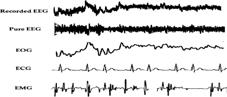
The artifacts are divided into two types physiologic and extra-physiologic. Extra-physiologic or Exterior Artifacts are created from outside the body like equipment, faulty electrodes, Power lines, ventilation, and digital artifacts (loose wiring, etc.). Physiological artifacts also called (Interior Artifacts) created from the body such as eye blinks, eye movements (EOG), heartbeat (ECG), muscular movement (EMG), Skin resistance, and power line interferences are also merged with brain signals, these types of artifacts are more muddled to eliminate. Figure 5 illustrate the EEG signal having physiological artifacts [48, 53, 61]. Types of artifacts in EEG signals are illustrated in Table 4.
Fig. 5.
EEG signal with physiological artifacts
Table 4.
Types of artifacts in EEG signals
| Interior Artifacts | Exterior Artifacts |
|---|---|
| Blinking of the eye (EOG) | Power line |
| Heartbeat (ECG) | Machine fault |
| Muscle movements (EMG) | Faulty electrode/poor placement |
| Skin resistance | ventilation |
| Subject’s movement | Digital artifacts (loose wiring, etc.) |
Several efficient techniques are used for artifact removal particularly for physiological artifacts, as demonstrated in Fig. 6. It demonstrates that the most generally use algorithms are BSS-based methods, particularly ICA (34%) [54]. Also, it is worthy to take note that because of the constraints of single strategies like single Regression and BSS, a lot of researchers incline toward the hybrid method (20%) to upgrade the exhibition of methods in recent years. Filtering (13%) and wavelet transform (9%) are also artifact removal algorithms. Despite the extensive research on artifact removal from EEG signals, there is no agreement ideal answer for all types of artifacts [53, 57, 104].
Fig. 6.
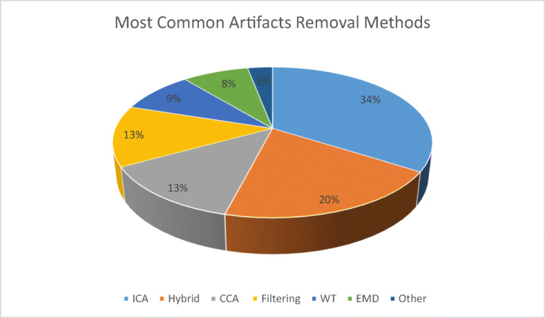
The percentages for the most common artifacts removal methods [53]
The constraints mean working on single methods sometimes not achieved acceptable results so the choice of using hybrid methods is good to achieve good performance on EEG seizure detection
Artifact removal takes place by using Common Average Referencing (CAR), Common Spatial Patterns (CSP), Principal Component Analysis (PCA), Surface Laplacian (SL), Independent Component Analysis (ICA), Frequency Normalization (Freq-Norm), Single Value Decomposition (SVD), Common Spatio-Spatial Patterns (CSSP), Common Spatial Subspace Decomposition (CSSD), Differential Window (DW), Local Averaging Technique (LAT), Robust Kalman Filtering, Butter worth filter, and simple classifier, etc. The most frequently used techniques are ICA, PCA, CAR, SL, CSP, and Adaptive Filtering [19, 80].
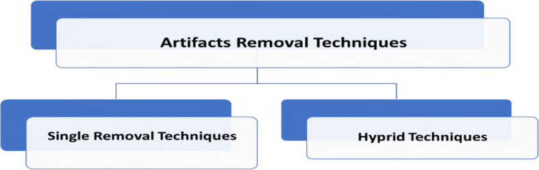
Artifact removal can be divided into two different categories. The first one is single removal techniques, and the other is the hybrid one as displayed in Figs. 7, 8, and 9.
Fig. 7.
Techniques of artifacts removal
Fig. 8.
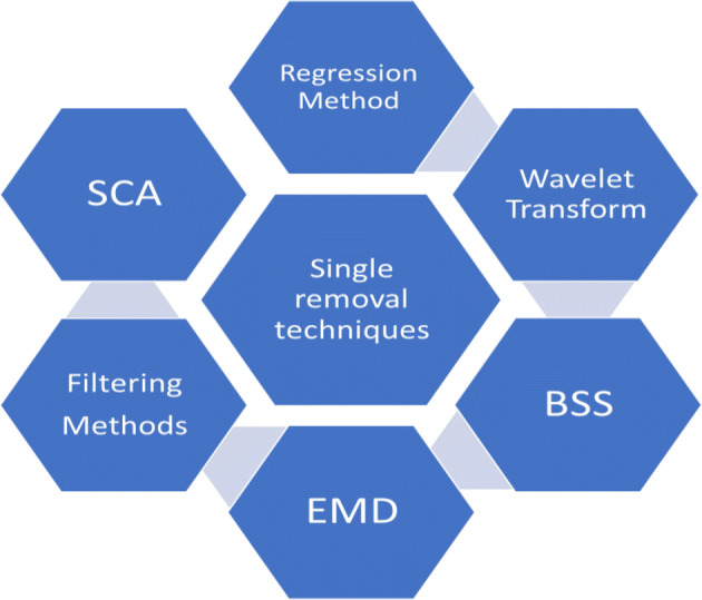
Single removal techniques
Fig. 9.

Basic hybrid methods
Tables 5 and 6 explore the techniques of artifact removal by providing their advantages and disadvantages [53, 104].
Table 5.
A Comparision between different artifacts removal techniques [53]
| Method | Additional Reference | Automatic | Online | Perform on Single Channel |
|---|---|---|---|---|
| Regression | √ | √ | X | X |
| Wavelet | X | √ | X | √ |
| ICA | X | X | √ | X |
| CCA | X | X | √ | X |
| Adaptive filter | √ | √ | √ | √ |
| Winner filter | X | √ | X | √ |
| Wavelet BSS | X | X | X | √ |
| EMD BSS | X | X | X | √ |
| BSS-SVM | X | √ | √ | X |
Table 6.
A comparison between different artifacts removal techniques [104]
| Method | Advantages | Disadvantages |
|---|---|---|
| ICA |
• Computationally efficient. • Shows High performance for large-sized data. • Decomposes signals into temporal independent and spatially fixed components |
• Can’t be applicable for underdetermined cases • Requires more computations for decomposition. |
| CAR |
• Outperforms all the reference methods • Yields improved SNR |
• Finite sample density and incomplete head coverage cause problems in calculating averages |
| SL |
• Robust against the artifacts generated at regions that are not covered by electrode cap. • It solves the electrode reference problem |
• Sensitive to artifacts • Sensitive to spline patterns |
| PCA |
• Helps in the reduction of feature dimensions • The ranking will be done and helps in the classification of data |
• Not well as ICA. |
| CSP | • Doesn’t require a priori selection of subspecific bands and knowledge of these bands |
• Requires use of many electrodes • Change in the position of the electrode may affect classification accuracies. |
Most of the practical applications of EEG are often needed real-time signal processing to be robust to artifacts. The artifact removal methods are required to be automatic and have a low computational cost. The automatic process indicates the chosen method can automatically identify and eliminate the artifact with no manual intervention. The regression and filtering approach can execute automatically. Furthermore, BSS methods will be automatic when there is a subsequent procedure, as cited in Table 5, above, like SVM.
Another factor is the number of channels used. Specifically for the home healthcare environment, less channel is often required. BBS algorithms cannot be applied in such conditions because the assumption of BSS is that more channels will lead to better accuracy. However, wavelet transform and EMD-based methods can be executed with a single channel.
ICA-based algorithms can deal with artifacts that appeared in EEG recordings. Regression and adaptive filters are more achievable choices when the reference channels for specific artifacts are available. Apart from ICA, CCA and its combinations of other methods appear to be a good choice for the removal of muscle artifacts. For application to a few channels, EMD, and its hybrid methods with BSS or WT could be an ideal choice. Wavelet transform fails to identify completely artifacts that overlap with spectral properties. EMD also suffers from the drawback of mode-mixing. Therefore, it is quite difficult to find a single method that is both efficient and accurate enough to satisfy all the conditions perfectly.
Feature extraction
After obtaining the (noise and artifacts)-free signals from the preprocessing phase, effective and meaningful features need to be extracted from the brain EEG signals. Different techniques for feature extraction from EEG signals such as Genetic Algorithms (GA), Wavelet Transformations (WT), Wavelet Packet Decomposition (WPD), Empirical Mode Decomposition (EMD), Fast Fourier Transformations (FFT), PCA, ICA, Adaptive Auto-Regressive parameters (AAR), bilinear AAR, and multivariate AAR. The most common techniques are ICA, PCA, WT, AR, WPD, and FFT. A comparison between these techniques is presented in Table 7 [48, 61, 149].
Table 7.
A Comparision between different feature selection techniques
| Method | Advantages | Disadvantages |
|---|---|---|
| ICA |
• Computationally efficient. • Shows High performance for large-sized data. • Decomposes signals into temporal independent and spatially fixed components |
• Can’t be applicable for underdetermined cases • Requires more computations for decomposition. |
| PCA | • A powerful tool for analyzing and reducing the dimensionality of data without important loss of information |
• Assumes data is linear and continuous. • For complicated manifold, PCA fails to process data. |
| WT |
• Capable to analyze signals with discontinuities through variable window sizes. • It can analyze signals both in time and frequency domains. • Can extract energy, distance, clusters, etc. |
• Lacking specific methodology to apply WT to the pervasive noise. • Performance limited by Heisenberg Uncertainty. |
| AR |
• Requires only a shorter duration of data records. • Reduces spectral loss problems and gives better frequency resolution. |
• Difficulties exist in establishing the model properties for EEG signals • Not applicable to non-stationary signals. |
| WPD | • Can analyze the non-stationary signals. | • Increased computation time. |
| FFT | • A powerful method of frequency analysis. |
• Applicable only to stationary signals and linear random processes. • Suffers from large noise sensitivity. • Poor time localization makes it not suiTable for all kinds of applications. |
Classification techniques
Once the preprocessing and features extraction phases of the detection framework are discussed, this is the time to separate between seizure and seizure-free. The quality of classification algorithms is largely dependent on the feature extracted that is fed to the classifier. The classifier is used to classify the signal into various classes so, the classifier is considered a decision-making system. A classifier can be separated into three types; linear classifiers, nonlinear classifiers, and nearest neighbor classifiers. The most used in BCI is linear and nonlinear classifiers. Table 5, above, shows a comparison among different types of classifiers [48, 61].
Two main steps can be carried out in the classification stage, that is, the training and testing phases. The extracted features are divided into those phases, and after training the classifier, the new data can be classified with the trained network this is called the testing phase. Clustering, machine learning, or, recently, deep neural networks are the classifier used for epileptic seizure detection systems.
Machine Learning Techniques
Artificial Intelligent (AI) algorithms are the most generally utilized classifiers in the automatic epilepsy detection framework. The conventional feature extraction methods are used to extract features and statistically analyze rank and select data that are used as input to AI classifiers. Several classification techniques have been proposed in the literature, such as k-nearest neighbor (k-NN), logistic regression, random forest, artificial neural networks (ANNs), fuzzy logic, and SVMs with various kernel functions. A list of studies utilizing machine learning algorithms with different feature extraction techniques is displayed in Table 8. And the comparison is applied to the most popular and commonly used classifier. This comparison takes into their consideration the advantages and disadvantages of each classifier.
Table 8.
A comparison between some classification techniques
| Method | Advantages | Disadvantages |
|---|---|---|
| LDA |
• It has low computational requirements • Simple to use. • It provides good results. |
• It fails when the discriminatory function is not in the mean but the variance of the features. • For non-Gaussian distributions, it may not preserve the complex structures. |
| SVM |
• It provides a good generalization. • Performance is more than another linear classifier. |
• Has high computational complexity. |
| ANN |
• Ease of use and implementation. • Robust in nature. • Simple computations are involved. • Small training set requirements are required. |
• Difficult to build. • Performance depends on the number of neurons in the hidden layer. |
| k-NN |
• Very simple to understand. • Easy to implement and debug. |
• Poor runtime performance if the training set is large. • Sensitive to irrelevant and redundant features. • On difficult classification tasks outperformed other classification methods. |
From Table 8, it can be noticed that the genetic algorithm, Bayesian net, and fuzzy clustering are not popular classifiers in EEG signal processing [51, 62, 84]. LDA, ANN, and KNN are quite promising classifiers with great accuracy [9, 55, 64]. But, SVM is the most commonly applied classifier [22, 101, 105].
The performance of various EEG detection algorithms from 2014 to 2021 is illustrated in Table 9. This table shows the performance of the classifier and the feature extraction technique. Upadhyay et al. [131] recorded an accuracy of 100% with the DWT and LS-SVM classifier. Sriraam et al. [106] achieve 96.66% accuracy using a new feature, Teager energy with supervised backpropagation. Some studies like the work of Saminu et al. [111] have employed multiple features with an ML technique. This study employed SVM with a feedforward neural network (FFNN) to detect and classify ictal and interictal signals. It was computationally less complex with high accuracy of 99.6%.
Table 9.
Reviewed works from 2014 to 2021 that used feature extraction techniques and machine learning classifiers
| Ref. | Extracted Features | Classifier | Metrics (Accuracy) | Year |
|---|---|---|---|---|
| [7] | Statistical, Non-linear | Linear Classifier | Acc = 99.85 | 2014 |
| [37] | Wavelet | Quadratic Classifier | Acc = 98.50 | 2014 |
| [59] | Wavelet entropy | SVM | Acc = 90.00 | 2014 |
| [91] | Morphological filters | ANN | Acc = 98.33 | 2015 |
| [51] | Wavelet, Morphological filters, EMD | Fuzzy Clustering | PI = 98.03, QV = 23.82 | 2015 |
| [21] | EWT, Focal and non-focal | EM, MEM, SVD | Acc = 90.00 | 2015 |
| [131] | DWT | LS-SVM | Acc = 100 | 2016 |
| [63] | DD-DWT | LS-SVM | Acc = 99.36 | 2016 |
| [12] | SRS, SFS | LS-SVM | Acc = 99.90 | 2016 |
| [62] | Entropy | GA-SVM | AUC = 0.97 | 2016 |
| [84] | Time, frequency domain features | Bayesian Net | Acc = 95.00 | 2016 |
| [83] | DTCWT | CVNN | Acc = 100 | 2016 |
| [50] | SpPCA, SubXPCA | SVM | Acc = 94.60 | 2016 |
| [100] | TQWT | LS-SVM + FD | Acc = 100 | 2017 |
| [64] | MODWT, LND | RFC | Acc = 100 | 2017 |
| [75] | ICFS | RFC | Acc = 100 | 2017 |
| [58] | Fuzzy relations, DWT | ANN | Acc = 99.90 | 2017 |
| [122] | LBP, Pyramid scheme | SVM | Acc = 99.89 | 2017 |
| [30] | Weighted complex network, time domain features | LS-SVM | Acc = 98.00 | 2017 |
| [81] | TQWT, Kraskov entropy | LS-SVM | Acc = 97.75 | 2017 |
| [126] | EMD | CSM-SVM | Acc = 96.40 | 2017 |
| [101] | MMSFL-OWFB-based KE | SVM | Acc = 100 | 2018 |
| [65] | GMM, GLCM, RFE-SVM | SVM | Acc = 100 | 2018 |
| [117] | Entropy-based features, FAWT | RELS-TSVM | Acc = 100 | 2018 |
| [26] | XHST | KNN | Acc = 100 | 2018 |
| [66] | WPT, KDE | LS-SVM | Acc = 99.60 | 2018 |
| [36] | WPD | SVM | Acc = 98.67 | |
| [114] | Sub-frequency band features | GRNN | Acc = 91.60 | 2018 |
| [111] | Teager energy feature |
Backpropagation Neural Network |
Acc = 96.66 | 2018 |
| [129] | Wavelet transform-based features |
Random Forest Classifier |
Acc = 95.00 | 2018 |
| [110] | DWT | ANN | Acc = 95.00 | 2018 |
| [28] | HRI features |
SVM + Adaptive Heuristic classifier |
Sen = 83.30 | 2018 |
| [60] | Hurst exponent | k-ANN | Acc = 100 | 2019 |
| [22] | Multifractal detrended fluctuation analysis | SVM | Acc = 100 | 2019 |
| [105] | Sigmoid entropy | SVM | Acc = 100 | 2019 |
| [35] | Time domain | Exponential Energy | Acc = 99.50 | 2019 |
| [96] | DWT, Entropies, Energy | SVM, FFANN | Acc = 99.00 | 2019 |
| [78] | SOM | RBFNN | Acc = 97.47 | 2019 |
| [136] | Symlet wavelet processing, grid search optimizer | Gradient Boosting Machine | Acc = 96.10 | 2019 |
| [116] | FAWT, FD | RELS-TSVM | Acc = 90.20 | 2019 |
| [23] | TQWT, IMFs, MEMD | SVM | Acc = 98.78 | 2020 |
| [15] | NA | Random Forest classifier | Acc = 97.08 | 2020 |
| [55] | Ensemble EMD | KNN | Acc = 97.00 | 2020 |
| [9] | DWT | ANN | Acc = 91.10 | 2020 |
| [31] | JPT | LS-SVM | Acc = between 88.75 and 100% | 2021 |
| [1] | DWT | KNN, FRNN | Acc = 92.79 | 2021 |
| [16] | Wavelet decomposition | LR, DT, NB, SVM, KNN, Ensemble, LD | Acc = 98 | 2021 |
| [33] | Statistical features | LR, DT, NB, SVM, KNN, Ensemble, LDA | Acc = 89, Sen = 100 | 2021 |
Mahjoub et al. [23] was a mix of linear and non-linear parameters and multiple features as its edge. It utilized feature extraction of epileptic EEGs with tunable-Q wavelet transform (TQWT) and intrinsic mode functions (IMFs) of multivariate empirical mode decomposition (MEMD) and directly from the EEG raw data. This approach provides an accuracy of 98.7% with SVM.
From Table 9, it is noticed that there are several algorithms that give high accuracy (equal to 100%) such as the algorithm in reference [65, 83, 100, 101, 131].
-
2-
Deep Learning Techniques
Shallow networks are a second name to conventional neural networks, CNN consists of many hidden layers (more than 2 layers to hundreds of layers). Due to the massive number of these hidden layers, the parameters of the network are increased. The feature extraction step in CNN architecture is made automatically in the process of classifying the EEG signal. While the max-pooling layer carries forward the significant feature decided/chosen by the convolutional layer. The fully connected layer simply compiles the extracted data for the SoftMax layer that conducts the binary classification, i.e., converting the data into probabilities between 0 and 1.
Nowadays, seizure detection systems employed deep learning algorithms to overcome the limitations associated with machine learning techniques. DL uses multilayer architecture so; features do not need to be extracted manually. it can manage huge datasets. Several models are proposed using DL techniques: long short-term memory (LSTM) and Gated-Recurrent-Units (GRU) are recurrent neural networks, convolutional neural networks (CNNs), AE, and DBN are employed as unsupervised learning. Table 9, above, shows the automated epilepsy detection works and analysis that utilized the deep learning method.
Table 10 summarized the performance of previously EEG detection and classification algorithms employed by researchers.
Table 10.
Summary of reviewed works that used deep learning techniques
| Ref. | Features | Metrics | Year |
|---|---|---|---|
| [87] | MCC-based R-SAE model | SEN = 100 | 2014 |
| [120] | CNN + RNN | SEN = 85.00 | 2016 |
| [18] | CNN | ACC = 87.51 | 2016 |
| [6] | CNN | ACC = 78.33 | 2016 |
| [137] | Multichannel CNN | ACC = 92.40 | 2016 |
| [42] | Semi-supervised stacked autoencoder | ACC = 96.90 | 2017 |
| [141] | STFT-Mssda | ACC = 93.82 | 2017 |
| [123] | CNN (1D and 2D) and/or LSTMs | SPE = 99.90 | 2018 |
| [143] | CNN | SEN = 86.29 | 2018 |
| [130] | P-1D-CNN | ACC = 99.90 | 2018 |
| [121] | CNN | ACC = 83.86 | 2018 |
| [46] | LSTM + FC | SPE = 100 | 2019 |
| [52] | Dual deep neural network | SEN = 100 | 2019 |
| [68] | CNN, LSTM, GRU | ACC = 96 | 2019 |
| [11] | WT-CNN | ACC = 99.40 | 2019 |
| [118] | DNN | ACC = 97.21 | 2019 |
| [38] | Deep CNN | ACC = 92.60 | 2020 |
| [25] | CNN, FCNN, RNN | ACC = 0.993 | 2020 |
| [76] | CNN | ACC = 98.50 | 2020 |
| [148] | 1D DNN | ACC = 99.52 | 2020 |
| [85] | CNN | ACC = 98.82 | 2020 |
| [2] | SDCAE+ Bi-LSTM | ACC =98.79 | 2021 |
| [45] | CNN+ LSTM | ACC =93–96 | 2021 |
| [77] | CNN | ACC =100 | 2021 |
From Table 10, it is noticed that the algorithm in reference [77] gives high accuracy (equal to 100%).
In the last years, there are several techniques for seizure classification. It is essential to review the statistical evaluation of the performance of these techniques. A summary of the different techniques, the database used, and their performance in terms of accuracy sensitivity, and specificity, is shown in Table 11.
Table 11.
Comparisons among different classifiers
| Classifier | Database | Performance metrics in % | ||
|---|---|---|---|---|
| Accuracy | Sensitivity | Specificity | ||
| Convolutional neural network [79] | SNUH-HYU | 73.7 | 74.5 | 72.9 |
| CHB-MIT | 68.9 | 65.8 | 72.2 | |
| Directed transfer function [135] | Ten patients from Jiaotong university | 98.45 | 93.36 | 98.42 |
| MAX(KNN + LDA + NB) [140] | Bonn University, Germany | 96.97 | 96.97 | 96.97 |
| k-means [98] | – | 94.6 | 93.8 | 92.3 |
| Multiplayer perceptron | 80.4 | 77.3 | 73.4 | |
| Linear regression | 78.9 | 72.2 | 69.6 | |
| Neural network [138] | CHB-MIT | 98.70 | – | – |
| k- Nearest Neighbor | 94.71 | – | – | |
| Naïve Bayes | 95.14 | – | – | |
| LDA [139] | PhysioNet | – | 88.06 | – |
| KNN | – | 79.38 | – | |
| SVM | – | 82.25 | – | |
| DT | – | 71.71 | – | |
| MLPNN | – | 70.15 | – | |
| Improved Approximate Entropy [10] | – | 83 | 78 | 88 |
| MLPNN [125] | Neurology Research left, india | 92.5 | 91.5 | 90.5 |
| LS_SVM | 95.6 | 94.5 | 93.2 | |
| Naïve Bayes | 84.4 | 85.2 | 85.3 | |
| KNN | 86.1 | 87.3 | 84.3 | |
| KNN [24] | Bonn University, Germany | 96.37 | 94.12 | 98.62 |
| LDA | 88.98 | 80.36 | 97.60 | |
| NB | 93.25 | 89.90 | 96.60 | |
| LR | 96.03 | 95.22 | 96.84 | |
| SVM | 97.24 | 96.14 | 98.34 | |
| SVM+ Wavelet ICA [94] | EEGLAB | 99.1 | 97.1 | 99.3 |
| SVM [34] | CHB-MIT | 93 | 95 | 90 |
| KNN [103] | Bonn University, Germany | 97.1 | – | – |
| NB | 95.8 | – | – | |
| LDA [56] | – | – | 91.032 | 94.210 |
| FEBANN [113] | – | 91 | 90.4 | 91.3 |
| SVM [115] | – | 99.25 | 100 | 98.5 |
| LDA | 86.87 | 100 | 73.75 | |
| LS-SVM [30] | Available online | 97.9 | 97 | 98 |
| NB | 91 | 91 | 90 | |
| KNN | 89.5 | 90 | 88 | |
| GMM [86] | Sri Ramakrishna Hospital, India | 97.73 | 96.45 | 96.73 |
| ANN [29] | Publicly available | 97.1 | 99.3 | 97.9 |
| ANN [145] | Bonn university | 87.2 | – | – |
| SVM [71] | Bonn university | 95.1 | 93.04 | 96.85 |
| KNN | 96.7 | 95.23 | 97.34 | |
| DT | 97 | 96.2 | 97.53 | |
| KNN [40] | Bonn university | 94.40 | 87 | 96.25 |
| LDA | 93.20 | 68 | 99.50 | |
| LR [89] | Sri Ramakrishna hospital, India | 95.88 | 96.04 | 95.71 |
| Ensemble [89] | 88.57 | – | – | |
| DT [134] | New York university | 91.3 | – | – |
| DT [90] | Sri Ramakrishna Hospital, India | 96.83 | 95.13 | 98.54 |
IoT-based automated epileptic seizure detection
Seizures are a very diverse group of disorders, with such a variety of epilepsy smart devices available which are not easily managed. it is difficult for a clinician to realize how to locate the best device for the seizure patient’s needs. Also, seizures can be varying for every individual; simply realizing that somebody has epilepsy doesn’t mention to you what their epilepsy is like, or what seizures they have. So, there is a need to develop an accurate and refined way of remotely monitoring patients at home.
Remote monitoring allows patients to use mobile medical devices to do routine tests at home and send the test information to a specialist in real-time. A challenge in this research is to blend the devices into the web environment and make them accessible, discoverable, and secure. A ‘seamless’ way of integrating these physical objects or things from the physical world to the cyber world is The Internet of Things [70].
Internet of Things (IoT) means the practice of designing and molding Internet-connected Things through computer networks. The term ‘IoT’ signifies that instead of using smaller effective gadgets like laptops, Tablets, and smartphones, it is better to use several efficient devices. The traditional methods used for epilepsy detection based on wired hospital monitoring systems are not appropriate for the recognition of long-term monitoring outdoors. Consequently, there must be a framework that can identify seizures in patients to alarm the members of the family or clinical staff for help, based on a wearable device, or a portable application in a framework dependent on the utilization of IoT and cloud computing.
There are many existing epilepsy devices smart and non-smart as in Table 12. it is difficult to know the best device to implement. For instance, the patient could be in the bath, in bed, or watching TV when the seizure occurs. For instance, a Smartwatch used for monitoring shaking may not be comfortable to wear in bed, so a Smart Mattress would take its place. Likewise, a Companion Monitor which monitors bed movement and sound but would not be fitting in the shower. Another variant is that various kinds of seizures will present themselves and there are not always solutions at the clinician’s fingertips; devices are just not reasonable for all the different environments we live in.
Table 12.
The epilepsy devices and factors
| Epilepsy Devices: Smart & Non-Smart | Sample factors associated with epilepsy devices | |||
|---|---|---|---|---|
| Placement | Use at Home | Purpose | Medical Training Needed | |
| EEG (Gold Standard) | Brain (real-time hospital setting) | x | Monitoring | √ |
| Smart Belt | Torso | √ | Detection | x |
| Vagus Nerve Stimulation (VNS) | Nerve in Neck | x | Diagnostic/ Prevention | √ |
| Brain Stethoscope | Brain | x | Monitoring | √ |
| Medpage™ Model MP5 | Mattress | √ | Alarm (for Nocturnal Seizures) | x |
| Emfit | Mattress | √ | Alarm Nocturnal Seizures) | x |
| SmartWatch™ | Wrist | √ | Monitoring/ Alarm | x |
| Embrace | Wrist | √ | Monitoring/ Alarm | x |
| Sami | Surveillance | √ | Camera/Video: Monitoring/ Alarm | x |
| EpiLert | Wrist/Foot | √ | Detection | x |
| Continuous Audio-Visual EEG Recording | Scalp (Electrodes) | x | Diagnostic | √ |
Epilepsy patients often preferred the use of smart devices in their daily routines, due to the recent advances in sensor development, processing, and displays that have enabled devices. Depending on the patient, some wearable devices can be placed on almost any part of the body: wrist, ankle, waist, chest, arm, legs, etc. Product design is another factor that affects patient-device suitability. A good design can reduce mental and physical stress, reduce the learning curve, improve user device operability in using the device and thus improve overall product quality. Recently, there are epilepsy devices and apps that track medication and send reminders to take medication, some are for an emergency with buttons to press before losing consciousness with a seizure and some detect a seizure based on the pattern of jerks. Therefore, it is vital to choose the most appropriate device to find evidence on device reliability for a seizure type, and specifically to avoid “false alarms which can be disruptive for the family.” Patients with epilepsy must “discuss with their doctors the pros and cons of each device. Table 13 presents a comparative study for different IoT methods. Finally, Table 14 is added to clarify the general difference among ML/DL in addition IoT system.
Table 13.
Comparative study for different IoT methods
| Ref | Description | Accuracy | Sensitivity | Specificity | Year |
|---|---|---|---|---|---|
| [133] | WD to be located on a wrist connected to a Smartphone, which in turn implements MCC services and has access to CC services | – | – | – | 2017 |
| [99] | smart seizure detection framework in the edge of the Internet of Things (IoT) using the discrete wavelet transform, statistical feature extraction, and a naive Bayes (NB) classifier | 98.65 | – | – | 2018 |
| [69] | IoT system is implemented by using ARM7LPC2138, RF modem, accelerometer sensor, sound detection sense, or, temperature sensor. | – | – | – | 2019 |
| [96] | employed two machine learning classifiers, support vector machine (SVM), feedforward neural network (FFNN) and DWT integrated with recent 5G network IoT devices for mobile applications, | 99.6% | 99.7 | 99.3 | 2019 |
| [27] | The EEG recording is measured via a wireless headset, then transmitted to the FPGA, where the deep learning algorithm is embedded | 96.1% | 97.41% | 94.8% | 2020 |
| [95] | The EEG data is segmented and filtered on the low-power device. Then, the time and frequency domain features are calculated. a logistic regression model is implemented. Then transmitted to the gateway to be used for XGBoost classification | 95.8% | 92.0% | 96.1% | 2018 |
| [132] | Two wearable devices, based on ECG and PPG, for long-term monitoring in daily life outside the hospital. | – | – | – | 2017 |
| [73] | Mobile multimedia healthcare framework, electroencephalogram signals from a head-mounted set are recorded and processed using CNN | 99.02% | 92.35% | – | 2018 |
| [14] | EEG signals are captured by a headset of electrodes. The headset acts as an IoT device, e EEG signals are transmitted to a mobile edge computing (MEC) server via a short-range communication protocol such as Wi-Fi or a local area network | 89.13% | 80.16% | 96.67% | 2019 |
Table 14.
Comparison among DL, ML and IoT based Systems
| Deep learning | Machine Learning | IoT Based | |
|---|---|---|---|
| Complexity | Quite complex | Less complexity, generally small models | IoT-based systems involve many decisions to be made in a short time, which in turn requires automation – not only in terms of infrastructure management, but also within the logic of the IoT applications themselves. |
| Amount of Data | Are best used on large volumes of data | Good results are obtained with small data and datasets | |
| Time | Takes longer time for training and execution (From hours to weeks) | Takes less time (From seconds to hours) | |
| Hardware | Require powerful resources like GPU | Standard PC | |
| Performance | High quality accuracy output | Less accuracy output than DL |
The basic building blocks of the automatic epileptic seizure detection system are shown in Fig. 10. The suggested framework consists of three key phases that collaborate to achieve the system objective. Each phase provides and delivers a specific task and operation in harmonizing with the other phases. The three phases can be as the following:
Stage 1: In this phase, IoT-based wearable medical sensors and smart-phone can be used for data acquisition in real-time. These will be connected to the patient’s heat to collect EEG data.
Stage 2: In this phase, the cloud will be used to provide a pool of processing and storage resources for receiving patients’ data from their smart phone over the internet to be sorted, and then it became available for doctor’s inspections. Besides, the data exploration and handling will be held in the cloud for any disorder detection in patient’s data, therefore, the abnormal changes in patient’s data will be classified based on patient status. All results in acquired data and extracted knowledge will be reported either to health-givers such as friends, family, and medical staff reliably and efficiently. In summary, the cloud enables collaboration and information sharing through its infrastructure which allows medical staff and experts to host information, analytics, and diagnostics. This discloses in earlier medicaments and real-time updates to the patient’s data and status.
Stage 3: In this phase, the medical staff uses a cloud-based web monitoring system to monitor a patient’s records and sensory EEG data. The staff will be able to inspect reports provided by the cloud-based analytical system and they able to take appropriate actions.
Fig. 10.

IoT-based cloud system for automatic epileptic seizure detection
Challenges and open research points
Working on the EEG based Seizure detection poses several challenges as the following:
The automated seizure registration techniques for improving the quality of seizure data.
An accurate professional database, because seizure monitoring is crucial for therapeutic decisions for patients or caretakers. Inaccurate seizure documentation has a bad effect on the patient’s treatment.
A real-time seizure detection system and subsequent evaluation by experts are still required.
The issue of obtaining longer recordings is due to technical reasons.
emergency call systems that require actively pushing an alarm button are inappropriate for most epilepsy patients and alternative approaches are needed.
Diagnostic accuracy is still missing so multimodal approaches which combine the measurement of autonomous parameters (e.g., heart rate, or muscular activity) will be required to detect these seizures.
The trade-off between seizure detection algorithm (SDA) accuracy vs. detection speed, such improvements may come at the expense of increased invasiveness of monitoring.
Dealing with non-stationarity and noise. EEG is prone to many different artifacts which obstruct the view of underlying brain activity.
Minimizing computational cost.
SDA Performance Assessment. Extensive prospective validation of any SDA is required to properly assess its performance.
Regarding this interesting topic, several open research points can lead the researcher in working with future work as the following:
Study the effect of the Corona Virus on epilepsy patients and how it affects the EEG brain signals.
Develop an efficient seizure detection model using spectrogram images for the EEG signals.
Using the Internet of Things for remotely monitoring patients who suffer from epilepsy.
Deep learning structures must be carefully chosen based on the problem’s peculiarities and include relevant datasets for real-time epilepsy detection. Likewise, hybrid deep learning techniques should be broadly investigated.
AI-based classifiers must be chosen carefully so that will not miss or skip all the relevant EEG channels and electrodes.
Develop mobile applications for remotely observing epilepsy patients by doctors and family members.
The epileptic seizure datasets that are with a large size and high dimension, it is necessary to utilize dimensional reduction methods to reduce the dataset dimension and still retain the important signal information need to be further explored. So, appropriate features that reduce the classifier’s computational complexity and time should be believed.
Conclusion
In recent years, using manual monitoring and studying EEG to diagnose epilepsy is a very difficult and challenging task of observing long recordings and decision-making through experience. With the growth of epilepsy patients, accurate identification becomes increasingly significant. Likewise, recognizing seizures accurately from an enormous amount of information becomes challenging. Besides, the machine and deep learning algorithms classifiers as a useful and suitable tool for accurate seizure identification due to the complexity of EEG signals in such datasets. Henceforward, there is a prerequisite for providing an automated seizure detection system that is a promising tool for neurologists in making epilepsy diagnoses. Further investigation must be thoroughly conducted on seizure detection techniques to improve the outcome. Therefore, this study investigated and reviewed various automated EEG epileptic seizure detection and classification techniques. Additionally. It highlighted both traditional feature extraction techniques and statistical and machine learning classifiers. Moreover, this work focused on the trends in the IoT framework for epilepsy detection. Finally, the challenges and open research points in EEG seizure detection are inspected.
Biographies
Athar A. Ein Shoka
received her B.Sc. from the Department of Computer Science and Engineering, Faculty of Electronic Engineering, Menoufia University, Egypt, in 2010. She received her M.Sc. From the Department of Computer Science and Engineering, Faculty of Electronic Engineering, Menoufia University, Egypt, in 2015. Her research area of interest includes; Image and Signal Processing, Virtualization, Cloud Computing, Internet of Things, Cryptography, Data Hiding, Digital Forensics, Routing, and Switching.

Mohamed M. Dessouky
is Assistant Professor at Department of Computer Science and Artificial Intelligence, Collage of Computer Science and Engineering (CCSE), University of Jeddah. He had received the B.Sc., (Hons.), M.Sc., Ph.D. degrees from the Faculty of Electronic Engineering (FEE), Menuofia University, Egypt, in 2006, 2011, and 2016, respectively. His research interests include artificial intelligence, medical image processing, Deep learning, and computer network security. He has published several indexed ISI Journal Papers, conference papers and book chapters. He supervised on more than 10 Ph.D. and M.Sc. students. on He is a CISCO Academy Curriculum Lead. He is a CISCO Certified Instructor and he had received an award from CISCO as a best instructor for more than 10 years.

Ayman El-Sayed
a Professor and Dean of the Faculty of Electronic Engineering, Menoufia University. He received a B.Sc. degree in computer science and engineering in 1994, a master’s degree in computer networks in 2000 from Menoufia University, Egypt, and a Ph.D. degree in a computer networks in 2004 from (Institute National De Poly Technique De Grenoble) INPG, France. He is specialized in soft computing, algorithms, data structure, cloud computing, and big data analysis. In addition, his interests include multicast routing protocols, application-level multicast techniques, multicast on both mobile network and mobile IP, and image processing techniques. In addition, there are other interesting topics such as bioinformatics, Biocomputing, and bio-computer. He is an approved supervisor for M.Sc. and Ph.D. programs in various universities. He has completed various projects in government and private organizations. He has published more than 165 research papers in international journals and two books about OSPF protocol and multicast protocols. Currently, he is serving as an editorial board member in various international Journals and conferences. He is a senior member of the IEEE.

Ezz El-Din Hemdan
received his B.Sc. from the Department of Computer Science and Engineering, Faculty of Electronic Engineering, Menoufia University, Egypt, in 2009. He received his M.Sc. From the Department of Computer Science and Engineering, Faculty of Electronic Engineering, Menoufia University, Egypt, in 2013. He received his Ph.D. degree in the Department of Computer Science, Mangalore University, India in 2018. He has several publications in national/international conferences and journals. His research area of interest includes; Cancelable Biometric, Blockchain, Digital Twins, Image Processing, Virtualization, Cloud Computing, Internet of Things/Nano-Things, Cryptography, Data Hiding, Digital Forensics, Cloud Forensics, Big Data Forensics, Data Science and Big Data Analytics.

Data availability
The data is available up to a request from the author.
Declarations
Competing interests
The author(s) declare(s) that they have no competing interests.
Footnotes
Publisher’s note
Springer Nature remains neutral with regard to jurisdictional claims in published maps and institutional affiliations.
Contributor Information
Athar A. Ein Shoka, Email: atharali@el-eng.menofia.edu.eg
Mohamed M. Dessouky, Email: mohamed.moawad@el-eng.menofia.edu.eg
Ayman El-Sayed, Email: ayman.elsayed@el-eng.menofia.edu.eg.
Ezz El-Din Hemdan, Email: ezzvip@yahoo.com.
References
- 1.Aayesha M, Qureshi B, Afzaal M, Qureshi MS, Fayaz M. Machine learning-based EEG signals classification model for epileptic seizure detection. Multimed Tools Appl. 2021;80(12):17849–17877. doi: 10.1007/s11042-021-10597-6. [DOI] [Google Scholar]
- 2.Abdelhameed A, Bayoumi M. A deep learning approach for automatic seizure detection in children with epilepsy. Front Comput Neurosci. 2021;15:29. doi: 10.3389/fncom.2021.650050. [DOI] [PMC free article] [PubMed] [Google Scholar]
- 3.Abualsaud K, Mohamed A, Khattab T, Yaacoub E, Hasna M, Guizani M (2018) Classification for imperfect EEG epileptic seizure in IoT applications: a comparative study. 2018 14th Int. Wirel. Commun. Mob. Comput. Conf. IWCMC 2018, pp 364–369. 10.1109/IWCMC.2018.8450279
- 4.Acharya UR, Fujita H, Sudarshan VK, Bhat S, Koh JEW. Application of entropies for automated diagnosis of epilepsy using EEG signals: a review. Knowl-Based Syst. 2015;88:85–96. doi: 10.1016/j.knosys.2015.08.004. [DOI] [Google Scholar]
- 5.Acharya UR, et al. Characterization of focal EEG signals: a review. Futur Gener Comput Syst. 2019;91:290–299. doi: 10.1016/j.future.2018.08.044. [DOI] [Google Scholar]
- 6.Achilles F, Tombari F, Belagiannis V, Loesch-Biffar A, Noachtar S, Navab N. Convolutional neural networks for real-time epileptic seizure detection. Comput Methods Biomech Biomed Eng Imaging Vis. 2016;6:1–6. doi: 10.1080/21681163.2016.1141062. [DOI] [Google Scholar]
- 7.Ahammad N, Fathima T, Joseph P. Detection of epileptic seizure event and onset using EEG. Biomed Res Int. 2014;2014:450573. doi: 10.1155/2014/450573. [DOI] [PMC free article] [PubMed] [Google Scholar]
- 8.Ahmad I, et al. EEG-based epileptic seizure detection via machine/deep learning approaches: a systematic review. Comput Intell Neurosci. 2022;2022:6486570. doi: 10.1155/2022/6486570. [DOI] [PMC free article] [PubMed] [Google Scholar] [Retracted]
- 9.Aileni RM, Pasca S, Florescu A. EEG-brain activity monitoring and predictive analysis of signals using artificial neural networks. Sensors (Basel) 2020;20(12):3346. doi: 10.3390/s20123346. [DOI] [PMC free article] [PubMed] [Google Scholar]
- 10.Akareddy S, Kulkarni P. EEG signal classification for epilepsy seizure detection using improved approximate entropy. Int J Public Health Sci. 2013;2:23–32. doi: 10.11591/ijphs.v2i1.1836. [DOI] [Google Scholar]
- 11.Akut R. Wavelet based deep learning approach for epilepsy detection. Heal Inf Sci Syst. 2019;7:1–9. doi: 10.1007/s13755-019-0069-1. [DOI] [PMC free article] [PubMed] [Google Scholar]
- 12.Al Ghayab HR, Li Y, Abdulla S, Diykh M, Wan X. Classification of epileptic EEG signals based on simple random sampling and sequential feature selection. Brain Inform. 2016;3(2):85–91. doi: 10.1007/s40708-016-0039-1. [DOI] [PMC free article] [PubMed] [Google Scholar]
- 13.Alam SM, Bhuiyan M. Detection of seizure and epilepsy using higher order statistics in the EMD domain. IEEE J Biomed Health Inform. 2013;17:312–318. doi: 10.1109/JBHI.2012.2237409. [DOI] [PubMed] [Google Scholar]
- 14.Alhussein M, Muhammad G, Hossain MS. EEG pathology detection based on deep learning. IEEE Access. 2019;7:27781–27788. doi: 10.1109/ACCESS.2019.2901672. [DOI] [Google Scholar]
- 15.Almustafa KM. Classification of epileptic seizure dataset using different machine learning algorithms. Inform Med Unlocked. 2020;21:100444. doi: 10.1016/j.imu.2020.100444. [DOI] [Google Scholar]
- 16.Alotaibi SM, Atta-ur-Rahman, Basheer MI, Khan MA. Ensemble machine learning based identification of pediatric epilepsy. Comput Mater Contin. 2021;68(1):149–165. doi: 10.32604/cmc.2021.015976. [DOI] [Google Scholar]
- 17.Alotaiby TN, Alshebeili SA, Alshawi T, Ahmad I, Abd El-Samie FE. EEG seizure detection and prediction algorithms: a survey. EURASIP J Adv Signal Process. 2014;2014(1):183. doi: 10.1186/1687-6180-2014-183. [DOI] [Google Scholar]
- 18.Antoniades A, Spyrou L, Took CC, Sanei S (2016) Deep learning for epileptic intracranial EEG data. In: 2016 IEEE 26th International Workshop on Machine Learning for Signal Processing (MLSP), pp 1–6. 10.1109/MLSP.2016.7738824
- 19.Bashashati A, Fatourechi M, Ward R, Birch G. A survey of signal processing algorithms in brain-computer interfaces based on electrical brain signals. J Neural Eng. 2007;4:R32–R57. doi: 10.1088/1741-2560/4/2/R03. [DOI] [PubMed] [Google Scholar]
- 20.Ben Slimen I, Boubchir L, Seddik H. Epileptic seizure prediction based on EEG spikes detection of ictal-preictal states. J Biomed Res. 2020;34(3):162–169. doi: 10.7555/JBR.34.20190097. [DOI] [PMC free article] [PubMed] [Google Scholar]
- 21.Bhattacharyya A, Sharma M, Pachori RB, Sircar P, Acharya UR. A novel approach for automated detection of focal EEG signals using empirical wavelet transform. Neural Comput Appl. 2018;29(8):47–57. doi: 10.1007/s00521-016-2646-4. [DOI] [Google Scholar]
- 22.Bose R, Pratiher S, Chatterjee S (2019) Detection of epileptic seizure employing a novel set of features extracted from multifractal spectrum of electroencephalogram signals. IET Signal Process 13(2):157–164
- 23.Chahira M, Le Bouquin Jeannès R, Lajnef T, Kachouri A (2020) Epileptic seizure detection on EEG signals using machine learning techniques and advanced preprocessing methods. Biomedical Engineering/Biomedizinische Technik 65(1):33–50 [DOI] [PubMed]
- 24.Chen G, Xie W, Bui TD, Krzyżak A. Automatic epileptic seizure detection in EEG using nonsubsampled wavelet–Fourier features. J Med Biol Eng. 2017;37(1):123–131. doi: 10.1007/s40846-016-0214-0. [DOI] [Google Scholar]
- 25.Cho K-O, Jang H-J. Comparison of different input modalities and network structures for deep learning-based seizure detection. Sci Rep. 2020;10:122. doi: 10.1038/s41598-019-56958-y. [DOI] [PMC free article] [PubMed] [Google Scholar]
- 26.Choudhury NR, Roy SS, Pal A, Chatterjee S, Bose R. 2018 Fourth International Conference on Research in Computational Intelligence and Communication Networks (ICRCICN) 2018. Epileptic seizure detection employing cross-hyperbolic stockwell transform; pp. 70–74. [Google Scholar]
- 27.Daoud H, Williams P, Bayoumi M. 2020 IEEE 6th World Forum on Internet of Things (WF-IoT) 2020. IoT based efficient epileptic seizure prediction system using deep learning; pp. 1–6. [Google Scholar]
- 28.De Cooman T, et al. Adaptive nocturnal seizure detection using heart rate and low-complexity novelty detection. Seizure. 2018;59:48–53. doi: 10.1016/j.seizure.2018.04.020. [DOI] [PubMed] [Google Scholar]
- 29.Dhif I, Hachicha K, Pinna A, Hochberg S, Garda P (2017) Epileptic seizure detection based on expected activity measurement and neural network classification, pp 2814–2817 [DOI] [PubMed]
- 30.Diykh M, Li Y, Wen P. Classify epileptic EEG signals using weighted complex networks based community structure detection. Expert Syst Appl. 2017;90:87–100. doi: 10.1016/j.eswa.2017.08.012. [DOI] [Google Scholar]
- 31.Djoufack Nkengfack LC, Tchiotsop D, Atangana R, Louis-Door V, Wolf D. Classification of EEG signals for epileptic seizures detection and eye states identification using Jacobi polynomial transforms-based measures of complexity and least-square support vector machine. Inform Med Unlocked. 2021;23:100536. doi: 10.1016/j.imu.2021.100536. [DOI] [Google Scholar]
- 32.Durai S, Vanathi P. EEG signal separation using improved EEMD - fast IVA algorithm. Asian J Res Soc Sci Humanit. 2017;7:1230. doi: 10.5958/2249-7315.2017.00239.8. [DOI] [Google Scholar]
- 33.Ein Shoka AA, Alkinani MH, El-Sherbeny AS, El-Sayed A, Dessouky MM. Automated seizure diagnosis system based on feature extraction and channel selection using EEG signals. Brain Inform. 2021;8(1):1. doi: 10.1186/s40708-021-00123-7. [DOI] [PMC free article] [PubMed] [Google Scholar]
- 34.Elgohary S, Eldawlatly S, Khalil MI. 2016 IEEE Conference on Computational Intelligence in Bioinformatics and Computational Biology (CIBCB) 2016. Epileptic seizure prediction using zero-crossings analysis of EEG wavelet detail coefficients; pp. 1–6. [Google Scholar]
- 35.Fasil OK, Rajesh R. Time-domain exponential energy for epileptic EEG signal classification. Neurosci Lett. 2019;694:1–8. doi: 10.1016/j.neulet.2018.10.062. [DOI] [PubMed] [Google Scholar]
- 36.Feng B, Zhao J, Fu W. 2018 IEEE 9th International Conference on Software Engineering and Service Science (ICSESS) 2018. Automated classification of epileptic eeg signals based on multi-feature extraction; pp. 382–386. [Google Scholar]
- 37.Gajic D, Djurovic Z, Di Gennaro S, Gustafsson F. Classification of EEG signals for detection of epileptic seizures based on wavelets and statistical pattern recognition. Biomed Eng Appl Basis Commun. 2014;26:1450021. doi: 10.4015/S1016237214500215. [DOI] [Google Scholar]
- 38.Gao Y, Gao B, Chen Q, Liu J, Zhang Y. Deep convolutional neural network-based epileptic electroencephalogram (EEG) signal classification. Front Neurol. 2020;11:375. doi: 10.3389/fneur.2020.00375. [DOI] [PMC free article] [PubMed] [Google Scholar]
- 39.Garcés Correa A, Orosco LL, Diez P, Laciar Leber E. Adaptive filtering for epileptic event detection in the EEG. J Med Biol Eng. 2019;39:1–7. doi: 10.1007/s40846-019-00467-w. [DOI] [Google Scholar]
- 40.Gill AF, et al. 2014 IEEE Symposium on Industrial Electronics & Applications (ISIEA) 2014. Time domain analysis of EEG signals for detection of epileptic seizure; pp. 32–35. [Google Scholar]
- 41.Giourou E, Stavropoulou-Deli A, Giannakopouou A, Kostopoulos G, Koutroumanidis M (2015) Introduction to epilepsy and related brain disorders, pp 11–38
- 42.Gogna A, Majumdar A, Ward R. Semi-supervised stacked label consistent autoencoder for reconstruction and analysis of biomedical signals. IEEE Trans Biomed Eng. 2017;64(9):2196–2205. doi: 10.1109/TBME.2016.2631620. [DOI] [PubMed] [Google Scholar]
- 43.Gómez C, Arbeláez P, Navarrete M, Alvarado-Rojas C, Le Van Quyen M, Valderrama M. Automatic seizure detection based on imaged-EEG signals through fully convolutional networks. Sci Rep. 2020;10(1):21833. doi: 10.1038/s41598-020-78784-3. [DOI] [PMC free article] [PubMed] [Google Scholar]
- 44.Gupta V, Nishad A, Pachori RB. 2018 IEEE International Conference on Bioinformatics and Biomedicine (BIBM) 2018. Focal EEG signal detection based on constant-bandwidth TQWT filter-banks; pp. 2597–2604. [Google Scholar]
- 45.Hussain W, Sadiq MT, Siuly S, Rehman AU. Epileptic seizure detection using 1 D-convolutional long short-term memory neural networks. Appl Acoust. 2021;177:107941. doi: 10.1016/j.apacoust.2021.107941. [DOI] [Google Scholar]
- 46.Hussein R, Palangi H, Ward RK, Wang ZJ. Optimized deep neural network architecture for robust detection of epileptic seizures using EEG signals. Clin Neurophysiol. 2019;130(1):25–37. doi: 10.1016/j.clinph.2018.10.010. [DOI] [PubMed] [Google Scholar]
- 47.Ibrahim F, Abd-Elateif el-Gindy S, el-Dolil SM, el-Fishawy AS, el-Rabaie ESM, Dessouky MI, Eldokany IM, Alotaiby TN, Alshebeili SA, Abd el-Samie FE. A statistical framework for EEG channel selection and seizure prediction on mobile. Int J Speech Technol. 2019;22(1):191–203. doi: 10.1007/s10772-018-09565-7. [DOI] [Google Scholar]
- 48.Iftikhar M, Khan SA, Hassan A. 2018 IEEE 9th annual information technology, electronics and mobile communication conference (IEMCON) 2018. A survey of deep learning and traditional approaches for EEG signal processing and classification; pp. 395–400. [Google Scholar]
- 49.Islam MS, Thapa K, Yang S-H (2022) Epileptic-net: an improved epileptic seizure detection system using dense convolutional block with attention network from EEG. Sensors 22(3):728 [DOI] [PMC free article] [PubMed]
- 50.Jaiswal AK, Banka H. Epileptic seizure detection in EEG signal using machine learning techniques. Australas Phys Eng Sci Med. 2018;41(1):81–94. doi: 10.1007/s13246-017-0610-y. [DOI] [PubMed] [Google Scholar]
- 51.Jaiswal P, Koushal R (2015) EEG signal classification using Modified Fuzzy Clustering algorithm. Int J Comput Sci Inform Technol 6(3):2031–2034
- 52.Jang H-J, Cho K-O. Dual deep neural network-based classifiers to detect experimental seizures. Korean J Physiol Pharmacol. 2019;23:131–139. doi: 10.4196/kjpp.2019.23.2.131. [DOI] [PMC free article] [PubMed] [Google Scholar]
- 53.Jiang X, Bian G-B, Tian Z. Removal of artifacts from EEG signals: a review. Sensors. 2019;19:987. doi: 10.3390/s19050987. [DOI] [PMC free article] [PubMed] [Google Scholar]
- 54.Jindal K, Upadhyay R, Singh HS. 2018 10th International Congress on Ultra Modern Telecommunications and Control Systems and Workshops (ICUMT) 2018. Eeg artifact removal and noise suppression using hybrid Glct -Ica technique; pp. 1–5. [Google Scholar]
- 55.Karabiber Cura O, Kocaaslan Atli S, Türe HS, Akan A. Epileptic seizure classifications using empirical mode decomposition and its derivative. Biomed Eng Online. 2020;19(1):10. doi: 10.1186/s12938-020-0754-y. [DOI] [PMC free article] [PubMed] [Google Scholar]
- 56.Khalid MI, Alotaiby T, Aldosari SA, Alshebeili SA, Al-hameed MH, Almohammed FSY. Epileptic MEG spikes detection using common spatial patterns and linear discriminant analysis. IEEE Access. 2016;4:4629–4634. doi: 10.1109/ACCESS.2016.2602354. [DOI] [Google Scholar]
- 57.Kim M, Kim S. 2018 6th International Conference on Brain-Computer Interface (BCI) 2018. A comparsion of artifact rejection methods for a BCI using event related potentials; pp. 1–4. [Google Scholar]
- 58.Kocadagli O, Langari R. Classification of EEG signals for epileptic seizures using hybrid artificial neural networks based wavelet transforms and fuzzy relations. Expert Syst Appl. 2017;88:419–434. doi: 10.1016/j.eswa.2017.07.020. [DOI] [Google Scholar]
- 59.Kumar Y, Dewal ML, Anand RS. Epileptic seizure detection using DWT based fuzzy approximate entropy and support vector machine. Neurocomputing. 2014;133:271–279. doi: 10.1016/j.neucom.2013.11.009. [DOI] [Google Scholar]
- 60.Lahmiri S, Shmuel A. Accurate classification of seizure and seizure-free intervals of intracranial EEG signals from epileptic patients. IEEE Trans Instrum Meas. 2019;68(3):791–796. doi: 10.1109/TIM.2018.2855518. [DOI] [Google Scholar]
- 61.Lakshmi MR, Prasad TV, Dr Prakash VC (2014) "Survey on EEG signal processing methods." International journal of advanced research in computer science and software engineering 4.1.
- 62.Li P, Karmakar C, Yan C, Palaniswami M, Liu C. Classification of 5-S epileptic EEG recordings using distribution entropy and sample entropy. Front Physiol. 2016;7:136. doi: 10.3389/fphys.2016.00136. [DOI] [PMC free article] [PubMed] [Google Scholar]
- 63.Li M, Chen W, Zhang T. Automatic epilepsy detection using wavelet-based nonlinear analysis and optimized SVM. Biocybern Biomed Eng. 2016;36:708–718. doi: 10.1016/j.bbe.2016.07.004. [DOI] [Google Scholar]
- 64.Li M, Chen W, Zhang T. Application of MODWT and log-normal distribution model for automatic epilepsy identification. Biocybern. Biomed. Eng. 2017;37(4):679–689. doi: 10.1016/j.bbe.2017.08.003. [DOI] [Google Scholar]
- 65.Li Y, Cui W, Luo M, Li K, Wang L. Epileptic seizure detection based on time-frequency images of EEG signals using gaussian mixture model and gray level co-occurrence matrix features. Int J Neural Syst. 2018;28(7):1850003. doi: 10.1142/S012906571850003X. [DOI] [PubMed] [Google Scholar]
- 66.Li M, Chen W, Zhang T. A novel seizure diagnostic model based on kernel density estimation and least squares support vector machine. Biomed Signal Process Control. 2018;41:233–241. doi: 10.1016/j.bspc.2017.12.005. [DOI] [Google Scholar]
- 67.Li F, Liang Y, Zhang L, Yi C, Liao Y, Jiang Y, Si Y, Zhang Y, Yao D, Yu L, Xu P. Transition of brain networks from an interictal to a preictal state preceding a seizure revealed by scalp EEG network analysis. Cogn Neurodyn. 2019;13(2):175–181. doi: 10.1007/s11571-018-09517-6. [DOI] [PMC free article] [PubMed] [Google Scholar]
- 68.Liu H, Xi L, Zhao Y, Li Z. Using deep learning and machine learning to detect epileptic seizure with electroencephalography (EEG) data. Mach Learn Res. 2019;4:39. doi: 10.11648/j.mlr.20190403.11. [DOI] [Google Scholar]
- 69.M NR, Prabu V, Kumar R, Ahamed MY. Iot based epilepsy monitoring and detection using ambulatory system. Int J Innov Res Adv Eng. 2019;6(03):123–125. [Google Scholar]
- 70.McHale SA, Pereira E, Weishmann U, Hall M, Fang H. 2017 14th International Symposium on Pervasive Systems, Algorithms and Networks & 2017 11th International Conference on Frontier of Computer Science and Technology & 2017 Third International Symposium of Creative Computing (ISPAN-FCST-ISCC) 2017. An IoT approach to personalised remote monitoring and management of epilepsy; pp. 414–418. [Google Scholar]
- 71.Mohammadpoory Z, Nasrolahzadeh M, Haddadnia J. Epileptic seizure detection in EEGs signals based on the weighted visibility graph entropy. Seizure. 2017;50:202–208. doi: 10.1016/j.seizure.2017.07.001. [DOI] [PubMed] [Google Scholar]
- 72.Mohammadpoory Z, Nasrolahzadeh M, Mahmoodian N, Sayyah M, Haddadnia J. Complex network based models of ECoG signals for detection of induced epileptic seizures in rats. Cogn Neurodyn. 2019;13(4):325–339. doi: 10.1007/s11571-019-09527-y. [DOI] [PMC free article] [PubMed] [Google Scholar]
- 73.Muhammad G, Masud M, Amin SU, Alrobaea R, Alhamid MF. Automatic seizure detection in a Mobile multimedia framework. IEEE Access. 2018;6:45372–45383. doi: 10.1109/ACCESS.2018.2859267. [DOI] [Google Scholar]
- 74.Munch Nielsen J, Zibrandtsen IC, Masulli P, Lykke Sørensen T, Andersen TS, Wesenberg Kjær T. Towards a wearable multi-modal seizure detection system in epilepsy: a pilot study. Clin Neurophysiol. 2022;136:40–48. doi: 10.1016/j.clinph.2022.01.005. [DOI] [PubMed] [Google Scholar]
- 75.Mursalin M, Zhang Y, Chen Y, Chawla NV. Automated epileptic seizure detection using improved correlation-based feature selection with random forest classifier. Neurocomputing. 2017;241:204–214. doi: 10.1016/j.neucom.2017.02.053. [DOI] [Google Scholar]
- 76.Nagappan I, Khan N, Shahina A. Deep learning approach to detect seizure using reconstructed phase space images. J Biomed Res. 2020;34:238. doi: 10.7555/JBR.34.20190043. [DOI] [PMC free article] [PubMed] [Google Scholar]
- 77.Nogay HS, Adeli H. Detection of epileptic seizure using Pretrained deep convolutional neural network and transfer learning. Eur Neurol. 2020;83(6):602–614. doi: 10.1159/000512985. [DOI] [PubMed] [Google Scholar]
- 78.Osman AH, Alzahrani AA. New approach for automated epileptic disease diagnosis using an integrated self-organization map and radial basis function neural network algorithm. IEEE Access. 2019;7:4741–4747. doi: 10.1109/ACCESS.2018.2886608. [DOI] [Google Scholar]
- 79.Park C, et al. 2018 International Conference on Electronics, Information, and Communication (ICEIC) 2018. Epileptic seizure detection for multi-channel EEG with deep convolutional neural network; pp. 1–5. [Google Scholar]
- 80.Parvez MZ, Paul M. Seizure prediction using undulated global and local features. IEEE Trans Biomed Eng. 2017;64(1):208–217. doi: 10.1109/TBME.2016.2553131. [DOI] [PubMed] [Google Scholar]
- 81.Patidar S, Panigrahi T. Detection of epileptic seizure using Kraskov entropy applied on tunable-Q wavelet transform of EEG signals. Biomed Signal Process Control. 2017;34:74–80. doi: 10.1016/j.bspc.2017.01.001. [DOI] [Google Scholar]
- 82.Peachap AB, Tchiotsop D. Epileptic seizures detection based on some new Laguerre polynomial wavelets, artificial neural networks and support vector machines. Informatics Med. Unlocked. 2019;16:100209. doi: 10.1016/j.imu.2019.100209. [DOI] [Google Scholar]
- 83.Peker M, Sen B, Delen D. A novel method for automated diagnosis of epilepsy using complex-valued classifiers. IEEE J Biomed Health Inform. 2016;20(1):108–118. doi: 10.1109/JBHI.2014.2387795. [DOI] [PubMed] [Google Scholar]
- 84.Pippa E, et al. Improving classification of epileptic and non-epileptic EEG events by feature selection. Neurocomputing. 2016;171:576–585. doi: 10.1016/j.neucom.2015.06.071. [DOI] [Google Scholar]
- 85.Pisano F, Sias G, Fanni A, Cannas B, Dourado A, Pisano B, Teixeira CA. Convolutional neural network for seizure detection of nocturnal frontal lobe epilepsy. Complexity. 2020;2020:4825767–4825710. doi: 10.1155/2020/4825767. [DOI] [Google Scholar]
- 86.Prabhakar SK, Rajaguru H (2017) Conceptual analysis of epilepsy classification using probabilistic mixture models. 2017 5th int. winter conf. brain-computer interface, pp 81–84. 10.1109/IWW-BCI.2017.7858166.
- 87.Qi Y, Wang Y, Zhang J, Zhu J, Zheng X. Robust deep network with maximum Correntropy criterion for seizure detection. Biomed Res Int. 2014;2014:703816. doi: 10.1155/2014/703816. [DOI] [PMC free article] [PubMed] [Google Scholar]
- 88.Rajaei H, Cabrerizo M, Janwattanapong P, Pinzon-Ardila A, Gonzalez-Arias S, Adjouadi M. 2016 38th Annual International Conference of the IEEE Engineering in Medicine and Biology Society (EMBC) 2016. Connectivity maps of different types of epileptogenic patterns; pp. 1018–1021. [DOI] [PubMed] [Google Scholar]
- 89.Rajaguru H (2017) Non linear ICA and logistic regression for classification of epilepsy from eeg signals. Int. Conf. Electron. Commun. Aerosp. Technol., pp 577–580
- 90.Rajaguru H (2017) Sparse PCA and soft decision tree classifiers for epilepsy classification from EEG signals. Int. Conf. Electron. Commun. Aerosp. Technol. ICECA, pp 581–584
- 91.Rajaguru H, Thangavel V. Wavelets and morphological operators based classification of epilepsy risk levels. Math Probl Eng. 2014;2014:813197–813113. doi: 10.1155/2014/813197. [DOI] [Google Scholar]
- 92.Rizzo C. In: EEG signal acquisition BT - clinical electroencephalography. Mecarelli O, editor. Cham: Springer International Publishing; 2019. pp. 53–73. [Google Scholar]
- 93.Rukasha T, Woolley SI, Kyriacou T, Collins T (2020) Evaluation of wearable electronics for epilepsy: A systematic review. Electronics 9(6):968
- 94.Sai CY, Mokhtar N, Arof H, Cumming P, Iwahashi M. Automated classification and removal of EEG artifacts with SVM and wavelet-ICA. IEEE J Biomed Health Inform. 2018;22(3):664–670. doi: 10.1109/JBHI.2017.2723420. [DOI] [PubMed] [Google Scholar]
- 95.Samie F, Paul S, Bauer L, Henkel J. 2018 Design, Automation & Test in Europe Conference & Exhibition (DATE) 2018. Highly efficient and accurate seizure prediction on constrained IoT devices; pp. 955–960. [Google Scholar]
- 96.Saminu S, Xu G, Zhang S, Isselmou AEK, Zakariyya RS, Jabire AH. 2019 15th International Conference on Electronics, Computer and Computation (ICECCO) 2019. Epilepsy detection and classification for smart iot devices using hybrid technique; pp. 1–6. [Google Scholar]
- 97.San-Segundo R, Gil-Martín M, D’Haro-Enríquez LF, Pardo JM. Classification of epileptic EEG recordings using signal transforms and convolutional neural networks. Comput Biol Med. 2019;109:148–158. doi: 10.1016/j.compbiomed.2019.04.031. [DOI] [PubMed] [Google Scholar]
- 98.Sareen S, Sood S, Gupta S. An automatic prediction of epileptic seizures using cloud computing and wireless sensor networks. J Med Syst. 2016;40:226. doi: 10.1007/s10916-016-0579-1. [DOI] [PubMed] [Google Scholar]
- 99.Sayeed A, Mohanty SP, Kougianos E, Yanambaka VP, Zaveri H. 2018 IEEE International Symposium on Smart Electronic Systems (iSES) (Formerly iNiS) 2018. A robust and fast seizure detector for IoT edge; pp. 156–160. [Google Scholar]
- 100.Sharma M, Pachori R. A novel approach to detect epileptic seizures using a combination of tunable-Q wavelet transform and fractal dimension. J Mech Med Biol. 2017;17:1740003. doi: 10.1142/S0219519417400036. [DOI] [Google Scholar]
- 101.Sharma M, Bhurane AA, Rajendra Acharya U. MMSFL-OWFB: a novel class of orthogonal wavelet filters for epileptic seizure detection. Knowl-Based Syst. 2018;160:265–277. doi: 10.1016/j.knosys.2018.07.019. [DOI] [Google Scholar]
- 102.Sharmila A. Epilepsy detection from EEG signals: a review. J Med Eng Technol. 2018;42(5):368–380. doi: 10.1080/03091902.2018.1513576. [DOI] [PubMed] [Google Scholar]
- 103.Sharmila A, Geethanjali P. DWT based detection of epileptic seizure from EEG signals using naive Bayes and k-NN classifiers. IEEE Access. 2016;4:7716–7727. doi: 10.1109/ACCESS.2016.2585661. [DOI] [Google Scholar]
- 104.Sheela P, Puthankattil SD. A hybrid method for artifact removal of visual evoked EEG. J Neurosci Methods. 2020;336:108638. doi: 10.1016/j.jneumeth.2020.108638. [DOI] [PubMed] [Google Scholar]
- 105.Shivarudhrappa R, Sriraam N, Temel Y, Rao S, Hegde A, Kubben P. Performance evaluation of DWT based sigmoid entropy in time and frequency domains for automated detection of epileptic seizures using SVM classifier. Comput Biol Med. 2019;110:127–143. doi: 10.1016/j.compbiomed.2019.05.016. [DOI] [PubMed] [Google Scholar]
- 106.Shoeibi A et al (2021) Epileptic seizures detection using deep learning techniques: A review. Int J Environ Res Public Health 18(11):5780 [DOI] [PMC free article] [PubMed]
- 107.Shoka A, Dessouky M, El-Sherbeny A, El-Sayed A. Literature review on EEG preprocessing, feature extraction, and classifications techniques. Menoufia J Electron Eng Res. 2019;28(ICEEM2019-Special Issue):292–299. doi: 10.21608/mjeer.2019.64927. [DOI] [Google Scholar]
- 108.Siddiqui MK, Islam M, Kabir A. A novel quick seizure detection and localization through brain data mining on ECoG dataset. Neural Comput Appl. 2019;31:1–14. doi: 10.1007/s00521-018-3381-9. [DOI] [Google Scholar]
- 109.Singh K, Malhotra J (2019) IoT and cloud computing based automatic epileptic seizure detection using HOS features based random forest classification. J Ambient Intell Humaniz Comput. 10.1007/s12652-019-01613-7
- 110.Singh G, Kaur M, Singh D. 2015 2nd International Conference on Recent Advances in Engineering & Computational Sciences (RAECS) 2015. Detection of epileptic seizure using wavelet transformation and spike based features; pp. 1–4. [Google Scholar]
- 111.Sriraam N, et al. Multichannel EEG based inter-ictal seizures detection using teager energy with backpropagation neural network classifier. Australas Phys Eng Sci Med. 2018;41:1047–1055. doi: 10.1007/s13246-018-0694-z. [DOI] [PubMed] [Google Scholar]
- 112.Stevenson NJ, Tapani K, Lauronen L, Vanhatalo S. A dataset of neonatal EEG recordings with seizure annotations. Sci. Data. 2019;6(1):190039. doi: 10.1038/sdata.2019.39. [DOI] [PMC free article] [PubMed] [Google Scholar]
- 113.Subasi A. Epileptic seizure detection using dynamic wavelet network. Expert Syst Appl. 2005;29(2):343–355. doi: 10.1016/j.eswa.2005.04.007. [DOI] [Google Scholar]
- 114.Sudalaimani C, Sivakumaran N, Elizabeth T, Rominus V (2018) Automated detection of the preseizure state in EEG signal using neural networks. Biocybern Biomed Eng 39. 10.1016/j.bbe.2018.11.007
- 115.Supriya S, Siuly S, Zhang Y. Automatic epilepsy detection from EEG introducing a new edge weight method in the complex network. Electron Lett. 2016;52(17):1430–1432. doi: 10.1049/el.2016.1992. [DOI] [Google Scholar]
- 116.Tanveer M, Pachori RB (eds.) (2018) Machine intelligence and signal analysis. vol 748. Springer
- 117.Tanveer M, Pachori RB, Angami NV. 2018 IEEE Symposium Series on Computational Intelligence (SSCI) 2018. Entropy based features in FAWT framework for automated detection of epileptic seizure EEG signals; pp. 1946–1952. [Google Scholar]
- 118.Thara DK, PremaSudha BG, Xiong F. Auto-detection of epileptic seizure events using deep neural network with different feature scaling techniques. Pattern Recognit Lett. 2019;128:544–550. doi: 10.1016/j.patrec.2019.10.029. [DOI] [Google Scholar]
- 119.Thara DK, Premasudha BG, Nayak RS, Murthy TV, Ananth Prabhu G, Hanoon N (2020) Electroencephalogram for epileptic seizure detection using stacked bidirectional LSTM_GAP neural network. Evol Intell. 10.1007/s12065-020-00459-9
- 120.Thodoroff P, Pineau J, Lim A (2016) Learning robust features using deep learning for automatic seizure detection. CoRR, vol abs/1608.0, [Online]. Available: http://arxiv.org/abs/1608.00220
- 121.Thomas J, Comoretto L, Jing J, Dauwels J, Cash S, Westover MB (2018) EEG classification via convolutional neural network-based interictal epileptiform event detection. In: 2018 40th Annual International Conference of the IEEE Engineering in Medicine and Biology Society (EMBC). IEEE [DOI] [PMC free article] [PubMed]
- 122.Tiwari AK, Pachori RB, Kanhangad V, Panigrahi BK. Automated diagnosis of epilepsy using key-point-based local binary pattern of EEG signals. IEEE J Biomed Health Inform. 2017;21(4):888–896. doi: 10.1109/JBHI.2016.2589971. [DOI] [PubMed] [Google Scholar]
- 123.Tjepkema-Cloostermans M, Carvalho R, van Putten M. Deep learning for detection of focal epileptiform discharges from scalp EEG recordings. Clin Neurophysiol. 2018;129:2191–2196. doi: 10.1016/j.clinph.2018.06.024. [DOI] [PubMed] [Google Scholar]
- 124.Torse D, Desai V, Khanai R. A review on seizure detection systems with Emphasis on multi-domain feature extraction and classification using machine learning. Brain-Broad Res Artif Intell Neurosci. 2017;8(4):109–129. [Google Scholar]
- 125.Torse D, Desai V, Khanai R (2018) A review on seizure detection systems with emphasis on multi-domain 752feature extraction and classification using machine learning
- 126.Torse D, Desai V, Khanai R (2019) Classification of EEG signals in seizure detection system using ellipse area features and support vector machine: ICDECT 2017, pp 87–96
- 127.Tsiouris KM, Konitsiotis S, Koutsouris DD, Fotiadis DI. 2019 IEEE EMBS International Conference on Biomedical & Health Informatics (BHI) 2019. Unsupervised seizure detection based on rhythmical activity and spike detection in EEG signals; pp. 1–4. [Google Scholar]
- 128.Tsipouras MG. Spectral information of EEG signals with respect to epilepsy classification. EURASIP J Adv Signal Process. 2019;2019(1):10. doi: 10.1186/s13634-019-0606-8. [DOI] [Google Scholar]
- 129.Tzimourta KD, Tzallas AT, Giannakeas N, Astrakas LG, Tsalikakis DG, Angelidis P, Tsipouras MG. A robust methodology for classification of epileptic seizures in EEG signals. Health Technol (Berl) 2019;9(2):135–142. doi: 10.1007/s12553-018-0265-z. [DOI] [Google Scholar]
- 130.Ullah I, Hussain M, Qazi E-H, Aboalsamh H. An automated system for epilepsy detection using EEG brain signals based on deep learning approach. Expert Syst Appl. 2018;107:61–71. doi: 10.1016/j.eswa.2018.04.021. [DOI] [Google Scholar]
- 131.Upadhyay R, Padhy PK, Kankar PK. A comparative study of feature ranking techniques for epileptic seizure detection using wavelet transform. Comput Electr Eng. 2016;53:163–176. doi: 10.1016/j.compeleceng.2016.05.016. [DOI] [Google Scholar]
- 132.Vandecasteele K, et al. Automated epileptic seizure detection based on wearable ECG and PPG in a hospital environment. Sensors (Basel) 2017;17(10):2338. doi: 10.3390/s17102338. [DOI] [PMC free article] [PubMed] [Google Scholar]
- 133.Vergara PM, de la Cal E, Villar JR, González VM, Sedano J. An IoT platform for epilepsy monitoring and supervising. J Sensors. 2017;2017:6043069–6043018. doi: 10.1155/2017/6043069. [DOI] [Google Scholar]
- 134.Wang S, Li Y, Wen P, Zhu G (2015) Analyzing EEG signals using graph entropy based principle component analysis and J48 decision tree. In: Proceedings of the 6th International Conference on Signal Processing Systems (ICSPS 2014). International Journal of Signal Processing Systems
- 135.Wang G, Ren D, Li K, Wang D, Wang M, Yan X. EEG-based detection of epileptic seizures through the use of a directed transfer function method. IEEE Access. 2018;6:47189–47198. doi: 10.1109/ACCESS.2018.2867008. [DOI] [Google Scholar]
- 136.Wang X, Gong G, Li N. Automated recognition of epileptic EEG states using a combination of symlet wavelet processing, gradient boosting machine, and grid search optimizer. Sensors (Basel) 2019;19(2):219. doi: 10.3390/s19020219. [DOI] [PMC free article] [PubMed] [Google Scholar]
- 137.Wei X, Zhou L, Chen Z, Zhang L, Zhou Y. Automatic seizure detection using three-dimensional CNN based on multi-channel EEG. BMC Med Inform Decis Mak. 2018;18(Suppl 5):111. doi: 10.1186/s12911-018-0693-8. [DOI] [PMC free article] [PubMed] [Google Scholar]
- 138.Yayik A, Yildirim E, Kutlu Y, Yildirim S. Epileptic state detection: pre-ictal, Inter-ictal, ictal. Int J Intell Syst Appl Eng. 2015;3(1):14. doi: 10.18201/ijisae.14531. [DOI] [Google Scholar]
- 139.Yildiz M, Bergil E, Oral C. Comparison of different classification methods for the preictal stage detection in EEG signals. Biomed Res. 2017;28(2):858–865. [Google Scholar]
- 140.Yol S, Ozdemir MA, Akan A, Chaparro LF. 2018 Medical Technologies National Congress (TIPTEKNO) 2018. Detection of epileptic seizures by the analysis of eeg signals using empirical mode decomposition; pp. 1–4. [Google Scholar]
- 141.Yuan Y, Xun G, Jia K, Zhang A (2017) A multi-view deep learning method for epileptic seizure detection using short-time fourier transform. In: Proceedings of the 8th ACM International Conference on Bioinformatics, Computational Biology, and Health Informatics
- 142.Yuan Y, Xun G, Jia K, Zhang A. A multi-view deep learning framework for EEG seizure detection. IEEE J Biomed Health Inform. 2019;23(1):83–94. doi: 10.1109/JBHI.2018.2871678. [DOI] [PubMed] [Google Scholar]
- 143.Yuvaraj R, Thomas J, Kluge T, Dauwels J. 2018 52nd Asilomar conference on signals, systems, and computers. 2018. A deep learning scheme for automatic seizure detection from long-term scalp EEG; pp. 368–372. [Google Scholar]
- 144.Zabihi M, Kiranyaz S, Ince T, Gabbouj M. IET Intelligent Signal Processing Conference 2013 (ISP 2013) 2013. Patient-specific epileptic seizure detection in long-term EEG recording in paediatric patients with intractable seizures; pp. 1–7. [Google Scholar]
- 145.Zahra A, Kanwal N, ur Rehman N, Ehsan S, McDonald-Maier KD. Seizure detection from EEG signals using multivariate empirical mode decomposition. Comput Biol Med. 2017;88:132–141. doi: 10.1016/j.compbiomed.2017.07.010. [DOI] [PubMed] [Google Scholar]
- 146.Zazzaro G, Pavone L (2022) Machine learning characterization of ictal and interictal states in EEG aimed at automated seizure detection. Biomedicines 10(7):1491 [DOI] [PMC free article] [PubMed]
- 147.Zhang G, et al. MNL-Network: a multi-scale non-local network for epilepsy detection from EEG signals. Front Neurosci. 2020;14:870. doi: 10.3389/fnins.2020.00870. [DOI] [PMC free article] [PubMed] [Google Scholar]
- 148.Zhao W, Zhao W, Wang W, Jiang X, Zhang X, Peng Y, Zhang B, Zhang G. A novel deep neural network for robust detection of seizures using EEG signals. Comput Math Methods Med. 2020;2020:9689821. doi: 10.1155/2020/9689821. [DOI] [PMC free article] [PubMed] [Google Scholar]
- 149.Zhou D, Li X (2020) Epilepsy EEG signal classification algorithm based on improved RBF. Front Neurosci 14:606. [Online]. Available: https://www.frontiersin.org/article/10.3389/fnins.2020.00606 [DOI] [PMC free article] [PubMed]
- 150.Zhou M, Tian C, Cao R, Wang B, Niu Y, Hu T, Guo H, Xiang J. Epileptic seizure detection based on EEG signals and CNN. Front Neuroinform. 2018;12:95. doi: 10.3389/fninf.2018.00095. [DOI] [PMC free article] [PubMed] [Google Scholar]
Associated Data
This section collects any data citations, data availability statements, or supplementary materials included in this article.
Data Availability Statement
The data is available up to a request from the author.