

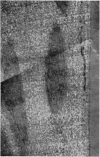
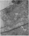
Full text
PDF


Images in this article


Selected References
These references are in PubMed. This may not be the complete list of references from this article.
- CAMERON D. A., ROBINSON R. A. Electron microscopy of cartilage and bone matrix at the distal epiphyseal line of the femur in the newborn infant. J Biophys Biochem Cytol. 1956 Jul 25;2(4 Suppl):253–260. doi: 10.1083/jcb.2.4.253. [DOI] [PMC free article] [PubMed] [Google Scholar]
- CAMERON D. A., ROBINSON R. A. Electron microscopy of epiphyseal and articular cartilage matrix in the femur of the newborn infant. J Bone Joint Surg Am. 1958 Jan;40-A(1):163–170. [PubMed] [Google Scholar]
- DALTON A. J., ZEIGEL R. F. A simplified method of staining thin sections of biolgical material with lead hydroxide for electron microscopy. J Biophys Biochem Cytol. 1960 Apr;7:409–410. doi: 10.1083/jcb.7.2.409. [DOI] [PMC free article] [PubMed] [Google Scholar]
- EKHOLM R. Nutrition of articular cartilage; a radioautographic study. Acta Anat (Basel) 1955;24(3-4):329–338. doi: 10.1159/000141049. [DOI] [PubMed] [Google Scholar]
- GLAUERT A. M., GLAUERT R. H., ROGERS G. E. A new embedding medium for electron microscopy. Nature. 1956 Oct 13;178(4537):803–803. doi: 10.1038/178803a0. [DOI] [PubMed] [Google Scholar]
- JACKSON S. F. The morphogenesis of avian tendon. Proc R Soc Lond B Biol Sci. 1956 Mar 13;144(917):556–572. doi: 10.1098/rspb.1956.0011. [DOI] [PubMed] [Google Scholar]
- LEVER J. D. A method of staining sectioned tissues with lead for electron microscopy. Nature. 1960 Jun 4;186:810–811. doi: 10.1038/186810a0. [DOI] [PubMed] [Google Scholar]
- LITTLE K., PIMM L. H., TRUETA J. Osteoarthritis of the hip: an electron microscope study. J Bone Joint Surg Br. 1958 Feb;40-B(1):123–131. doi: 10.1302/0301-620X.40B1.123. [DOI] [PubMed] [Google Scholar]
- MacCONAILL M. A. The movements of bones and joints; the mechanical structure of articulating cartilage. J Bone Joint Surg Br. 1951 May;33B(2):251–257. [PubMed] [Google Scholar]
- RICHARDSON K. C., JARETT L., FINKE E. H. Embedding in epoxy resins for ultrathin sectioning in electron microscopy. Stain Technol. 1960 Nov;35:313–323. doi: 10.3109/10520296009114754. [DOI] [PubMed] [Google Scholar]
- SCOTT B. L., PEASE D. C. Electron microscopy of the epiphyseal apparatus. Anat Rec. 1956 Dec;126(4):465–495. doi: 10.1002/ar.1091260405. [DOI] [PubMed] [Google Scholar]
- WATSON M. L. Staining of tissue sections for electron microscopy with heavy metals. II. Application of solutions containing lead and barium. J Biophys Biochem Cytol. 1958 Nov 25;4(6):727–730. doi: 10.1083/jcb.4.6.727. [DOI] [PMC free article] [PubMed] [Google Scholar]
- ZBINDEN G. Uber Feinstruktur und Altersveranderungen des hyalinen Knorpels im elektronenmikroskopischen Schnittpraparat und Beitrag zur Kenntnis der Verfettung der Knorpelgrundsubstanz. Schweiz Z Pathol Bakteriol. 1953;16(2):165–189. [PubMed] [Google Scholar]
- ZELANDER T. Ultrastructure of articular cartilage. Z Zellforsch Mikrosk Anat. 1959;49(6):720–738. doi: 10.1007/BF00342718. [DOI] [PubMed] [Google Scholar]