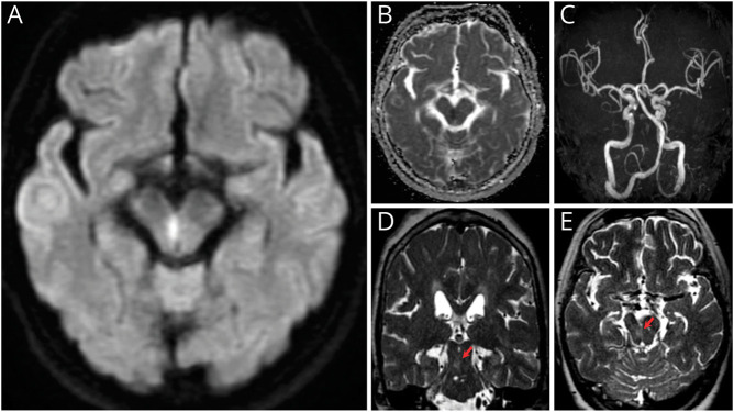
Figure. Brain MRI.
(A, B) Axial brain MRI showed hyperintensity and hypointensity in the right paramedian midbrain on diffusion-weighted imaging and apparent diffusion coefficient map, respectively. (C) Magnetic resonance angiography showed no obvious major artery stenosis on day 1. (D, E) Axial and coronal T2-weighted images on day 27 showed a faint high-signal lesion at the same site (arrow). The inferior olivary nucleus showed no signal change.

