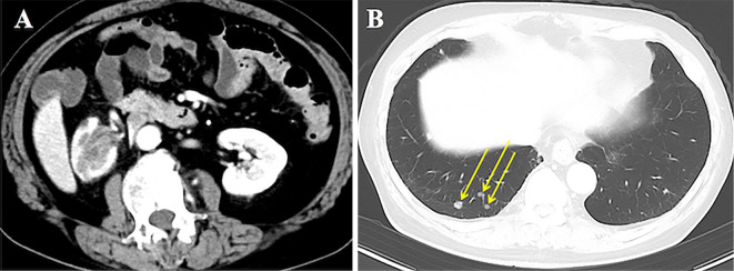
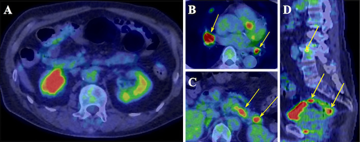
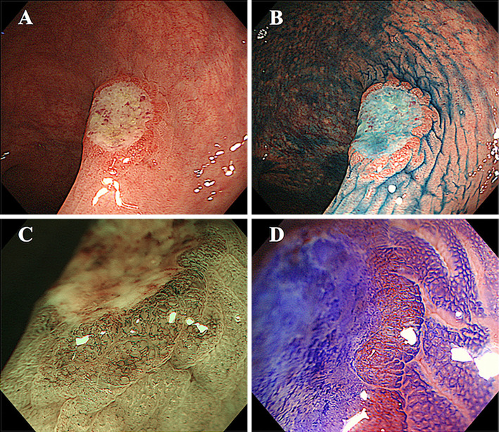
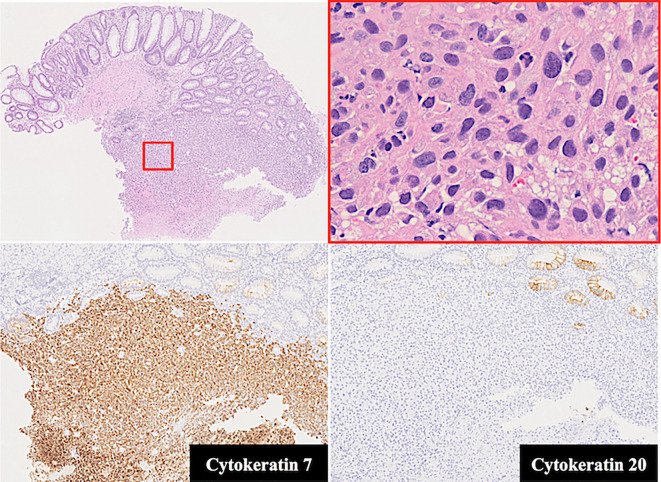
A 73-year-old man was referred for the investigation of positive urine cytology and right lumbar pain. Contrast-enhanced computed tomography (CT) showed a tumor in the upper right kidney (Picture 1A) and lung masses (Picture 1B, arrows). Positron emission tomography-CT showed an increased radionuclide uptake in the kidney (Picture 2A), lymph nodes (Picture 2B, arrows), pancreas (Picture 2C, arrows), lungs, bones, and rectum (Picture 2D, arrows). Colonoscopy showed a 10-mm type 0-IIa+IIc rectal tumor. The IIa part was erythematous with a dilated tortuous vessel pattern but magnifying endoscopy did not show an irregular pit pattern (Picture 3). A histological examination of a biopsy specimen from the tumor margin suggested urothelial cancer; immunohistochemical results were positive for cytokeratin 7 but negative for cytokeratin 20 (Picture 4). These findings yielded a final diagnosis of Stage IV renal pelvic cancer. To our knowledge, this is the first report of metastasis of renal pelvic cancer to the rectum mimicking primary colorectal cancer (1,2).
Picture 1.
Picture 2.
Picture 3.
Picture 4.
The authors state that they have no Conflict of Interest (COI).
References
- 1. Shinagare AB, Ramaiya NH, Jagannathan JP, et al. Metastatic pattern of bladder cancer: correlation with the characteristics of the primary tumor. Am J Roentgenol 196: 117-122, 2011. [DOI] [PubMed] [Google Scholar]
- 2. Ii Y, Munakata S, Honjo K, et al. Rectal metastasis from bladder urothelial carcinoma: a case report. Surg Case Rep 7: 100, 2021. [DOI] [PMC free article] [PubMed] [Google Scholar]


