Abstract
Abstract—Nowadays the possible influence of the coronavirus infection on cartilage degeneration and synovial membrane inflammation during chronic joint pathology—osteoarthritis—remains largely unelucidated. The aim of the presented work is to analyze the TGFB1, FOXO1, and COMP gene expression and free radical generation intensity in blood of patients suffering from osteoarthritis after beating the SARS-CoV2 infection. The work was carried out using molecular genetics and biochemistry methods. The decrease of the TGFB1 and FOXO1 expression level was shown to be more evident in the osteoarthritis patients after COVID-19 if compared to the group with knee osteoarthritis during simultaneous and more prominent diminishing of both superoxide dismutase and catalase activity (possibly indicating cell redox state disruption and TGF- P1-FOXO1 signaling attenuation) in patients with osteoarthritis after SARS-CoV2 disease. At the same time, the more prominent decrease of COMP gene expression level was demonstrated in patients with osteoarthritis after COVID-19 compared to the group with knee osteoarthritis and more intense increase of the COMP concentration in patients with osteoarthritis after the SARS-CoV2 infection was revealed. These data indicate more significant activation of cell destructive processes after the infection as well as further pathology progression.
Keywords: Keywords: SARS-CoV-2, gene expression of TGFB1, FOXO1, COMP, osteoarthritis, COMP, catalase, superoxide dismutase
INTRODUCTION
Coronavirus infections are mainly characterized by respiratory symptoms, but complications associated with musculoskeletal dysfunction have been identified. In particular, it was established that arthralgia is present in 14.9% of patients with coronavirus infection (Joob et al., 2020; Gasparotto et al., 2021). There is presently insufficient information on the potential impact of this infection on cartilage degeneration and synovial inflammation in chronic joint disease–osteoarthritis (OA) (Lai et al., 2020; Lauwers et al., 2022). At the same time, the investigation of the molecular and genetic mechanisms of the occurrence of this disease and its possible exacerbation due to SARS-CoV2 infection is extremely important (Lai et al., 2020; Gasmi et al., 2021; Lauwers et al., 2022).
It is known that OA is characterized by changes in the levels of cartilage oligomeric collagen, aggrecan, and cartilage oligomeric matrix protein (COMP, also known as thrombospondin-5; encoded by the COMP gene) (Huet et al., 2019; Mishra et al., 2019; Jaabar et al., 2022). It was shown that the mutations of the COMP gene in humans can lead to dysplasia, while a lack of oligomeric matrix protein can cause arthritis of various etiologies as well as other joint diseases (Huet et al., 2019; Jaabar et al., 2022).
A multifunctional cytokine is transforming growth factor-β1 (TGF-β1, coded by the TGFB1 gene), which activates the eponymous signaling cascade, and is a critical regulator of articular cartilage tissue protection and chondrocyte homeostasis (Mishra et al., 2019; Wang et al., 2020). However, the regulatory networks and downstream signaling pathways that govern the chondroprotective function of TGF-β in the OA are not fully defined. Recently, it was proposed that the transcription factor FOXO1 (forkhead box protein O1, encoded by the FOXO1 gene) is an important mediator of the TGF-β signaling pathway and may protect against OA by maintaining articular cartilage homeostasis (Wang et al., 2020). Thus, a correlation between the decrease in the expression level of and FOXO1 and OA-like pathologies in humans and in animal models was found (Matsuzaki et al., 2018; Lee et al., 2019; Wang et al., 2020).
It is known that the development of osteoarthritis is also associated with oxidative stress and excessive production of reactive oxygen species (ROS). Under physiological conditions, ROS regulate intracellular signaling processes, aging and apoptosis of chondrocytes, synthesis and degradation of the extracellular matrix, and, under pathological conditions, they are involved in the synovial inflammation and dysfunction of subchondral bone due to inhibition of the activity of enzymes of antioxidant protection of cells (Wu et al., 2016; Abusarah et al., 2017; Lee et al., 2019).
In view of the above, the aim of the study was the analysis of the TGFB1, FOXO1, and COMP gene expression and the intensity of free radical processes in the blood of osteoarthritis patients after a SARS-CoV2 infection.
MATERIALS AND METHODS
Forming experimental and control groups of patients–volunteers for further research. Sixty men between 40 and 45 years took part in the study. The selection of volunteers and the diagnosis of patients with “stage II and III knee osteoarthritis” was carried out at the Orthoklinika specialized medical center, the city of Ternopil, Ukraine. During the selection stage all patients underwent radiography of the knee joints in anterior-posterior and lateral projections. The assessment of pain intensity and the functional state of the patients' knee joints was carried out using the calculation of the WOMAC index (Western Ontario and McMaster Universities Osteoarthritis Index). The WOMAC index is calculated as a result of each patient’s self-administered test (McConnell et al., 2001), which includes 24 questions that reflect the severity of pain sensations (five questions), stiffness (two questions), and functional activity (17 questions). The volunteers were divided into the following groups: the first group (n = 20) were conditionally healthy people; the second group (n = 20) were patients with stage II and III knee osteoarthritis; the third group (n = 20) were patients with stage II and III knee osteoarthritis, who suffered a mild and moderate-severe form of COVID-19 6 months previous. The diagnosis of COVID-19 was confirmed by molecular analysis (RT-PCR) of a nasopharyngeal swab. The collection of biological material (blood, blood serum) was carried out at the Orthoklinika specialized medical center, Ternopil, Ukraine.
Determination of superoxide dismutase and catalase activity in blood serum of donors of all experimental groups. Superoxide dismutase (SOD) activity was assessed by the ability of SOD to compete with nitroblue tetrazolium for superoxide radicals (Durak et al., 1993). Catalase activity was measured by the amount of undecomposed hydrogen peroxide in the sample (Gyth, 1991). Protein content was measured according to the Lowry method (Lowry et al., 1951).
Quantitative reverse transcription PCR (RT-qPCR). RNA was obtained from blood using the Chomczynski method (Chomczynski et al., 1987). cDNA synthesis and quantitative PCR (qPCR) were carried out using the Thermo Scientific Verso SYBR Green 1-Step qRT-PCR ROX Mix commercial kit (Thermo Scientific, Lithuania), according to temperature conditions recommended by the manufacturer.
The following sequences of primers were used in the reactions: for TGFB1: direct TTG—AGCCGTGGAGGGGAAATT and reverse—AGGCCGGTTCATGCCATGAAT; FOXO1: direct—GGCGGGCTGGAAGAATTCAAT and reverse—CTCTTGCCACCCTCTGGATT-GA; COMP, direct—AAGTGGGCTACATCA-GGGTG and reverse—GTGTCATTGCAGC-GGTAACG; for ACTB (β‑actin gene used as an internal reaction control due to constitutive expression): forward—CTTCCAGCTCCTCCCTGGAG and reverse— CCACAGGACTCCATGCCCAG. The reproducibility of the amplification results was checked in parallel experiments by repeating RT-qPCR on RNA samples from all volunteers with each primer at least three times. The relative amount of mRNA was calculated according to the ΔΔCT Method comparative CT method (Livak et al., 2001), the efficiency of PCR reactions was the same (Ex = (10–1/slope) – 1), slope <0.1. The relative expression level of the specified genes was normalized to the expression level of the ACTB.
ELISA. The concentration of COMP was measured in blood serum (in triplicates for each sample) using a Human COMP DuoSet ELISA enzyme-linked immunosorbent assay kit (Catalog # DY3134, Novus Biologicals, United States) according to the manufacturer’s recommendations (wavelength 450 nm) using the STAT FAX 2100 device (Awareness Technology Inc., United States).
Statistical processing of results. The obtained data were tested for normal distribution by the Shapiro-Wilk test using the GraphPad Prism 8.4.3 software package (GraphPad Software Inc., United States). Further calculation was performed using one-way ANOVA with Tukey’s test. The obtained results are shown as the arithmetic mean ± standard deviation (SD). Results were considered significant at p ≤ 0.05.
RESULTS
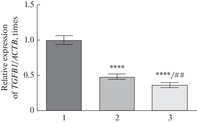
Expression level of TGFB1 and FOXO1 genes. As a result of our experimental studies, it was shown that the level of the expression of the TGFB1 gene in the blood of patients with OA was 2.1 times lower (p ≤ 0.0001) compared to the group of conditionally healthy people. In patients with knee OA after COVID-19, this indicator decreased by 2.8 times (p ≤ 0.0001) compared to the group of conditionally healthy people and by 1.3 times (p ≤ 0.01) compared to the group of patients with OA (Fig. 1).
Fig. 1.

Gene expression level of the TGFB1 in the blood of patients with osteoarthritis. 1—Healthy people; 2—patients with osteoarthritis; 3—osteoarthritis + COVID-19; ****p ≤ 0.0001 relative to healthy people; ##p ≤ 0.01 relative to the group with osteoarthritis.
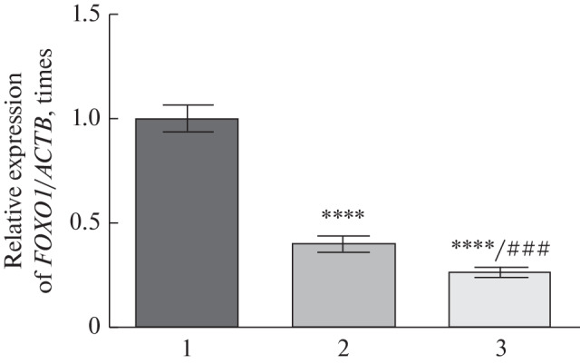
As a result of further studies, it was found that the level of the expression of the FOXO1 gene in the blood of patients with OA was 2.5 times lower (p ≤ 0.0001) compared to the group of conditionally healthy people. In patients with knee OA after COVID-19, this indicator decreased by 3.8 times (p ≤ 0.0001) compared to the group of conditionally healthy people and by 1.5 times (p ≤ 0.001) compared to the group of patients with OA (Fig. 2).
Fig. 2.

Gene expression level of the FOXO1 in the blood of patients with osteoarthritis. 1—Healthy people; 2—patients with osteoarthritis; 3—osteoarthritis + COVID-19; ****p ≤ 0.0001 relative to healthy people; ### p ≤ 0.001 relative to the group with osteoarthritis.
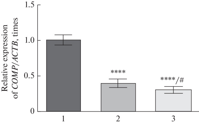
Expression level of the COMP gene. We established that the level of the expression of the COMP gene in the blood of patients with OA was 2.4 times lower (p ≤ 0.0001) compared to the group of conditionally healthy people. In patients with knee OA after COVID-19, this indicator decreased by 3.3 times (p ≤ 0.0001) compared to the group of conditionally healthy people and by 1.4 times (p ≤ 0.05) compared to the group patients with OA (Fig. 3).
Fig. 3.

Gene expression level of the COMP in the blood of patients with osteoarthritis. 1—Healthy people; 2—patients with osteoarthritis; 3—osteoarthritis + COVID-19; **** p ≤ 0.0001 relative to healthy people; # p ≤ 0.05 relative to the group with osteoarthritis.
Concentration of COMP. As a result of our research, it was shown that the concentration of cartilage oligomeric matrix protein was 2.6 times higher in the blood serum of patients with knee OA compared to a group of conditionally healthy people (Table 1). An analysis of this indicator in the blood serum of patients with knee OA after COVID-19 revealed that the concentration of COMP increased three times compared to the group of conditionally healthy people and the 1.2 times compared to the group of patients with OA (Table 1).
Table 1. .
Concentration of COMP in blood serum of experimental groups (M ± SD, n = 60)
| Groups (men) | ng/mL |
|---|---|
|
Conditionally healthy (n = 20) |
54.1 ± 3.8 |
|
Osteoarthritis (n = 20) |
142.1 ± 13.2**** |
|
Osteoarthritis + COVID-19 (n = 20) |
164.4 ± 15.6****/# |
*** p ≤ 0.0001, relative to healthy people; #p ≤ 0.05 relative to the group with osteoarthritis.
Superoxide dismutase and catalase activity. As a result of further experiments, it was found that superoxide dismutase and catalase activities were 1.3 and 1.7 times lower, respectively, in the blood serum of patients with knee OA compared to a group of conditionally healthy people (Table 2). Superoxide dismutase and catalase activity in the blood serum of patients with knee OA after COVID-19 decreased by 1.8 and 2.2 times, respectively, compared to the group of conditionally healthy people, and decreased by 1.4 and 1.3 times, respectively, compared to the group of patients with OA (Table 2).
Table 2. .
Superoxide dismutase and catalase activity in blood serum of experimental groups (M ± SD, n = 60)
| Groups (men) | Indicator | |
|---|---|---|
| Superoxide dismutase, U × min–1 × mg of protein–1 |
Catalase, μMx × min–1 × mg of protein–1 |
|
| Conditionally healthy (n = 20) | 3.23 ± 0.29 | 12.21 ± 1.43 |
| Osteoarthritis (n = 20) | 2.50 ± 0.23* | 7.28 ± 0.69*** |
| Osteoarthritis + COVID-19 (n = 20) | 1.80 ± 0.15***/# | 5.65 ± 0.48***/# |
*** p ≤ 0.001, *p ≤ 0.05 relative to healthy people; #p ≤ 0.05 relative to the group with osteoarthritis.
DISCUSSION
Molecular genetic studies of three FoxO (1, 3, and 4) have shown that TGF-β signaling (specifically, the TGF-β1 signaling pathway) regulates FOXO1 exclusively through TGF-β-activated TAK1 kinase. This kinase (another name is MAP3K7, “mitogen-activated protein kinase kinase kinase 7" is a cytosolic serine/threonine protein kinase of the MAP3K family, a component of several signaling pathways that regulate inflammation and the immune response of the body. Thus, it was found that TGF-β1 stably induced both expression of the FOXO1 gene and its product in articular chondrocytes (Wang et al., 2020).
At the same time, it was found in animal models that the postnatal loss of FOXO1 in cartilage led to OA-like pathologies: the initial cartilage thickening progressed to its degeneration (Matsuzaki et al., 2018; Lee et al., 2019; Wang et al., 2020). Similar results were obtained in patients with OA and for the aging cartilage (Verdonk et al., 2016). On the contrary, in mice, hyperexpression of the FOXO1 prevented the development of OA. Thus, it has been proposed that FOXO1 is an important downstream effector of TGF-β signaling in the regulation of postnatal cartilage homeostasis (Cheng et al., 2017; Wang et al., 2020).
We found a decrease in the level of the expression the TGFB1 and FOXO1 genes (indicating a possible silencing of TGF-b1-FoxO1 signaling) to a greater extent in osteoarthritis patients after COVID-19 compared to the group of knee osteoarthritis patients. This may be due to an increase in systemic inflammation caused by the response of the body to viral infection.
Loss of FoxOl was shown to lead to increased ROS, which were efficiently eliminated by the activation of autophagy. On the contrary, in vitro introduction of a vector with FoxOl inhibited H2O2-induced cell apoptosis and activated the action of antioxidant defense enzymes. Similar results were obtained in vivo in mice with induced OA (Peng et al., 2017; Wang et al., 2020). It is known that translocation of FoxOs from the cytoplasm into the nucleus occurs under conditions of oxidative stress with the involvement of SIRT1 deacetylase, which leads to the activation of FoxOs (Klotz et al., 2015; Wang et al., 2020). Therefore, it has been proposed that FOXO1 maintains articular chondrocyte homeostasis by modulating autophagy, and overexpression of the FOXO1 in chondrocytes prevents the development of OA caused by the loss of the TGF-β1 signaling cascade. However, the mechanism by which TGF-β1/FOXO1 regulates articular chondrocyte proliferation and homeostasis requires further investigation (Wang et al., 2020).
In our study, the above-mentioned decrease in the level of the expression of the TGFB1 and FOXO1 genes in the second and third groups of patients was shown against the background of a decrease in both superoxide dismutase and catalase activity, which indicates a violation of the redox status of cells. Similarly to the analysis of gene expression levels, inhibition of the activity of antioxidant cell protection enzymes was more significant in osteoarthritis patients after COVID-19 compared to the group of knee osteoarthritis patients. The impairment of the oxidative-antioxidant balance towards the intensification of free radical processes plays an important role in the pathogenesis of various pathological conditions. In particular, in the synovial fluid of the joint, free radical processes play a significant role in the development of the inflammatory response, lipoperoxidation, and aging of articular cartilage cells (Wu et al., 2016; Abusarah et al., 2017; Lee et al., 2019).
In addition, in animal models of OA, hyperexpression FOXO in chondrocytes caused an increase in the level of expression of genes that encode components of the extracellular matrix, Comp, Col1a1 (the gene encoding α1-chain of type I collagen), etc. as well as genes of antioxidant protection (Lee et al., 2019).
Thus, we found a decrease in the level of the expression of the COMP gene in the second and third groups of patients against the background of increased concentration of COMP. This is consistent with our data demonstrating the disruption of the redox status of cells and a decrease in the level of the expression of the TGFB1 and FOXO1 genes.
The increase in the level of ROS (against the background of a decrease in the level of the expression of the FOXO) can induce catabolic signaling, cause oxidative stress, increase the expression of both genes involved in the development of inflammation and those encoding matrix metalloproteinases (MMP), which, in turn, affect matrix components, including collagen, aggrecan, COMP (Huet et al., 2019; Mishra et al., 2019; Yamamoto et al., 2021). In this case, COMP fragments are released into the synovial fluid even in the early stages of OA, and, thus, COMP is considered to be a marker of cartilage degradation (Huet et al., 2019; Mishra et al., 2019; Jaabar et al., 2022).
In general, the obtained results coincide with the above-mentioned literature data, which demonstrated the connection of the TGF-β1-FoxO1 system with both the activity of the enzymes of the antioxidant system of the body and the state of the extracellular matrix of chondrocytes in the pathogenesis of OA (Matsuzaki et al., 2018; Lee et al., 2019; Wang 2020 et al., 2020). A more significant decrease in the level of the expression of the COMP gene in patients with osteoarthritis after COVID-19, in comparison with the patients with knee osteoarthritis joints, against the background of a more intensive increase in the concentration of COMP, indicates a more significant activation of destructive processes in cells after the infection and further progression of the disease, which is consistent with the literature data demonstrating the potential impact of COVID-19 on joint aging and OA due to the inflammatory process (Joob et al., 2020; Lai et al., 2020; Gasparotto et al., 2021; Lauwers et al., 2022).
CONCLUSIONS
Using the molecular biological approaches, a more significant decrease in the level of the expression of the TGFB1 and FOXO1 genes was shown in patients with osteoarthritis after COVID-19 compared to the patients with knee osteoarthritis against the background of a more significant decrease in both superoxide dismutase and catalase activity (which indicates an impairment of the redox status of cells and possible silencing of TGF-β1-FOXO1 signaling) in patients with osteoarthritis after SARS-CoV2 infections. This may be associated with an increase in systemic inflammation due to the response of the body to the viral invasion. At the same time, a more significant decrease in the expression of the COMP gene was found in patients with osteoarthritis after COVID-19 in comparison with the patients with knee osteoarthritis against the background of a more intense increase in the concentration of COMP in patients with osteoarthritis after SARS-CoV2 infection. Such data indicate a more significant activation of destructive processes in cells after an infection and further progression of the disease. Understanding the clear mechanisms of the formation of a more severe course of osteoarthritis and the development of complications in patients with a post-COVID-19 syndrome using the example of the functioning of the TGF-β1-FoxO1 signaling cascade requires further investigation.
COMPLIANCE WITH ETHICAL STANDARDS
Conflict of interest. The authors declare that they have no conflicts of interest.
Statement of compliance with standards of research involving humans as subjects. All procedures performed in studies involving human participants were in accordance with the ethical standards of the institutional and/or national research committee and with the 1964 Helsinki Declaration and its later amendments or comparable ethical standards. All procedures performed in studies were approved by orders of the Ministry of Health of Ukraine no. 690 of September 23, 2009, no. 944 of December 14, 2009, and no. 616 of August 3, 2012; the bioethics commission of the Orthoclinic Medical Center (protocol no. 1 dated January 17, 2022, Ternopil, Ukraine); and the Educational and Scientific Center Institute of Biology and Medicine of Taras Shevchenko National University of Kyiv (protocol no. 3 dated October 3, 2022, Kyiv, Ukraine). Informed consent was obtained from all individual participants involved in the study. All necessary measures were taken to ensure patient anonymity.
REFERENCES
- 1.Abusarah J., Bentz M., Benabdoune H. An overview of the role of lipid peroxidation-derived 4-hydroxynonenal in osteoarthritis. Inflammation Res. 2017;66:637–651. doi: 10.1007/s00011-017-1044-4. [DOI] [PubMed] [Google Scholar]
- 2.Cheng N.T., Meng H., Ma L.F. Role of autophagy in the progression of osteoarthritis: The autophagy inhibitor, 3-methyladenine, aggravates the severity of experimental osteoarthritis. Int. J. Mol. Med. 2017;39:1224–1232. doi: 10.3892/ijmm.2017.2934. [DOI] [PMC free article] [PubMed] [Google Scholar]
- 3.Chomczynski P., Sacchi N. Single-step method of RNA isolation by acid guanidinium thiocyanate-phenol-chloroform extraction. Anal. Biochem. 1987;162:156–159. doi: 10.1006/abio.1987.9999. [DOI] [PubMed] [Google Scholar]
- 4.Durak I., Yurtarslanl Z., Canbolat O. A methodological approach to superoxide dismutase (SOD) activity assay based on inhibition of nitroblue tetrazolium (NBT) reduction. Clin. Chim. Acta. 1993;214:103–104. doi: 10.1016/0009-8981(93)90307-p. [DOI] [PubMed] [Google Scholar]
- 5.Gasmi A., Tippairote T., Mujawdiya P.K. Neurological Involvements of SARS-CoV2. Infect. Mol. Neurobiol. 2021;58:944–949. doi: 10.1007/s12035-020-02070-6. [DOI] [PMC free article] [PubMed] [Google Scholar]
- 6.Gasparotto M., Framba V., Piovella C., Doria A., Iaccarino L. Post-COVID-19 arthritis: a case report and literature review. Clin. Rheumatol. 2021;40:3357–3362. doi: 10.1007/s10067-020-05550-1. [DOI] [PMC free article] [PubMed] [Google Scholar]
- 7.Góth L. A simple method for determination of serum catalase activity and revision of reference range. Clin. Chim. Acta. 1991;196:143–151. doi: 10.1016/0009-8981(91)90067-m. [DOI] [PubMed] [Google Scholar]
- 8.Huet A., Dvorshchenko K., Korotkyi O. Expression of Nos2 and acan genes in rat knee articular cartilage in osteoarthritis. Cytol. Genet. 2019;53:481–488. doi: 10.3103/S0095452719060021. [DOI] [Google Scholar]
- 9.Jaabar, I.L., Cornette, P., Miche, A., et al., Deciphering pathological remodelling of the human cartilage extracellular matrix in osteoarthritis at the supramolecular level, Nanoscale, 2022, no. 24. 10.1039/D2NR00474G [DOI] [PubMed]
- 10.Joob B., Wiwanitkit V. Arthralgia as an initial presentation of COVID-19: observation. Rheumatol. Int. 2020;40:823. doi: 10.1007/s00296-020-04561-0. [DOI] [PMC free article] [PubMed] [Google Scholar]
- 11.Klotz L.O., Sánchez-Ramos C., Prieto-Arroyo I. Redox regulation of FoxO transcription factors. Redox Biol. 2015;6:51–72. doi: 10.1016/j.redox.2015.06.019. [DOI] [PMC free article] [PubMed] [Google Scholar]
- 12.Lai Q., Spoletini G., Bianco G. SARS-CoV2 and immunosuppression: A double-edged sword. Transpl. Infect. Dis. 2020;22:e13404. doi: 10.1111/tid.13404. [DOI] [PMC free article] [PubMed] [Google Scholar]
- 13.Lauwers M., Au M., Yuan S. COVID-19 in joint ageing and osteoarthritis: current status and perspectives. Int. J. Mol. Sci. 2022;23:720. doi: 10.3390/ijms23020720. [DOI] [PMC free article] [PubMed] [Google Scholar]
- 14.Lee K.I., Choi S., Matsuzaki T. FOXO1 and FOXO3 transcription factors have unique functions in meniscus development and homeostasis during aging and osteoarthritis. Proc. Natl. Acad. Sci. U. S. A. 2020;117:3135–3143. doi: 10.1073/pnas.1918673117. [DOI] [PMC free article] [PubMed] [Google Scholar]
- 15.Livak K., Schmittgen T. Analysis of relative gene expression data using real-time quantitative PCR and the 2−ΔΔCT Method. Methods. 2001;25:402–408. doi: 10.1006/meth.2001.1262. [DOI] [PubMed] [Google Scholar]
- 16.Lowry O.H., Rosebrough N.J., Farr A.L. Protein measurement with the Folin phenol reagent. J. Biol. Chem. 1951;193:265–275. doi: 10.1016/S0021-9258(19)52451-6. [DOI] [PubMed] [Google Scholar]
- 17.Matsuzaki, Ò., Alvarez-Garcia, O., Mokuda, S., et al., FoxO transcription factors modulate autophagy and proteoglycan 4 in cartilage homeostasis and osteoarthritis, Sci. Transl. Med., 2018, vol. 10, p. eaan0746. 10.1126/scitranslmed.aan0746 [DOI] [PMC free article] [PubMed]
- 18.McConnell S., Kolopack P., Davis A.M. The Western Ontario and McMaster universities osteoarthritis index (WOMAC): a review of its utility and measurement properties. Arthritis Care Res. 2001;45:453–461. doi: 10.1002/1529-0131(200110)45:5<453::aid-art365>3.0.co;2-w. [DOI] [PubMed] [Google Scholar]
- 19.Mishra A., Awasthi S., Raj S. Identifying the role of ASPN and COMP genes in knee osteoarthritis development. J. Orthop. Surg. Res. 2019;14:337. doi: 10.1186/s13018-019-1391-7. [DOI] [PMC free article] [PubMed] [Google Scholar]
- 20.Peng Q., Qin J., Zhang Y. Autophagy maintains the stemness of ovarian cancer stem cells by FOXA2. J. Exp. Clin. Cancer Res. 2017;36:171. doi: 10.1186/s13046-017-0644-8. [DOI] [PMC free article] [PubMed] [Google Scholar]
- 21.Verdonk R., Madry H., Shabshin N. The role of meniscal tissue in joint protection in early osteoarthritis. Knee Surg., Sports Traumatol. Arthroscopy. 2016;24:1763–1774. doi: 10.1007/s00167-016-4069-2. [DOI] [PubMed] [Google Scholar]
- 22.Wang C., Shen J., Ying J. FoxO1 is a crucial mediator of TGF-β/TAK1 signaling and protects against osteoarthritis by maintaining articular cartilage homeostasis. PNAS. 2016;117:30488–30497. doi: 10.1073/pnas.2017056117. [DOI] [PMC free article] [PubMed] [Google Scholar]
- 23.Wu Q., Zhong Z.M., Zhu S.Y. Advanced oxidation protein products induce chondrocyte apoptosis via receptor for advanced glycation end products-mediated, redox-dependent intrinsic apoptosis pathway. Apoptosis. 2016;21:36–50. doi: 10.1007/s10495-015-1191-4. [DOI] [PubMed] [Google Scholar]
- 24.Yamamoto K., Wilkinson D., Bou-Gharios G. Targeting dysregulation of metalloproteinase activity in osteoarthritis. Calcif. Tissue Int. 2021;109:277–290. doi: 10.1007/s00223-020-00739-7. [DOI] [PMC free article] [PubMed] [Google Scholar]


