Abstract
Background
The purpose of this study was to determine if adding a reconstructed superior acromioclavicular (AC) joint ligament adds significant biomechanical stability to the AC joint over anatomic coracoclavicular (CC) ligament reconstruction alone.
Methods
Fourteen cadaver shoulders were used for the comparison of biomechanical stability among the anatomic CC ligament reconstruction alone, CC and AC ligament reconstruction, and the intact groups by measuring the displacement under cyclic loads. A load to failure test was then performed in the vertical direction at a loading rate of 2 mm /sec to determine surgical-repair joints’ tolerance to the maximum failure load.
Results
The average peak-to-peak displacement induced by cyclic load in the sagittal axis and vertical axis direction was not significantly different between CC ligament reconstruction, CC and AC ligament reconstruction, and intact groups. The maximum failure load for the CC reconstruction (224.9 ± 91.8 N (Mean ± SEM)) was lower than CC/AC reconstruction groups (326.2 ± 123.3 N). The CC/AC reconstruction group failed at a significantly higher load (t test, p = 0.016) than the CC reconstruction group.
Conclusion
CC/AC reconstruction surgical technique yielded a better shoulder stability than CC ligament alone reconstruction that may better maintain reduction of the AC joint.
Level of Evidence: Level II.
Keywords: acromioclavicular joint, coracoclavicular ligament, injury, reconstruction, biomechanics
Introduction
Injuries to the AC joint are common in young athletes, especially in contact sports such as rugby, wrestling, football and hockey. Treatment is generally directed by the Rockwood classification of the injury. 1 Type I and II are almost universally treated nonoperatively because the coracoclavicular (CC) ligaments remain intact. Uncertainty surrounds the treatment of type III acromioclavicular (AC) joint injuries, but nonoperative treatment is favoured in most cases.2–4 Types IV-VI are generally treated operatively due to the significant soft tissue disruption and large displacement of the distal clavicle from the acromion.
Many surgical techniques have been suggested for treatment of AC separation with variations of fixating the clavicle to the coracoid or stabilising the AC joint. In 1972, Weaver and Dunn described their technique in which they resected a portion of the distal clavicle then transferred the coracoacromial (CA) ligament from the acromion to the clavicle. 5 Modifications to this technique have been added to reinforce the CA ligament transfer. Recent biomechanical studies have suggested that anatomic repair may improve stability of the lateral clavicle when compared to the modified Weaver-Dunn procedure, but do not fully restore the strength of the injured shoulder to the intact state. 6 Only a Bosworth screw with bicortical coracoid fixation has been shown to match the tensile strength of the intact CC and AC ligaments, but this offers no biologic advantages and may be complicated by hardware irritation, screw pullout or breakage or infection. 7
In this study, we conducted a biomechanical evaluation of two novel reconstruction techniques. In the first technique we anatomically reconstructed the CC ligament alone using an allograft semitendinosus. In the second technique we anatomically reconstructed CC ligaments and reconstructed the AC ligament. The purpose of this study was to determine the efficiency of two surgical repairing techniques in restoring joint stability by comparing the stiffness and peak-to-peak elongation between surgical repairing and normal groups. The injury tolerance of the repaired AC joints was studied be comparing their maximum failure load.
Materials and methods
Fourteen fresh-frozen cadaveric shoulders were obtained from seven donors (3 female, 4 male). These shoulders were randomised to either the anatomic CC reconstruction alone group or anatomic CC reconstruction with AC reconstruction group. The specimens (n = 7) were used in the anatomic CC reconstruction group with an average age in the group of 59.3 years (range 48–69). The average age in anatomic CC reconstruction with AC reconstruction group (n = 7) was 62.7 years (range 48–73).
Biomechanical testing
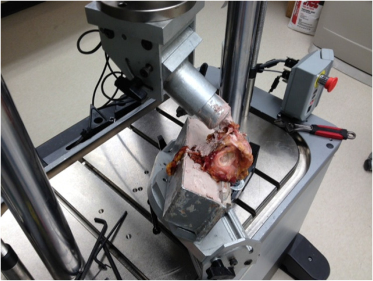
All soft tissue was removed from the shoulder leaving only the CC and AC ligaments intact. The scapula and medial end of the clavicle were potted in a polyester resin material (Bondo, 3 M, St Paul, MN). Sixty millimetres of lateral clavicle was left exposed so the reconstruction could be performed after testing with intact ligaments. The clavicle was then positioned anatomically and the potted scapula and clavicle were rigidly secured to the base of a materials testing machine (ElectroPuls E10000, Instron, Norwood, MA) with the glenoid used as a reference, being horizontal for posterior load testing and vertical for superior load testing (Figure 1). This setup was similar to one published previously. 8
Figure 1.
Biomechanical testing setup of a cadaveric shoulder mounted on the instron material testing machine. The scapula and medial end of the clavicle were potted in two adjustable fixtures respectively using a polyester resin material. The glenoid was used as a reference being horizontal for posterior load testing and vertical for superior load testing.
The shoulder was first positioned for superior loading. A 40 N load was applied in the superior direction through the clavicle with the scapula secured to the Instron base. One-hundred cycles of a 40 N superior load was applied. This force was used as it has previously been reported to be the amount of force due to the weight of the arm at varying arm positions. 9 The shoulder was then positioned for posterior loading. A 40 N load was applied in the posterior direction through the clavicle with the scapula secured to the Instron base. One-hundred cycles of a 40 N posterior load was applied.
The CC and AC ligaments were transected while leaving the clavicle and scapula secured to the Instron to maintain its position. Each shoulder was then reconstructed with one of the two reconstruction techniques in the following manner.
Anatomic CC reconstruction
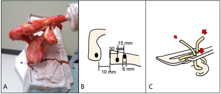
Measurements were taken from the distal end of the clavicle medially to 45 mm and 30 mm for the conoid and trapezoid attachments, respectively. Holes were drilled to 5.5 mm with the conoid drill hole positioned along the posterior aspect of the clavicle and directed to the coracoid and the trapezoid drill hole positioned slightly anterior on the clavicle and directed to the coracoid. A semitendinosus autograft was passed down through the conoid drill hole, around the undersurface of the coracoid then up through the trapezoid drill hole (Figure 2). The conoid portion of the graft was fixed with a 5.5 mm PEEK interference screw (Arthrex, Naples, FL). The clavicle was then reduced, and the trapezoid portion of the graft was tensioned and secured with another 5.5 mm PEEK interference screw (Arthrex, Naples, FL).
Figure 2.
A shows the experimental setting of anatomic CC ligament reconstruction. B shows the dimension of holes drilled for reconstruction. C shows the schematic illustration of anatomic CC ligament reconstruction pathway.
Anatomic CC reconstruction with AC reconstruction
Measurements were taken from the distal end of the clavicle medially to 45 mm and 30 mm for the conoid and trapezoid attachments, respectively. The conoid drill hole was drilled to 5.5 mm, positioned along the posterior aspect of the clavicle and directed to the coracoid. The trapezoid drill hole was drilled to 6.0 mm, positioned slightly anterior on the clavicle and directed to the coracoid. A third hole was drilled through the acromion to a 5.5 mm diameter. This was positioned in line with the midportion of the clavicle, leaving a minimum 5 mm bone bridge to the edge of the acromion. A semitendinosus autograft was passed down through the conoid drill hole, around the undersurface of the coracoid then up through the trapezoid drill hole. The long tail of the trapezoid portion of the graft was then passed over the AC joint and down through the acromion drill hole, passed back across the undersurface of the AC joint and up through the trapezoid drill hole (Figure 3). The conoid portion of the graft was secured with a 5.5 mm PEEK interference screw (Arthrex, Naples, FL). The clavicle was then reduced, and the graft was tensioned assuring adequate tension of the graft both around the coracoid and through the acromion. The trapezoid portion of the graft was then fixed with another PEEK interference screw (Arthrex, Naples, FL).
Figure 3.
A shows the experimental setting of anatomic CC plus AC ligament reconstruction. B shows the dimension of holes drilled for reconstruction. C shows the schematic illustration of anatomic CC and AC ligament reconstruction pathway.
Biomechanical testing
After reconstruction, testing was repeated in both the posterior and superior direction. Following superior cyclic load testing, a superior load to failure was done, with 2 cm considered failure, approximating a complete AC dislocation.
Force and displacement were recorded by the computer. Peak-to-peak displacement was the average difference between the highest and the lowest displacement of the last three cycles. The peak load was defined as the superior load to failure for all specimens. The stiffness was calculated for each specimen after testing by calculating the slope of the line in the linear portion of the force/displacement curve along the superior load direction.
Statistical analysis was performed with SPSS statistical software version 26 (IBM, Armonk, NY, USA). One-way ANOVA test or Univariate with PostHoc LSD was performed to determine the statistical difference of Peak-to-peak displacement and stiffness between AC reconstruction, AC plus CC reconstruction, and normal groups. The t test was performed to determine the statistical difference of maximum failure load between AC reconstruction group and AC plus CC reconstruction group. Statistical significance was defined as a P value smaller than 0.05.
Results
Peak-to-peak displacement
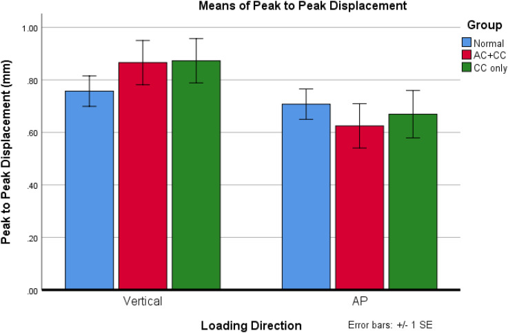
The average peak-to-peak displacement in the posterior direction was 0.70 ± 0.23 mm (Mean ± SD) intact and 0.67 ± 0.19 mm after the CC reconstruction. This value was 0.62 ± 0.20 mm in intact group and 0.62 ± 0.23 mm) in the CC plus AC reconstruction in the anterior-posterior (AP) direction (Figure 1).
The average peak-to-peak displacement in the superior direction (vertical direction) was 0.75 ± 0.23 mm (Mean ± SD) in intact group, 0.87 ± 0.28 mm in the CC reconstruction group (p = 0.22), and 0.87 ± 0.29 mm after the CC/AC reconstruction in the superior direction.
There was not a statistical difference of peak-to-peak displacement between groups (Univariate PostHoc LSD, p = 0.865). There was a statistical difference of peak-to-peak displacement between vertical load direction and AP load direction (Univariate PostHoc LSD, p = 0.01) under 40 N of load. There was more displacement along superior and inferior direction than AP direction (Figure 4).
Figure 4.
Peak to peak displacement under cyclic load. Vertical load caused more displacement than anterior-posterior (AP) direction load.
Ultimate load to failure
The ultimate load to failure for the CC reconstruction and CC/AC reconstruction groups was 224.9 N (224.9 ± 91.8 N) (Mean ± SD) and 326.2 N (326.2 ± 123.3 N) respectively. The CC/AC reconstruction group failed at a significantly higher load (t test, p = 0.016) than the CC reconstruction group.
Stiffness
The average stiffness at vertical direction load for the CC reconstruction group was 27.5 N/mm (SEM 11 N/mm) and 26.2 N/mm (SEM 9.9 N) for the CC/AC reconstruction group (t test, p = 0.35).
Discussion
The purpose of our study was to compare the biomechanical properties of anatomic reconstruction of the CC ligaments alone to anatomic reconstruction of both the CC and AC ligaments. We found that when AC ligaments were reconstructed along with the CC ligaments, there was a significant increase in load to failure.
Bietzel et al. in a review of AC joint injuries, noted very few clinical studies evaluating anatomic and nonanatomic reconstruction techniques. 7 Only two clinical studies were found directly comparing anatomic to nonanatomic reconstruction techniques. Fraschini et al. evaluated anatomic reconstruction with a synthetic graft compared to a polyester vascular prosthesis fixated around the clavicle and coracoid. 10 Anatomic reconstruction techniques were found to have a satisfactory outcome in 93% of patients versus 53% in the nonanatomic group. 10 Tauber et al. evaluated anatomic AC reconstruction with semitendinosus allograft vs nonanatomic reconstruction using a semitendinosus allograft around the clavicle and coracoid to reduce the AC joint. 6 They found significantly improved ASES and constant scores in the anatomic reconstruction group.
Grutter and Peterson found the tensile strength of an intact AC joint to be 815 N. 11 They also found that the Bosworth screw was the only fixation method that had a higher load to failure than the intact state. 11 However, the Bosworth screw is associated with a unique group of complications including screw pullout, screw breakage, hardware irritation and the need for a second surgery to remove the screw.12,13
Harris et al. isolated the CC ligaments and found their tensile strength to be 500 N. In the same study, the tensile strength of several other reconstructions was evaluated. 14 None of the reconstructions, with the exception of the Bosworth screw were able to match this strength.
Mazzocca et al. evaluated the biomechanics of a modified Weaver-Dunn reconstruction compared with an arthroscopic reconstruction using nonabsorbable suture and an anatomic CC ligament reconstruction using a semitendinosus graft. 15 They assessed both anterior-posterior translation and superior-inferior translation as well as load to failure. The modified Weaver-Dunn reconstruction was found to have significantly higher posterior translation. 15 They found the anatomic reconstruction technique resulted in improved restoration of AC joint biomechanics and inferred that it may decrease recurrent subluxation at the AC joint and improve patient outcomes. 15 They concluded that maximal clinical outcomes after surgical treatment for chronic AC joint dislocations should focus on controlling both superior translation of the distal clavicle and anterior-posterior stability at the AC joint level. They suggested that re-creation of the anatomical orientation of the conoid and trapezoid ligaments with a technique that also controls excessive anterior-posterior translation would potentially achieve this goal.
Morikawa et al. in a recent study assessed the contribution of the three segments of the acromioclavicular ligament complex (ACLC) to posterior translation and rotational stability. 16 This biomechanical study of cadaveric shoulders tested the anterior, superior and posterior segments of the ACLC when intact, sectioned in different combinations, and repaired in different combinations. The most important finding of this study was that superior segment of the ACLC was the most important contributor to posterior translation and rotational stability of the AC joint suggesting that in chronic cases of AC joint injury, reconstruction of the entire superior ACLC may be needed.
Hislop et al. applied a similar suture technique for AC joint repair construct using Nitinol wires, cortical buttons (Dog-Bone; Arthrex Inc) 2 strands of 2-mm suture tape (FiberTape; Arthrex Inc). 17 Their repair constructs included CC ligament along and CC plus AC constructs. Stiffness of vertical load was 11.80 ± 4.57 and 13.36 ± 4.20 for CC construct and CC plus AC construct. Our autograft tendon suture technique yielded 27.5 ± 11.0 N/mm stiffness for CC construct and 26.2 ± 9.9 N/mm stiffness for CC plus AC construct, demonstrating a higher stiffness than Nitinol wires. There was not a statistical difference of the peak-to-peak displacement between CC and CC plus AC constructs under 40N cyclic loads in our study and under 70 N cyclic load in Hislop's study. Hislop reported the vertical direction load-to-failure to be 361.8 N and 387.7 N for CC and CC plus AC constructs respectively, which appeared to be higher than our tendon repair construct. Further clinical studies are required to compare the merits of two methods.
In our study we compared two anatomic AC reconstructions, one with CC and AC ligament reconstruction and one with CC reconstruction alone. We found that AC joint reconstruction that includes reconstruction of the AC ligaments was significantly stronger than AC joint reconstruction with only reconstruction of the CC ligaments, having load-to-failure strengths of 326 N and 224 N, respectively. We believe reconstruction of the AC ligaments along with the CC ligaments provides for a stronger reconstruction that may better maintain reduction of the AC joint and lead to fewer failures.
There are several limitations in this study. The failure loads were somewhat lower than other studies testing anatomic reconstruction strength.11,14,15,18 We propose 3 possible explanations. First, we used 2 cm as our definition of failure. This may have caused a halt to testing prior to the graft itself completely failing. However, the displacement of 2 cm would be considered a clinical failure. Second, we fixed the medial end of the clavicle on the materials testing machine. To perform anatomic reconstruction, we needed 60 mm of the distal clavicle exposed. This may have allowed some flexion of the clavicle during superior load testing. Although this more likely recreates the forces that would be placed on the graft in vivo, not all of the 2 cm of superior displacement would have been realised at the AC joint, as some would have been taken up within the clavicle itself. Third, we performed cyclic testing first, which may have weakened the graft prior to load to failure testing. Other limitations include the variability of the autograft tendons and cadaveric specimens regarding bone and joint morphology, degenerative joint disease, and bone mineralisation.
This study compared the biomechanical behaviours of surgical repair constructs for AC join injury, the AC join injury tolerance including the maximum load under faster loading rates has not been investigated. Hence the future studies will be performed to understand the AC injury tolerance to high-speed loads as occurs in falling, sport collisions, and car accidents, as well as to document the load to the distal clavicle during the activity of daily life.
Conclusion
In this study we found significantly higher load to failure of an AC reconstruction that includes reconstruction of the AC ligaments when directly compared to reconstructing the CC ligaments using identical testing techniques. The reconstruction of the AC ligaments along with the CC ligaments provides a stronger reconstruction that may better maintain reduction of the AC joint and lead to fewer failures.
Footnotes
The author(s) declared no potential conflicts of interest with respect to the research, authorship, and/or publication of this article.
ORCID iD: Chaoyang Chen https://orcid.org/0000-0003-0838-2245
References
- 1.Rockwood CJ, Williams G, Young D. Disorders of the acromioclavicular join. In: Rockwood CJ, Matsen FA, eds. The Shoulder. Philadelphia: WB Saunders, 1998: 483–553. [Google Scholar]
- 2.Murena L, Canton G, Vulcano E, et al. Scapular dyskinesis and SICK scapula syndrome following surgical treatment of type III acute acromioclavicular dislocations. Knee Surg Sports Traumatol Arthrosc 2013; 21: 1146–1150. [DOI] [PubMed] [Google Scholar]
- 3.Tamaoki MJ, Belloti JC, Lenza M, et al. Surgical versus conservative interventions for treating acromioclavicular dislocation of the shoulder in adults. Cochrane Database Syst Rev 2010; 8: CD007429. [DOI] [PMC free article] [PubMed] [Google Scholar]
- 4.Trainer G, Arciero RA, Mazzocca AD. Practical management of grade III acromioclavicular separations. Clin J Sport Med 2008; 18: 162–166. [DOI] [PubMed] [Google Scholar]
- 5.Weaver JK, Dunn HK. Treatment of acromioclavicular injuries, especially complete acromioclavicular separation. J Bone Joint Surg Am 1972; 54: 1187–1194. [PubMed] [Google Scholar]
- 6.Tauber M, Gordon K, Koller H, et al. Semitendinosus tendon graft versus a modified Weaver-Dunn procedure for acromioclavicular joint reconstruction in chronic cases: a prospective comparative study. Am J Sports Med 2009; 37: 181–190. [DOI] [PubMed] [Google Scholar]
- 7.Beitzel K, Cote MP, Apostolakos J, et al. Current concepts in the treatment of acromioclavicular joint dislocations. Arthroscopy 2013; 29: 387–397. [DOI] [PubMed] [Google Scholar]
- 8.Corteen DP, Teitge RA. Stabilization of the clavicle after distal resection: a biomechanical study. Am J Sports Med 2005; 33: 61–67. [DOI] [PubMed] [Google Scholar]
- 9.Lee SJ, Keefer EP, McHugh MP, et al. Cyclical loading of coracoclavicular ligament reconstructions: a comparative biomechanical study. Am J Sports Med 2008; 36: 1990–1997. [DOI] [PubMed] [Google Scholar]
- 10.Fraschini G, Ciampi P, Scotti C, et al. Surgical treatment of chronic acromioclavicular dislocation: comparison between two surgical procedures for anatomic reconstruction. Injury 2010; 41: 1103–1106. [DOI] [PubMed] [Google Scholar]
- 11.Grutter PW, Petersen SA. Anatomical acromioclavicular ligament reconstruction: a biomechanical comparison of reconstructive techniques of the acromioclavicular joint. Am J Sports Med 2005; 33: 1723–1728. [DOI] [PubMed] [Google Scholar]
- 12.Galpin RD, Hawkins RJ, Grainger RW. A comparative analysis of operative versus nonoperative treatment of grade III acromioclavicular separations. Clin Orthop Relat Res 1985; 193: 150–155. [PubMed] [Google Scholar]
- 13.Guy DK, Wirth MA, Griffin JL, et al. Reconstruction of chronic and complete dislocations of the acromioclavicular joint. Clin Orthop Relat Res 1998; 347: 138–149. [PubMed] [Google Scholar]
- 14.Harris RI, Wallace AL, Harper GD, et al. Structural properties of the intact and the reconstructed coracoclavicular ligament complex. Am J Sports Med 2000; 28: 103–108. [DOI] [PubMed] [Google Scholar]
- 15.Mazzocca AD, Santangelo SA, Johnson ST, et al. A biomechanical evaluation of an anatomical coracoclavicular ligament reconstruction. Am J Sports Med 2006; 34: 236–246. [DOI] [PubMed] [Google Scholar]
- 16.Morikawa D, Dyrna F, Cote MP, et al. Repair of the entire superior acromioclavicular ligament complex best restores posterior translation and rotational stability. Knee Surg Sports Traumatol Arthrosc 2019; 27: 3764–3770. [DOI] [PubMed] [Google Scholar]
- 17.Hislop P, Sakata K, Ackland DC, et al. Acromioclavicular joint stabilization: a biomechanical study of bidirectional stability and strength. Orthop J Sports Med 2019; 7: 2325967119836751. [DOI] [PMC free article] [PubMed] [Google Scholar]
- 18.Costic RS, Labriola JE, Rodosky MW, et al. Biomechanical rationale for development of anatomical reconstructions of coracoclavicular ligaments after complete acromioclavicular joint dislocations. Am J Sports Med 2004; 32: 1929–1936. [DOI] [PubMed] [Google Scholar]


