PLOS ONE logoLink to PLOS ONE
. 2023 Apr 6;18(4):e0283773. doi: 10.1371/journal.pone.0283773

In vitro spermatogenesis in isolated seminiferous tubules of immature mice

Xuemin Feng 1,#, Takafumi Matsumura 1,2,#, Yuki Yamashita 2,#, Takuya Sato 1,2, Kiyoshi Hashimoto 3, Hisakazu Odaka 3, Yoshinori Makino 4, Yuki Okada 4, Hiroko Nakamura 5, Hiroshi Kimura 5, Teruo Fujii 6, Takehiko Ogawa 1,2,*
Editor: Suresh Yenugu7
PMCID: PMC10079070  PMID: 37023052

Abstract

Mouse spermatogenesis, from spermatogonial stem cell proliferation to sperm formation, can be reproduced in vitro by culturing testis tissue masses of neonatal mice. However, it remains to be determined whether this method is also applicable when testis tissues are further divided into tiny fragments, such as segments of the seminiferous tubule (ST), a minimal anatomical unit for spermatogenesis. In this study, we investigated this issue using the testis of an Acrosin-GFP/Histone H3.3-mCherry (Acr/H3) double-transgenic mouse and monitored the expression of GFP and mCherry as indicators of spermatogenic progression. Initially, we noticed that the cut and isolated stretches of ST shrunk rapidly and conglomerated. We therefore maintained the isolation of STs in two ways: segmental isolation without truncation or embedding in soft agarose. In both cases, GFP expression was observed by fluorescence microscopy. By whole-mount immunochemical staining, meiotic spermatocytes and round and elongating spermatids were identified as Sycp3-, crescent-form GFP-, and mCherry-positive cells, respectively. Although the efficiency was significantly lower than that with tissue mass culture, we clearly showed that spermatogenesis can be induced up to the elongating spermatid stage even when the STs were cut into short segments and cultured in isolation. In addition, we demonstrated that lowered oxygen tension was favorable for spermatogenesis both for meiotic progression and for producing elongating spermatids in isolated STs. Culturing isolated STs rather than tissue masses is advantageous for explicitly assessing the various environmental parameters that influence the progression of spermatogenesis.

Introduction

Most organs can be divided into smaller anatomical subunits that can be regarded as a minimal functional unit. These functional units derived from particular organs can be maintained in culture as explants for a certain period of time. Organoids, which are self-organizing systems of stem cells and their progeny cells, are in many cases composed of such functional units, providing a useful tool for biomedical research [1].

Spermatogenesis takes place in the seminiferous tubules (STs), which are anatomical structures consisting of Sertoli cells on the inside, peritubular myoid cells on the outside, and basal lamina between them. Several different types of cells outside STs, collectively called interstitial cells, also play important roles in controlling spermatogenesis [2]. Approximately 11 STs are packed inside a testis of an adult mouse, and the individual STs have an average length of 140 mm [3]. Therefore, the functional minimum unit of the testis is presumed to be an ST segment of certain length with germ cells inside and interstitial cells around.

In 2011, we developed a culture system that reproduced the complete process of spermatogenesis, from spermatogonial stem cell proliferation to sperm formation, in an explanted mouse testis tissue. Offspring were obtained by micro-insemination with the haploid cells produced in the explants [4]. The explanted testis tissue pieces were about 1 mm3 or larger in size and consisted of numbers of the functional units. In experimental systems, manipulating a single or a small number of functional units could serve to clarify the mechanistic details of physiological phenomena and pathological disorders. Namely, interactions between functional units as well as between a unit and the cells surrounding it could be examined experimentally. However, it has proven quite difficult to dissect the functional unit of spermatogenesis. In fact, only a few studies have reported the use of isolated segments of STs to promote or maintain spermatogenesis in culture. Some of these studies, using the testis of adult rats of 60 to 120 days of age, demonstrated the progression of a limited span of spermatogenesis lasting for several days [57]. Another recent study showed the progression of spermatogenesis for 40 days using a culture of STs in soft agar [8]. On the other hand, other investigations have tried to reconstruct testis tissue architecture from singly isolated testicular cells. These studies showed different extents of successful spermatogenesis [911]. Based on these previous studies and our tissue mass culture experience, we expected that spermatogenesis in an isolated ST would be possible.

However, our early attempts were hampered by the stubborn nature of the STs themselves, which contracted and shrunk rapidly upon culturing. We overcame this problem using two methods and demonstrated that the isolated STs could support spermatogenesis up to the stage of elongating spermatids. This result showed experimentally for the first time that a limited ST segment can be a functional unit for spermatogenesis. In addition, using this method, we demonstrated that spermatogenesis in the isolated STs responded sensitively to oxygen concentration. This was not fully demonstrated in previous studies culturing tissue masses. The ST culture method, therefore, is useful for investigating various factors and conditions that influence the progression and maintenance of spermatogenesis.

Materials and methods

Animals

Acrosin (Acr)-GFP transgenic mice [12, 13] (genetic background: ICR, C57BL/6, and their mixture) were provided by RIKEN BRC through the National Bio-Resource Project of MEXT, Japan. Histone H3.3-mCherry (H3) transgenic mice [14] were provided by Makino & Okada. Acr-GFP/Histone H3.3.3-mCherry double (Acr/H3) transgenic mice were generated by crossing an Acr-GFP homogeneous female mouse with an H3 homogeneous male mouse. Mice aged 1–7 days postpartum (dpp) were euthanized by an anesthetic overdose using a medetomidine/midazolam/butorphanol (0.3/4/5) cocktail. Their testes were then collected. Mice were housed in specific pathogen-free, air-conditioned rooms at 24±1°C and 55±5%, with a 13-h light/11-h dark lighting cycle. They were fed ad libitum with commercially available hard pellets (MF; Oriental Yeast). Drinking water was acidified to pH 2.8–3.0 by HCl. All animal experiments were performed in accordance with the Guidelines for Proper Conduct of Animal Experiments (Science Council of Japan) and were approved by the Institutional Committee of Laboratory Animal Experimentation (Animal Research Center of Yokohama City University; protocol no. F-A-20-038).

Culture media and reagents

The culture medium used was α-minimum essential medium (α-MEM) (12000–022; Gibco) supplemented with AlbuMAX I (11020–021; Thermo Fisher Scientific) at a final concentration of 40 mg/mL. NaHCO3 (7%) was then added (0.026 ml/ml medium) to achieve a final concentration of 1.82 g/L (0.0182 g for 10 ml of medium). Antibiotic-antimycotic (15240062; Thermo Fisher Scientific) was added at a 1/100 volume to achieve a final concentration of 100 IU/mL for penicillin, 100 μg/mL for streptomycin, and 250 ng/mL for amphotericin. Sterilization was performed with Millipore filtration and followed by storage in a refrigerator before use.

Agarose gel preparation

To make the agarose gel block for organ culture, agarose powder (Dojindo Molecular Technologies) was dissolved in water purified with Milli-Q (35–1251; Merck) at 1.5% (w/v) and autoclaved. While cooling, 33 mL of the agarose solution was poured into 10 cm dishes to form a 5 mm thick gel. The gel was cut into squares of approximately 10 × 10 mm size and these were used as a stand for testis tissue placement. The gel blocks were submerged in the culture medium in 12-well culture plates for more than 6 h before use. Testes tissue fragments or isolated STs were transferred to the surface of agarose gel blocks, the lower half of which were soaked in 0.5 mL medium in individual wells. A medium change was performed once a week. The culture incubator was supplied with 5% carbon dioxide in air (20% O2), unless otherwise described in the text, and maintained at 34°C.

PDMS ceiling chip

The PDMS (polydimethyl-siloxane) ceiling (PC) chip was fabricated by conventional photolithography and soft lithography methods [15]. The PC chip was produced by mixing PDMS prepolymer and curing reagent (Silpot 184; Dow Corning) at a 10:1 weight ratio. The mixture was poured into a mold master made by the photolithography method, and the mold master was placed in a vacuum chamber for degassing. After 30–45 min, the mold master was moved to an oven and baked for 2 h at 70°C. After cooling down, the solidified PDMS was peeled off from the master and this PDMS disk was cut into individual chips with a cutting knife. The thickness of the chips was dependent on the amount of PDMS mixture poured in. The depth of dent, the tissue setting area, ranged from 150 μm to 190 μm depending on the mold [16]. The modified PC chip, named the STPC chip, had a row of pillars in the tissue set region. The pillars were 0.1 mm wide squares, equal in height to the depth of the set region (150–190 μm), and arranged in straight rows at 0.1 mm intervals (Fig 1D). A thin PDMS membrane (75 μm thick, ASAHI Rubber Inc.) was used in some cases for pressing testis tissues against the base agarose gel by placing them under the PDMS ceiling to hold the tissue in place.

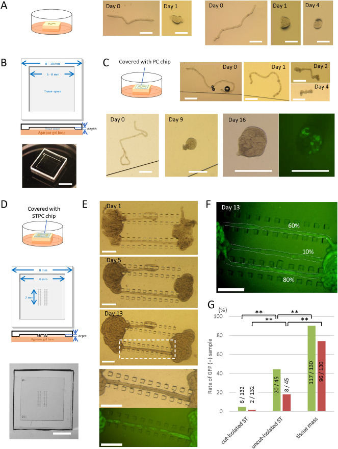
Fig 1. Culturing of isolated ST segments.

Fig 1

(A) According to an air-liquid interphase method, cut-isolated STs of a 5 dpp mouse were placed on an agarose gel block half-soaked in the medium. The STs shrunk and conglomerated in a day. (B) Schematic diagram of a PC chip, top and cross-sectional views, and photos of a PC chip. The depth of dent for accommodating culture samples was between 150 and 190 μm. (C) A schematic drawing of a culture experiment using a PC chip, and a stereomicroscopic view of a cut-isolated ST of a 5 dpp mouse covered with a PC chip. The ST shrunk in several days. Another cut-isolated ST cultured under a PC chip conglomerated but showed GFP expression on culture day 16. (D) A schematic drawing of a culture experiment using STPC, and a schematic diagram of a STPC chip, top and cross-sectional views. The size of each pillar was 0.1 × 0.1 mm. Four parallel pillar rows made of 11 pillars each were arranged in the center of the tissue space. The depth of the tissue space ranged from 150 to 190 μm. A photo of an STPC chip from above is shown in the bottom pannel. (E) Stereomicroscopic view of an uncut-isolated ST cultured under an STPC chip on days 1, 5, and 13. The dashed rectangular area is enlarged in the bottom two panels. Fluorescence microscopy revealed Acr-GFP expression in the stretched portion of the ST. (F) A photo of the GFP-expressing portion of three uncut-isolated STs, taken on culture day 13. The percentages of GFP-expressing area were visually measured as 60%, 10%, and 80%, respectively. (G) The percentages of GFP-positive area in the samples of three ST states: cut-isolated STs, uncut-isolated STs, and tissue mass. Samples with GFP-expressing areas over 10% (green bar) and 50% (red bar) during culture days 31 to 40 were counted as GFP-positive. Numerators and denominators in or over each bar are the numbers of GFP-positive tissue samples and total examined tissue samples, respectively. The experiments were performed under a 20% O2 concentration. **P<0.01. Scale bars: 5 mm (B), 2 mm (D), 0.5 mm (A, C, E).

Culture methods–PDMS ceiling (PC) method

The testes of mice were decapsulated—i.e., the capsule (tunica albuginea) was removed by forceps. The remaining tissue mass was mostly composed of seminiferous tubules, which lightly adhered to each other via the intervening microvasculature. Each testis tissue was gently separated into 6–8 pieces with forceps. For tissue mass culture, these clamps were simply placed on the agarose gel block. For the culture of isolated STs, the testis tissue mass was soaked in the medium to loosen and disperse the STs. The STs were then gently separated with forceps and cut with spring scissors as needed, depending on the desired state of the STs. These STs in the medium were sucked up with a micropipette and poured in the hollow space of a PC chip (Fig 1B). The PC chip was then inverted and placed on an agarose gel. In case of the uncut-isolated ST method—i.e., to separate the STs without cutting—the tissue mass was placed in the hollow portion of the STPC chip (Fig 1D). Forceps were used to gently dissociate the tissue mass into STs, and then to isolate an ST segment with both ends connected to the tissue mass so that the ST segment would fit between the pillar rows. The STPC chip was then inverted and set on an agarose gel.

Culture methods—Soft agarose method

After preparing cut-isolated STs in the culture medium, an equivalent volume of low melting point Agarose-L (319–01181; Nippon Gene) solution (1% w/v) was added and gently mixed. After cooling down at 4°C for about 15 min, the soft agarose gel containing the cut-isolated STs was cut into squares of 8 × 8 mm size and laid on a 1.5% agarose gel block (base gel). Medium was poured to half the height of the gel block (S3 Fig, Method A in S1 File and S1 Video). The PDMS ceiling chip was optional but was used in many cases. As an alternative approach, we adopted the protocol of a soft agar culture system from a previous study [8]. In brief, the soft agarose (0.5% w/v) containing the cut-isolated STs was poured onto the solid bottom gel layer. To make the bottom gel layer, 1.5% agarose solution was poured into 5 cm or 3 cm dishes. After cooling down and solidification, in order to replace the water in the gel with medium, an identical volume of culture medium was added to the dish and left for more than 6 h until use. The dish with its 2 layers of gel was left at 4°C for about 15 min until the soft agar layer became solid. The upper soft gel layer was adjusted to a thickness of around 0.4 mm (exactly 0.395, 0.443, 0.435, 0.220, and 0.361 mm when measured). Then the two-layer agarose gel was cut into squares of about 10 × 10 mm size, each of which was moved to a well in a 12-well plate with 0.5 mL medium in each well (S3 Fig, Method B in S1 File and S1 Video). Medium change was performed once a week. The culture incubator was supplied with 5% carbon dioxide in air and maintained at 34°C.

Observations

Cultured tissues and ST segments were observed at least once a week under a stereomicroscope equipped with an excitation light for GFP (LeicaM205 FA; Leica, Germany). Acr-GFP begins to be expressed in mice at around 15 dpp in vivo, but its expression could be delayed by several days in vitro. In addition, GFP emission can fluctuate at intervals of several days. Therefore, to avoid false-negative results, GFP positivity was not necessarily determined on a single day; in most cases, two observations with a 7-day interval were taken, resulting in an observation period of greater than 7 days. Samples showing GFP expression during those periods were considered GFP-positive. The GFP-positive portions in each stretch of ST were measured by visual approximation as 0%, 1–10%, 11–20%, 21–40%, 41–60%, 61–80% or 81–100%.

H3.3 mCherry appears beginning at around 28 dpp in vivo. In vitro, however, its expression is delayed days to weeks, depending on the sample. For the reliable identification of mCherry, each sample tissue was removed from the culture well, placed on a slide glass and observed with an inverted microscope (IX73; Olympus) or a confocal microscope (FV1000–MPE; Olympus). The observation timing was carefully determined in each case, taking the status of preceding GFP expression into account.

Histological and immunohistochemical examinations

In histological examinations, specimens were fixed with Bouin’s fixative and embedded in paraffin. One section showing the largest cut surface was made for each specimen and stained with hematoxylin and eosin (H&E) or periodic acid Schiff (PAS). In immunofluorescence staining, tissues were fixed with 4% of paraformaldehyde in PBS at 4°C overnight. Tissues were then soaked in solutions of 10%, 15%, and 20% (w/v) sucrose in PBS for 1 h each in succession for cryoprotection. They were cryo-embedded in OCT compound (Sakura Finetek Japan) and cut into 7-μm-thick sections. Antibodies used as primary antibody were anti-GFP (ab13970, 1:1000; abcam), anti-synaptonemal complex protein 3 (SCP3) (ab97672, 1:100; abcam), and anti-GFRα1 (1:200, AF560; Bio-Techne, MN, USA). Lectin PNA from Arachis hypogea (peanut), Alexa Fluor 568 Conjugate (L32458, 1:1000; Thermo Fischer Scientific, MA, USA) was used to identify the acrosome. Antibodies used for secondary antibody were Alexa Fluor 488-conjugated goat anti-chicken antibody (A-11039, 1:200; Thermo Fischer Scientific), Alexa Fluor 555-conjugated goat anti-mouse antibody (A-21424, 1:200; Thermo Fischer Scientific), Alexa Fluor 555-conjugated goat anti-rabbit antibody (A-21428, 1:200; Thermo Fischer Scientific), Alexa Fluor 647-conjugated goat anti-rat antibody (A-21247, 1:200; Thermo Fischer Scientific), and Alexa Fluor 647-conjugated donkey anti-goat antibody (A-21447, 1:200; Thermo Fischer Scientific). Nuclei were counterstained with Hoechst33342 dye. Observation of immunostained samples were performed with a confocal laser microscope (FV1000-MPE; Olympus).

Whole-mount immunohistochemical staining

The cultured seminiferous tubules were fixed with 4% paraformaldehyde in PBS at 4°C overnight. After washing with PBS for 3 min, the tubules were dehydrated in 100% methanol for 30 min at room temperature. The dehydrated tubules were washed with PBS containing 1% Triton-X100 (1% PBST) 4 times for 10 min each, and then blocked with Image-iT™ FX Signal Enhancer (Thermo Fisher Scientific) for 1 h and incubated with primary antibody diluted in blocking buffer at 4°C overnight. After washing with 1% PBST 4 times for 10 min each, the tubules were reacted with secondary antibody diluted in blocking buffer at room temperature for 1 h. After washing with 1% PBST 4 times for 10 min each, the nuclei were counterstained with Hoechst 33342 dye. Specimens were observed with a confocal laser microscope (Olympus FV-1000D). The primary antibodies used were chicken anti-GFP antibody (1:1000; Abcam), rabbit anti-RFP polyclonal antibody (1:1000; MBL), mouse anti-SYCP3 antibody (1:500; Abcam), goat anti-GFRα1 antibody (1:200; R&D Systems), rabbit anti-STRA8 antibody (1:250; Abcam), rabbit anti-γH2AX (1:500; Abcam), and rabbit anti-Mouse HSD3B antibody (1:250; Trans Genic Inc.). The secondary antibodies used were goat anti-chicken IgG, goat anti-rabbit IgG, and goat anti-mouse IgG, conjugated with Alexa 488, Alexa 555 or Alexa 647 (1:200; Invitrogen).

Statistical analysis

Fisher’s exact test was used for statistical analyses. The Holm method was used to adjust the family-wise error rate in multiple comparisons.

Results

Acr/H3 double-Tg mouse testis as a system for monitoring spermatogenesis

The GFP expression in the spermatogenic cells of Acr-GFP transgenic mice starts from pachytene spermatocytes at stage 4 onward [12]. In Histone H3.3-mCherry transgenic mice, mCherry is expressed in spermatids from step 11 onward [14]. Then, we first examined if Acr/H3 double-Tg mouse testis faithfully express GFP and mCherry as expected in their progression of spermatogenesis. Testes of mice aged 10, 20, and 40 days postpartum (dpp) were collected and prepared for immunohistochemical observation (S1A Fig in S1 File). The testis of 10 dpp was negative for both GFP and mCherry. In the 20 dpp testis, GFP-positive cells and peanut agglutinin (PNA)-positive dots were observed in the center of STs, indicating haploid cells were emerging. In the 40 dpp testis, GFP, mCherry and PNA were clearly observed. At higher magnification, the combination of GFP and mCherry, which respectively localized to the acrosome and nucleus, was identified as a unique figure that served as a faithful discriminative marker of elongating spermatids at step 11 onward (S1B Fig in S1 File) [14]. These results confirmed that the Acr/H3 double-Tg mouse testis was an excellent system for monitoring the progression of spermatogenesis and spermiogenesis.

Cut and isolated ST segments shrunk in cultivation

After removing the tunica albuginea, STs in the testis of a neonatal Acr-GFP Tg or an Acr/H3 double-Tg mouse were loosened and dispersed, in order to randomly cut and isolate ST segments (cut-isolated STs). The cut-isolated STs were cultured on an agarose gel block, which was half-soaked in culture medium, and the GFP expression was monitored with a stereomicroscope. However, these isolated stretches of ST shrunk and conglomerated in a day (Fig 1A). We suspected that this may have been due to the surface tension of the medium, and we therefore used a ceiling chip made of an air-permeable silicone, polydimethylsiloxane (PDMS), to cover the ST segments [16, 17]. This PDMS ceiling (PC) chip is a small PDMS plate having a shallow dent/hollow on one side which serves as a space for testis tissues and STs (Fig 1B). By placing the PC chip over the ST, we expected it could alleviate the surface tension and reduce the shrinkage. This strategy worked to some extent, but within several days the cut-isolated STs beneath the chip had gradually contracted and shrunk (Fig 1C). Proper spermatogenesis could not be expected in such shrunken STs. In fact, among the 132 cut-isolated ST samples that exhibited shrinkage in culture, GFP-expression was observed in only 7 (5.3%) (Fig 1C and 1G). We then produced a new PC chip, named the seminiferous tubule PC (STPC) chip. In the dent space of the STPC chip, we designed four rows of aligned pillars to hold the ST in place, hoping to prevent or reduce ST shrinkage (Fig 1D and S2A Fig in S1 File). However, we found that the cut-isolated STs set between the pillar rows contracted to almost the same degree as those without pillar rows (S2B Fig in S1 File). Even among several STs that we set in ways to hold them, such as meandering between the pillars, shrinkage still occurred (S2C, S2D Fig in S1 File). Such ST shrinkage would be expected to disturb and obstruct the progression of spermatogenesis.

Uncut and segmentally isolated ST method

In order to prevent the shrinkage of STs, we gave up cutting and detaching the STs completely, and instead isolated a portion of the ST, 1–2 mm in length, leaving both ends connected to the tissue mass. The ST segment isolated in this manner, which we designated the uncut and segmentally isolated ST (uncut-isolated ST), was placed in the STPC, and the tissue masses were placed on both sides of the edge of the pillar rows (Fig 1E). Although the tissue as a whole again showed a tendency to contract, the segmentally isolated part of the ST remained stretched. After 2 weeks of culture, GFP expression was observed in the uncut-isolated STs by fluorescence microscopy (Fig 1E). Photographs were taken weekly and the ratio of the GFP-expressing portion to the total uncut-isolated ST was measured visually (Fig 1F). The uncut-isolated STs whose GFP-expressing portion constituted more than 10% or 50% of the total length were regard as GFP-positive. We then compared STs in 3 different states: cut-isolated STs, uncut-isolated STs and tissue masses (Fig 1G). For cut-isolated (conglomerated) STs, only 5.3% of samples were GFP-positive with a 10% positive area threshold, as stated above. With a 50%-positive area threshold, only 1.5% (2 out of 132) of samples were positive. The uncut-isolated STs showed GFP-positivity rates of 53.3% (24 out of 45 samples) and 31.1% (14 out of 45 samples) for the 10% and 50% positive-area thresholds, respectively. The tissue masses showed 90.0% (117 out of 130 samples) and 73.8% (96 out of 130 samples) positivity rates, respectively. These results indicated that although spermatogenesis was induced in cut-isolated and uncut-isolated STs, at least up to the Acr-GFP-expressing stage—i.e., the early pachytene stage [13]—the efficiency of spermatogenesis was lower than that in the tissue mass.

Long-term culture of uncut-isolated STs

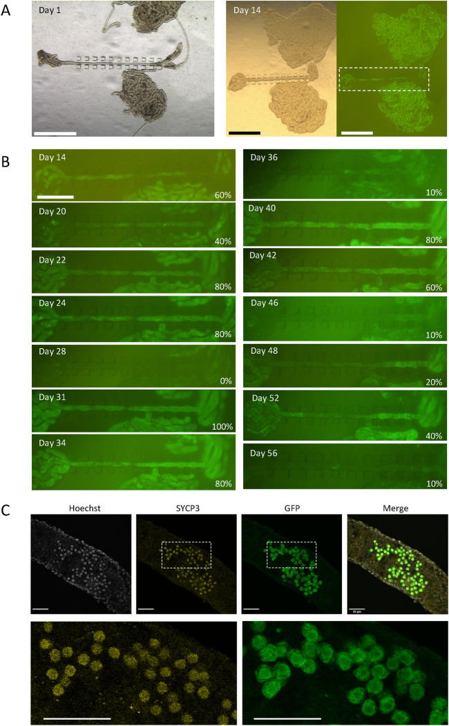
When we performed a culture experiment of uncut-isolated STs for a long period with frequent observations, Acr-GFP expression in a ST showed unique oscillatory fluctuation (Fig 2A and 2B). The same phenomenon was observed and reported previously in our study with a tissue mass culture experiment using a microfluidic device [18]. We conjectured that this fluctuation reflected an incomplete spermatogenesis in which degenerated and dead Acr-GFP expressing cells were cleaned up by phagocytotic Sertoli cells, followed by the reappearance of Acr-GFP expressing cells from among the remaining precursor cells in the ST. This finding thus indicated that the isolated ST can support spermatogenesis, although incompletely, including not only the so-called first wave but also subsequent successive spermatogenesis. To determine how far spermatogenesis had proceeded in the uncut-isolated STs, we performed whole-mount immunochemistry on STs from a 5 dpp mouse cultured for 34 days, with antibodies to SYCP3, GFP, and mCherry. Cells positive for SYCP3 and GFP, representing meiotic spermatocytes at the pachytene stage, were observed as the most advanced differentiating cells (Fig 2C).

Fig 2. A long-term culturing of uncut-isolated STs using STPC chips.

Fig 2

(A) A testis of an Acr-GFP mouse of 3 dpp were dissected and STs were set under STPC chips for culturing. On culture day 14, GFP expression was observed. The dashed rectangular region is enlarged in B. (B) Photographs taken sequentially on each culture day under excitation light for GFP recognition were aligned. GFP expression rates are noted in the lower right corner of each panel. (C) Confocal microscopic photos of whole-mount immunochemical staining of an uncut-isolated ST were taken on culture day 34, using a 5 dpp mouse testis. SYCP3- and GFP-positive cells represent meiotic spermatocytes. Scale bar: 1 mm (A), 0.5 mm (B), 50 μm (C).

Cultivation of cut-isolated STs using a soft agarose method

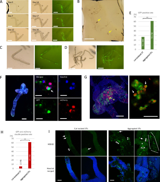
As a second approach to prevent the shrinkage of STs, we adopted a soft agarose method. Initially, 0.35% soft agarose was used as described in the literature [8, 19] and the cut-isolated STs mixed in the gel were set in the STPC chip and placed on the base agarose gel (1.5%). Under this condition, however, the shrinkage was not sufficiently suppressed. We therefore raised the concentration of the soft agarose in the gel to 0.5%. A square thin piece of soft agarose containing the cut-isolated STs was moved onto the base gel, with or without a PC chip covering (S3 Fig, Method A in S1 File and S1 Video). However, the soft agarose gel was too fragile to handle by itself, and the gel was prone to breakage during the transfer. We therefore adopted Gholami’s method [8]. The cut-isolated STs suspended in 0.5% soft agarose solution were poured on the bottom layer of a 1.5% agarose gel, and the resultant two-layered gel was cut out for cultivation (S3 Fig, Method B in S1 File and S1 Video). The cut-isolated STs in 0.5% soft agarose gel did not shrink and maintained their original form (Fig 3A). In this procedure, in addition to cut-isolated STs, small clumps of aggregated STs were observed in the gel. These mostly resulted from the insufficient isolation of STs and were denoted as aggregated STs when 3 or more STs clustered together (Fig 3B).

Fig 3. Culture of cut-isolated and aggregated STs in the soft agarose.

Fig 3

(A) Series photos of a cut-isolated ST embedded in soft agarose using method B, showing the maintenance of its form without shrinkage and the induction of GFP expression from day 14. (B) Low-magnification view of cut-isolated STs embedded in the soft agarose. Yellow arrows indicate aggregated STs. (C) A cut-isolated ST in the soft agarose with GFP expression at culture day 22, derived from a 7 dpp mouse testis. (D) Aggregated STs in the soft agarose with GFP expression at culture day 22, derived from a 7 dpp mouse testis. (E) GFP expression was examined by fluorescence microscopy in samples from 7 experiments and assessed during culture days 20 to 25. Numerators and denominators in or over each bar are the numbers of GFP-positive tissue samples and total examined tissue samples, respectively. The experiments were performed under a 20% O2 concentration. ** P < 0.01. (F) Whole-mount immunostaining of a cut-isolated ST with anti-GFP antibody (green) and anti-RFP antibody (red). Nuclei were counterstained with Hoechst 33342 dye (blue). The box indicated by the dotted lines in the left panel is enlarged in the right panels. Germ cells with both GFP and mCherry expressions, indicating elongating spermatids at or beyond the step 11, were observed (white arrow). (G) Whole-mount immunostaining of aggregated STs demonstrated several elongating spermatids, expressing both GFP and mCherry. The rectangular region surrounded by a dotted line is enlarged in the right panel. (H) GFP and mCherry expressions were confirmed by whole-mount immunohistochemical staining in samples from a single experiment. The tissues were fixed either on culture day 45, 52, or 59. The experiment was performed under a 20% O2 concentration. ** P < 0.01. (I) Whole-mount immunostaining of cut-isolated and aggregated STs with HSD3β antibody (Green), along with counterstaining with Hoechst. Within aggregated STs, faint dot-like GFP expression derived from Acr-GFP was observed (*). An HSD3β signal was observed in cells adhering to the outside of the STs, either singly (arrows) or as clusters (arrowheads). In these pictures, the numbers of such HSD3β-positive cells were counted to be 5 in cut-isolated and 41 in aggregated STs. In addition, in the case of aggregated STs, a clumpy interstitial tissue containing about 20 HSD3β-positive cells was also observed (surrounded by braces). Scale bars: 200 μm (A, C, F left, G left), 2 mm (B), 0.5 mm (D), 50 μm (G right, I) and 10 μm (F right 4 panels).

Within two weeks, GFP expression was confirmed in 72 out of 268 cut-isolated STs (26.9%) under fluorescence microscopic observation (Fig 3A, 3C and 3E). As for aggregated STs, GFP expression was observed in 55 out of 90 samples (61.1%) (Fig 3D and 3E). This result suggested that STs under the aggregated condition were somehow favored for spermatogenic progression. In 40–55 days, STs from an experiment were examined by whole-mount immunochemical staining. In 2 of the 41 samples of cut-isolated STs examined, cells positive for both GFP and mCherry were observed (Fig 3F and 3H). This result showed that cut-isolated STs can support spermatogenesis up to the formation of elongating spermatids. As for aggregated STs, GFP and mCherry double-positive cells were observed in 37 out of 60 samples examined (Fig 3G and 3H). Again, it appeared that aggregation of STs confers an advantage for the promotion of spermatogenesis.

We speculated that interstitial cells that remain attached to the aggregated STs could be a reason for the abundant and more advanced spermatogenesis. To investigate this possibility, we stained Leydig cells with anti-HSD3β antibody under a whole-mount condition. In this instance, HSD3β-positive cells were detected abundantly in aggregated STs and only marginally in isolated ST samples (Fig 3I). The relationship between spermatogenic efficiency and the amount of interstitial cells around the ST would be an important issue to scrutinize in future studies.

Lowered oxygen concentration was favorable for spermatogenesis

In our previous study using rat testis tissues, we found that an O2 concentration lower than the atmospheric concentration of 20% induced spermatogenesis with higher efficiency [20]. Then, in a pilot study using testis tissues of Acr-GFP Tg mice, we compared two different concentrations of O2, i.e., 20% and 10%, in the incubator (S4 Fig in S1 File). In 20% O2, GFP expression was observed evenly throughout the tissue. On the other hand, in 10% O2, GFP expression was limited exclusively to the peripheral regions, leaving the central region completely negative. This expression pattern was reasonable, considering the limited diffusion of O2 into the tissue mass in the case of 10% O2. However, round spermatid formation, identified by GFP aggregation into a cap-shape, was intensively observed in the GFP-expressing area of 10% O2 samples. Round spermatids were also observed in 20% O2 samples, but rather sporadically and as smaller foci. Because the physiological O2 concentration in a body is around 1% to 8% [21], a concentration of 20% O2 must be too high for many, if not all, cellular activities.

The above results suggested that there was a benefit in culturing the testis tissue in 10% rather than 20% O2. However, the regional difference in spermatogenic progression in a single tissue mass made it difficult to interpret the effect of oxygen concentration. After all, the oxygen tension can vary significantly from one location to another within a tissue mass, and the tension can continuously change in accordance with the oxygen permeation throughout the tissue and consumption by the tissue.

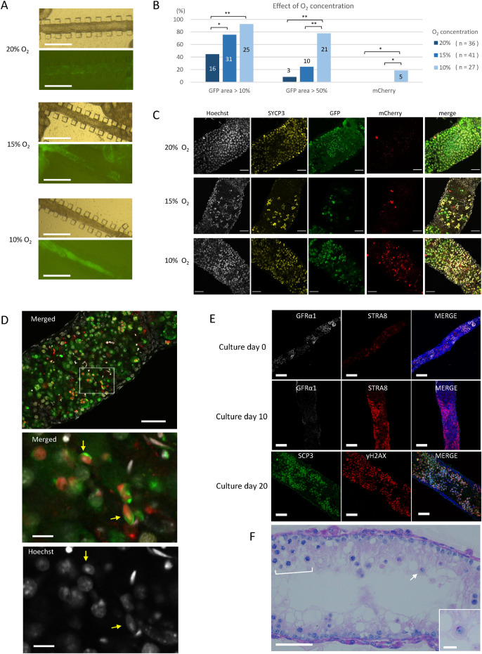
We therefore adopted the uncut-isolated ST culture method to examine the effect of O2 concentrations of 20%, 15% and 10% on the spermatogenesis (Fig 4A). Among the 36 samples cultured in 20% O2, the number having an Acr-GFP-expressing region rate over 10% and 50% in a stretch of ST were 16 (44.4%) and 3 (8.3%), respectively. The corresponding numbers among the 41 samples cultured in 15% O2 were 31 (75.6%) and 10 (24.4%), respectively. Among the 27 samples cultured in 10% O2, the respective values were 25 (92.6%) and 21 (77.8%) (Fig 4B). The number of samples varied among the three groups due to technical difficulties in the preparation of uncut-isolated STs. Namely, some samples were lost prior to evaluation due to fragmentation or slipping of the ST apart from the frame of the PDMS pillars. Nonetheless, this experiment clearly demonstrated that lower O2 concentrations were associated with a higher rate of GFP expression in a larger area. Interestingly, such a difference was not obvious among tissue masses cultured in the same respective STPC chips. The tissue masses showed Acr-GFP expression in most of their regions regardless of O2 concentration (S5 Fig in S1 File). This relative insensitivity of the tissue masses to O2 concentration could have been caused by reduced O2 tension in a large area of tissue mass due to the consumption of O2 by the tissue itself.

Fig 4. Effect of lower oxygen concentrations on spermatogenesis evaluated with uncut-isolated STs.

Fig 4

(A) Stereomicroscopic view of uncut-isolated STs cultured at different O2 concentrations. GFP expression was observed at culture day 25 by a fluorescence microscope. (B) Data from nine experiments are summarized. Samples with GFP-expressing areas over 10% and 50%, respectively, during culture days 20 to 30 were counted as GFP-positive. The mCherry expression was confirmed on culture day 30 or 36. Numbers in or over the bar are the numbers of marker-positive samples among total samples examined in each O2 concentration; the numbers of total sample are shown in parenthesis. * P < 0.05, ** P < 0.01. (C) Whole-mount immunostaining with anti-SYCP3 antibody (yellow), anti-GFP antibody (green), and anti-RFP (mCherry) antibody (red) was performed on culture day 34, using a 5dpp Acr/H3 double-Tg mouse testis. (D) A 5dpp Acr/H3 double-Tg mouse testis was cultured in 10% O2 for 34 days and examined by whole-mount immunohistochemistry. A merged image of GFP (green), mCherry (red), and Hoechst (gray) was shown in the top panel. The dashed rectangular area is enlarged in the panels below. An acrosome cap (green) and mCherry-positive nuclei (red) of elongated spermatids (yellow arrows) were observed. (E) Uncut-isolated STs cultured under 10% O2 for 0, 10 and 20 days were subjected to whole-mount immunostaining. (F) An uncut-isolated ST cultured under 10% O2 for 20 days was sectioned and stained with PAS, revealing the regional layers of spermatocytes (bracket) and an isolated round spermatid (arrow and enlarged in the inset) having a cap-shaped acrosome stained in red. Scale bars: 500 μm (A), 50 μm (C, D top, E, F), and 10 μm (D middle and bottom, F inset).

After culturing about 30 days, 6 GFP-positive samples, 2 in each O2 concentration group, were examined with whole-mount immunochemical staining. Sycp3-positive cells (meiotic cells) and GFP-positive cells were confirmed in all the O2 concentration groups. However, mCherry-positive cells were sparse in samples cultured in 20% and 15% O2, but the samples cultured in 10% O2 contained many such cells (Fig 4C). Germ cells expressing both GFP and mCherry, representing elongating spermatids at step 11 onward, were confirmed only in the samples in 10% O2, and only in 5 of 27 samples (Fig 4D and 4B).

To ensure that this condition—namely uncut-isolated ST under 10% O2—recapitulates the normal development of the first wave spermatogenesis, a time-course examination using whole-mount immunohistochemistry with stage-specific markers was performed (Fig 4E). The STs of a 5 dpp mouse contained undifferentiated spermatogonia, which were stained positive with GFRα1, as the most abundant germ cell at explantation (day 0). In 10 days, STRA8-positive cells, probably preleptotene spermatocytes, emerged and increased in number, while GFRα1-positive undifferentiated spermatogonia became more sparse. In 20 days, spermatocytes positive for both SCP3 and γH2AX appeared and the STs became markedly thicker. A sectioned histological examination with PAS staining showed spermatocytes and round spermatids (Fig 4F). Taken together, these results demonstrated that the 10% O2 concentration was more favorable than higher O2 concentrations for promoting spermatogenesis up to the production of elongating spermatids. Of note, it is even possible that O2 lower than 10% would be optimal for isolated STs to promote spermatogenesis. In conclusion, culturing isolated STs is a sensitive and reliable means for elucidating the microenvironmental signals that affect or control spermatogenesis.

Discussion

In the history of mammalian in vitro spermatogenesis studies, culturing isolated segments of ST can be categorized as a separate technique from culturing tissue as a mass [5]. The first studies using the former culture technique were performed in the 1970s [22]. However, Parvinen and colleagues would be the first to report the progression of spermatogenesis in a defined segment of adult rat STs [6]. Specifically, STs containing late pachytene and diakinetic primary spermatocytes were cultured in a chemically defined medium for 6 days. In those STs, meiotic divisions were completed and the newly formed spermatids, which had acrosonic systems characteristic of step 5, were observed [6, 7]. In a subsequent study, Toppari et al. extended these findings. They excised segments of rat STs from stages II to III and cultured them in the same way. The round spermatids at steps 2–3 developed into step 7 spermatids within 7 days, replicating the progression in vivo. Moreover, the spermatogonia and spermatocytes in that segment developed in correspondence with those in vivo [23].

We initiated the present study as an extension of our previous studies of testis tissue cultivation, which succeeded in the production of fertile sperm from spermatogonial stem cells [4, 24]. To our surprise, the STs underwent drastic shrinking and lost their original tubular appearance in only a couple of days. However, the explanted tissues themselves would certainly have experienced drastic changes in environmental conditions, and this would be expected to elicit various reactions in the tissues. In fact, we recently observed that testis tissues exhibited a significant level of inflammation within a mere two days after explantation [25]. After trying several approaches, we resorted to isolating STs segmentally without cutting them in order to keep them from shrinkage. This was an incomplete method of isolation, but it was an effective method for inducing spermatogenesis at a higher efficiency. Having settled on this uncut-isolation method, we then explicitly evaluated the effect of oxygen concentration on spermatogenesis. In our pilot study we cultured tissue masses under different O2 concentrations, and the results suggested that lower oxygen concentration might be superior in inducing efficient spermatogenesis. However, because the oxygen tension at any point in a tissue is the result of the sequential oxygen supply and consumption at that point as well as at points nearby, the value fluctuates and is difficult to measure precisely. The results of the pilot study were thus hard to interpret in a straightforward manner. In contrast, the segmentally isolated portion of STs should be bathed in a constant O2 concentration that is close to the concentration set in the incubator throughout the cultivation period. Therefore, culturing STs, instead of tissue masses, seemed to be a superior way to directly evaluate the effect of environmental factors.

In addition to performing uncut-isolated ST experiments, we also successfully minimized the shrinkage of cut-isolated STs by adopting the soft agarose method reported by Gholami and colleagues [8]. The soft agar culture method was initially developed to characterize clonal expansion of bone marrow cells [2628]. Then, it was widely applied in different ways, and eventually became the gold-standard assay for cellular transformation in vitro [2932]. In 2008, Stukenborg et al. used the soft agar method to cultivate spermatogonia for proliferation. They also intended to induce differentiation of spermatogonia by coculturing with somatic cells [19]. Gholami’s report was the first trial, to our best knowledge, to culture STs in soft agar [8]. Although they reported the progression of spermatogenesis, their evaluations were incomplete, and thus further studies were warranted. In the present study, we used Acr/H3 double-Tg mice which faithfully express GFP and mCherry, respectively, as reliable markers of meiotic pachytene and step 11 spermatids onward. In particular, the combination of GFP accumulation in the acrosome and mCherry expression in the nucleus, which were located closely side by side, was a strong marker for the identification of elongating spermatids. Thus, it was demonstrated faithfully for the first time that in vitro spermatogenesis in the isolated STs proceeded up to the stage of elongating spermatids.

Although our experiments demonstrated that culturing ST segments without shrinkage is possible, the efficiency was much lower than that by culturing tissue masses or aggregated STs. The GFP-positive rates for each isolation method were summarized as 5.3%, 26.9%, 53.3%, 61.1%, and 90% for cut-isolated conglomerated, cut-isolated in soft agarose, uncut-isolated, aggregated in soft agarose, and tissue mass, respectively, under 20% O2 concentration (S6 Fig in S1 File). This indicates that STs that remained adherent rather than isolated singly showed better performance in inducing spermatogenesis. On the other hand, when the uncut-isolated method was performed under 20%, 15%, and 10% O2, the GFP positive rates were 44.4%, 75.6%, and 92.6%, respectively. This result suggests that the local O2 tension, which should be reduced in aggregated STs and tissue masses, contributes significantly to spermatogenesis performance. However, there are two other possible explanations for the favorable performance of aggregated STs and tissue masses. First, interstitial cells attaching to the STs could be essential or at least influential. Aggregated STs and tissue masses would be expected to contain interstitial cells more abundantly than isolated STs. Indeed, our whole mount immunohistochemical study showed that the aggregated STs contained many Leydig cells, while the cut-isolated STs contained only a few. In addition to Leydig cells, there are several different types of cells outside the STs in the testis, including macrophages, lymphatic endothelial cells and so forth. Numerous studies have described the pivotal roles of these cells in spermatogenesis. Leydig cells produce and secrete androgens and other factors, such as colony-stimulating factor 1 (CSF-1) [33]. The testicular macrophages also produce CSF-1 and enzymes involved in retinoic acid (RA) biosynthesis [34]. Lymphatic endothelial cells secrete fibroblast growth factors (FGFs), which maintain stem cell populations for spermatogenesis [35]. All these factors and others not yet identified could contribute to the promotion and maintenance of spermatogenesis and would be necessary for efficient spermatogenesis in vitro. It was even reported recently that bone marrow-derived mesenchymal cells co-cultured with testis tissue promoted in vitro spermatogenesis, suggesting that an optimal culture condition for spermatogenesis could be achieved via such a paracrine effect from cells from an extra-testicular source [36].

A second possibility is the paracrine effect between STs. Each ST should secrete various molecules; most of these would be metabolites, and some of these metabolites could directly or indirectly promote spermatogenesis. In organ culture experiments, a testicular tissue mass with a certain volume may be able to create its own internal state through such a paracrine effect. This issue, i.e., the difference between isolated and aggregated STs, could be investigated experimentally in the future by improving the culture technique for isolated STs. In line with such research progress, trials culturing reaggregated testis cells rather than ST segments would be a fruitful technique for in vitro spermatogenesis [10, 11].

We expect that the isolated ST culture method would open a new platform for precision culture experiments, especially when combined with microfluidic technologies. It has been reported that microfluidic systems can produce a continuous microflow of culture medium to produce a desired concentration gradient of a biochemical [37, 38]. Combined with such technologies, isolated ST culture experiments could reveal the regulatory mechanisms of spermatogenesis, which have been difficult to elucidate to date.

Conclusion

Spermatogenesis does not proceed by germ cells alone, but requires close support from several types of testicular somatic cells. This suggests that an organ culture method would be the best approach for in vitro spermatogenesis. However, in order to accurately elucidate the regulatory mechanisms of spermatogenesis, it is advantageous and preferable to culture only a small number of functional units—or one functional unit, if possible—rather than large tissue masses. In this study, we showed that in vitro spermatogenesis was possible in an isolated ST up to the elongating spermatid stage. There are certainly many limitations and unanswered questions in regard to this technique, including technical difficulty in handling STs, low efficiency of spermatogenesis, unknown effects of interstitial cells, fertility competence of the elongating spermatids, and so forth. Nonetheless, this method allows us to evaluate various microenvironmental parameters involved in the progression of spermatogenesis and to elucidate the regulatory mechanisms of spermatogenesis.

Supporting information

S1 File. Six figures and a table.

(PDF)

S1 Video. Procedure of the soft agarose method.

(MP4)

Acknowledgments

We thank Mayuka Nishida and Shino Nagata for creating the figure illustrations.

Data Availability

All relevant data are within the paper and its Supporting Information files.

Funding Statement

This work was supported by a Grant-in-Aid for Scientific Research on the Innovative Area “Ensuring integrity in gametogenesis” (18H05546), CREST by JST (JPMJCR21N1), a Grant for Strategic Research Promotion of Yokohama City University (SK2811) to T.O., and a KAKENHI grant (JP19J01276) from Japan Society for the Promotion of Science (JSPS) to T.M. The funders had no role in study design, data collection and analysis, decision to publish, or preparation of the manuscript.

References

  • 1.Yin X, Mead BE, Safaee H, et al. Engineering Stem Cell Organoids. Cell Stem Cell 2016;18:25–38. doi: 10.1016/j.stem.2015.12.005 [DOI] [PMC free article] [PubMed] [Google Scholar]
  • 2.Chojnacka K, Zarzycka M, Mruk DD. Biology of the Sertoli Cell in the Fetal, Pubertal, and Adult Mammalian Testis. Results Probl Cell Differ 2016;58:225–251. doi: 10.1007/978-3-319-31973-5_9 [DOI] [PubMed] [Google Scholar]
  • 3.Nakata H, Wakayama T, Sonomura T, et al. Three-dimensional structure of seminiferous tubules in the adult mouse. Journal of anatomy 2015;227:686–694. doi: 10.1111/joa.12375 [DOI] [PMC free article] [PubMed] [Google Scholar]
  • 4.Sato T, Katagiri K, Gohbara A, et al. In Vitro Production of Functional Sperm in Cultured Neonatal Mouse Testes. Nature 2011;471:504–507. doi: 10.1038/nature09850 [DOI] [PubMed] [Google Scholar]
  • 5.Komeya M, Sato T, Ogawa T. In Vitro Spermatogenesis: A century-long research journey, still half way around. Reproductive medicine and biology 2018;17:407–420. doi: 10.1002/rmb2.12225 [DOI] [PMC free article] [PubMed] [Google Scholar]
  • 6.Parvinen M, Wright WW, Phillips DM, et al. Spermatogenesis in vitro: completion of meiosis and early spermiogenesis. Endocrinology 1983;112:1150–1152. doi: 10.1210/endo-112-3-1150 [DOI] [PubMed] [Google Scholar]
  • 7.Toppari J, Parvinen M. In vitro differentiation of rat seminiferous tubular segments from defined stages of the epithelial cycle morphologic and immunolocalization analysis. J Androl 1985;6:334–343. doi: 10.1002/j.1939-4640.1985.tb03289.x [DOI] [PubMed] [Google Scholar]
  • 8.Gholami K, Pourmand G, Koruji M, et al. Organ culture of seminiferous tubules using a modified soft agar culture system. Stem cell research & therapy 2018;9:249. doi: 10.1186/s13287-018-0997-8 [DOI] [PMC free article] [PubMed] [Google Scholar]
  • 9.Yokonishi T, Sato T, Katagiri K, Komeya M, Kubota Y, Ogawa T. In Vitro Reconstruction of Mouse Seminiferous Tubules Supporting Germ Cell Differentiation. Biol Reprod. 2013. Jul;89(1):15. doi: 10.1095/biolreprod.113.108613. Epub 2013 Jun 12. [DOI] [PubMed] [Google Scholar]
  • 10.Baert Y, Dvorakova-Hortova K, Margaryan H, Goossens E. Mouse in vitro spermatogenesis on alginate-based 3D bioprinted scaffolds. Biofabrication. 2019. Apr 26;11(3):035011. doi: 10.1088/1758-5090/ab1452 . [DOI] [PubMed] [Google Scholar]
  • 11.AbuMadighem A, Shuchat S, Lunenfeld E, Yossifon G, Huleihel M. Testis on a chip-a microfluidic three-dimensional culture system for the development of spermatogenesis in-vitro. Biofabrication. 2022. Apr 20;14(3). doi: 10.1088/1758-5090/ac6126 . [DOI] [PubMed] [Google Scholar]
  • 12.Nakanishi T, Ikawa M, Yamada S, et al. Real-time observation of acrosomal dispersal from mouse sperm using GFP as a marker protein. FEBS Lett 1999;449:277–283. doi: 10.1016/s0014-5793(99)00433-0 [DOI] [PubMed] [Google Scholar]
  • 13.Ventelä S, Mulari M, Okabe M, et al. Regulation of acrosome formation in mice expressing green fluorescent protein as a marker. Tissue & cell 2000;32:501–507. doi: 10.1016/s0040-8166(00)80006-3 [DOI] [PubMed] [Google Scholar]
  • 14.Makino Y, Inoue E, Hada M, et al. Generation of a dual-color reporter mouse line to monitor spermatogenesis in vivo. Frontiers in cell and developmental biology 2014;2:30. doi: 10.3389/fcell.2014.00030 [DOI] [PMC free article] [PubMed] [Google Scholar]
  • 15.McDonald JC, Whitesides GM. Poly(dimethylsiloxane) as a material for fabricating microfluidic devices. Accounts of chemical research 2022;35:491–499. [DOI] [PubMed] [Google Scholar]
  • 16.Kojima K, Nakamura H, Komeya M, et al. Neonatal testis growth recreated in vitro by two-dimensional organ spreading. Biotechnol Bioeng 2018;115:3030–3041. doi: 10.1002/bit.26822 [DOI] [PMC free article] [PubMed] [Google Scholar]
  • 17.Komeya M, Yamanaka H, Sanjo H, et al. In Vitro Spermatogenesis in Two-Dimensionally Spread Mouse Testis Tissues. Reproductive medicine and biology 2019;18:362–369. doi: 10.1002/rmb2.12291 [DOI] [PMC free article] [PubMed] [Google Scholar]
  • 18.Komeya M, Kimura H, Nakamura H, et al. Long-term Ex Vivo Maintenance of Testis Tissues Producing Fertile Sperm in a Microfluidic Device. Scientific reports 2016;6:21472. doi: 10.1038/srep21472 [DOI] [PMC free article] [PubMed] [Google Scholar]
  • 19.Stukenborg JB, Wistuba J, Luetjens CM, et al. Coculture of spermatogonia with somatic cells in a novel three-dimensional soft-agar-culture-system. J Androl 2008;29:312–329. doi: 10.2164/jandrol.107.002857 [DOI] [PubMed] [Google Scholar]
  • 20.Matsumura T, Sato T, Abe T, et al. Rat in vitro spermatogenesis promoted by chemical supplementations and oxygen-tension control. Scientific Reports 2021;11:3458. doi: 10.1038/s41598-021-82792-2 [DOI] [PMC free article] [PubMed] [Google Scholar]
  • 21.Jež M, Rožman P, Ivanović Z, Bas T. Concise review: the role of oxygen in hematopoietic stem cell physiology. J Cell Physiol 2015;230:1999–2005. doi: 10.1002/jcp.24953 [DOI] [PubMed] [Google Scholar]
  • 22.Eddy EM, Kahri AI. Cell association and surface features in cultures of juvenile rat seminiferous tubules. Anat Rec 1976;185:333–358. doi: 10.1002/ar.1091850307 [DOI] [PubMed] [Google Scholar]
  • 23.Toppari J, Mali P, Eerola E. Rat spermatogenesis in vitro traced by quantitative flow cytometry. The journal of histochemistry and cytochemistry: official journal of the Histochemistry Society 1986;34:1029–1035. doi: 10.1177/34.8.3734416 [DOI] [PubMed] [Google Scholar]
  • 24.Sato T, Katagiri K, Yokonishi T, et al. In vitro production of fertile sperm from murine spermatogonial stem cell lines. Nature Communications 2011;2:472. doi: 10.1038/ncomms1478 [DOI] [PubMed] [Google Scholar]
  • 25.Abe T, Nishimura H, Sato T, et al. Transcriptome analysis reveals inadequate spermatogenesis and immediate radical immune reactions during organ culture in vitro spermatogenesis. Biochemical and Biophysical Research Communications 2020;530:732–738. [DOI] [PubMed] [Google Scholar]
  • 26.Bradley TR, Metcalf D. The growth of mouse bone marrow cells in vitro. The Australian journal of experimental biology and medical science 1966;44:287–299. doi: 10.1038/icb.1966.28 [DOI] [PubMed] [Google Scholar]
  • 27.Pluznik DH, Sachs L. The cloning of normal "mast" cells in tissue culture. J Cell Physiol 1965;66:319–324. doi: 10.1002/jcp.1030660309 [DOI] [PubMed] [Google Scholar]
  • 28.Senn JS, McCulloch EA, Till JE. Comparison of colony-forming ability of normal and leukaemic human marrow in cell culture. Lancet (London, England) 1967;2:597–598. doi: 10.1016/s0140-6736(67)90742-8 [DOI] [PubMed] [Google Scholar]
  • 29.Borowicz S, Van Scoyk M, Avasarala S, et al. The soft agar colony formation assay. Journal of visualized experiments: JoVE, 2014; e51998. doi: 10.3791/51998 [DOI] [PMC free article] [PubMed] [Google Scholar]
  • 30.Lin HS, Kuhn C, Kuo T. Clonal growth of hamster free alveolar cells in soft agar. The Journal of experimental medicine 1975;142:877–886. doi: 10.1084/jem.142.4.877 [DOI] [PMC free article] [PubMed] [Google Scholar]
  • 31.Quaroni A, Wands J, Trelstad RL, Isselbacher KJ. Epithelioid cell cultures from rat small intestine. Characterization by morphologic and immunologic criteria. J Cell Biol 1979;80:248–265. doi: 10.1083/jcb.80.2.248 [DOI] [PMC free article] [PubMed] [Google Scholar]
  • 32.Rotem A, Janzer A, Izar B, et al. Alternative to the soft-agar assay that permits high-throughput drug and genetic screens for cellular transformation. Proc Natl Acad Sci U S A 2015; 112: 5708–5713. doi: 10.1073/pnas.1505979112 [DOI] [PMC free article] [PubMed] [Google Scholar]
  • 33.Oatley JM, Oatley MJ, Avarbock MR, et al. Colony stimulating factor 1 is an extrinsic stimulator of mouse spermatogonial stem cell self-renewal. Development 2009;136:1191–1199. doi: 10.1242/dev.032243 [DOI] [PMC free article] [PubMed] [Google Scholar]
  • 34.DeFalco T, Potter SJ, Williams AV, et al. Macrophages Contribute to the Spermatogonial Niche in the Adult Testis. Cell Rep 2015; 12: 1107–1119. doi: 10.1016/j.celrep.2015.07.015 [DOI] [PMC free article] [PubMed] [Google Scholar]
  • 35.Kitadate Y, Jörg DJ, Tokue M, et al. Competition for Mitogens Regulates Spermatogenic Stem Cell Homeostasis in an Open Niche. Cell Stem Cell 2019; 24: 79-92.e76. [DOI] [PMC free article] [PubMed] [Google Scholar]
  • 36.Önen S, Köse S, Yersal N, Korkusuz P. Mesenchymal stem cells promote spermatogonial stem/progenitor cell pool and spermatogenesis in neonatal mice in vitro. Sci Rep. 2022. Jul 7;12(1):11494. doi: 10.1038/s41598-022-15358-5 [DOI] [PMC free article] [PubMed] [Google Scholar]
  • 37.Qasaimeh MA, Ricoult SG and Juncker D. Microfluidic probes for use in life sciences and medicine. Lab on a Chip 2013; 13: 40–50. doi: 10.1039/c2lc40898h [DOI] [PubMed] [Google Scholar]
  • 38.Shinha K, Nihei W and Kimura H. A Microfluidic Probe Integrated Device for Spatiotemporal 3D Chemical Stimulation in Cells. Micromachines 2020;11:691. doi: 10.3390/mi11070691 [DOI] [PMC free article] [PubMed] [Google Scholar]

Decision Letter 0

Suresh Yenugu

15 Aug 2022

PONE-D-22-19509Mouse in vitro spermatogenesis in isolated seminiferous tubulesPLOS ONE

Dear Dr. Ogawa,

Thank you for submitting your manuscript to PLOS ONE. After careful consideration, we feel that it has merit but does not fully meet PLOS ONE’s publication criteria as it currently stands. Therefore, we invite you to submit a revised version of the manuscript that addresses the points raised during the review process. There are some concerns on the efficiency and extent of spermatogenesis and culturing the tissues in environment that lack the factors that are crucial in an in vivo system. Further, there are a number of issues with methodology. Presentation of the manuscript should be improved significantly, especially in providing strong rationale and citing up to date literature; clear mention of the methodologies used; statistical analyses; appropriately describing the results for easy understanding; and providing the discussion that is relevant to the results observed backed by strong literature survey.

Please submit your revised manuscript by Sep 29 2022 11:59PM. If you will need more time than this to complete your revisions, please reply to this message or contact the journal office at plosone@plos.org. When you're ready to submit your revision, log on to https://www.editorialmanager.com/pone/ and select the 'Submissions Needing Revision' folder to locate your manuscript file.

Please include the following items when submitting your revised manuscript:

  • A rebuttal letter that responds to each point raised by the academic editor and reviewer(s). You should upload this letter as a separate file labeled 'Response to Reviewers'.

  • A marked-up copy of your manuscript that highlights changes made to the original version. You should upload this as a separate file labeled 'Revised Manuscript with Track Changes'.

  • An unmarked version of your revised paper without tracked changes. You should upload this as a separate file labeled 'Manuscript'.

If you would like to make changes to your financial disclosure, please include your updated statement in your cover letter. Guidelines for resubmitting your figure files are available below the reviewer comments at the end of this letter.

If applicable, we recommend that you deposit your laboratory protocols in protocols.io to enhance the reproducibility of your results. Protocols.io assigns your protocol its own identifier (DOI) so that it can be cited independently in the future. For instructions see: https://journals.plos.org/plosone/s/submission-guidelines#loc-laboratory-protocols. Additionally, PLOS ONE offers an option for publishing peer-reviewed Lab Protocol articles, which describe protocols hosted on protocols.io. Read more information on sharing protocols at https://plos.org/protocols?utm_medium=editorial-email&utm_source=authorletters&utm_campaign=protocols.

We look forward to receiving your revised manuscript.

Kind regards,

Suresh Yenugu

Academic Editor

PLOS ONE

Journal Requirements:

When submitting your revision, we need you to address these additional requirements.

1. Please ensure that your manuscript meets PLOS ONE's style requirements, including those for file naming. The PLOS ONE style templates can be found at 

https://journals.plos.org/plosone/s/file?id=wjVg/PLOSOne_formatting_sample_main_body.pdf and 

https://journals.plos.org/plosone/s/file?id=ba62/PLOSOne_formatting_sample_title_authors_affiliations.pdf

2. To comply with PLOS ONE submissions requirements, in your Methods section, please provide additional information regarding the experiments involving animals and ensure you have included details on (1) methods of sacrifice, (2) methods of anesthesia and/or analgesia, and (3) efforts to alleviate suffering.

3. We note that the grant information you provided in the ‘Funding Information’ and ‘Financial Disclosure’ sections do not match. 

When you resubmit, please ensure that you provide the correct grant numbers for the awards you received for your study in the ‘Funding Information’ section.

4. Please include captions for your Supporting Information files at the end of your manuscript, and update any in-text citations to match accordingly. Please see our Supporting Information guidelines for more information: http://journals.plos.org/plosone/s/supporting-information.

[Note: HTML markup is below. Please do not edit.]

Reviewers' comments:

Reviewer's Responses to Questions

Comments to the Author

1. Is the manuscript technically sound, and do the data support the conclusions?

The manuscript must describe a technically sound piece of scientific research with data that supports the conclusions. Experiments must have been conducted rigorously, with appropriate controls, replication, and sample sizes. The conclusions must be drawn appropriately based on the data presented.

Reviewer #1: Partly

Reviewer #2: No

Reviewer #3: Yes

**********

2. Has the statistical analysis been performed appropriately and rigorously?

Reviewer #1: Yes

Reviewer #2: I Don't Know

Reviewer #3: Yes

**********

3. Have the authors made all data underlying the findings in their manuscript fully available?

The PLOS Data policy requires authors to make all data underlying the findings described in their manuscript fully available without restriction, with rare exception (please refer to the Data Availability Statement in the manuscript PDF file). The data should be provided as part of the manuscript or its supporting information, or deposited to a public repository. For example, in addition to summary statistics, the data points behind means, medians and variance measures should be available. If there are restrictions on publicly sharing data—e.g. participant privacy or use of data from a third party—those must be specified.

Reviewer #1: Yes

Reviewer #2: No

Reviewer #3: Yes

**********

4. Is the manuscript presented in an intelligible fashion and written in standard English?

PLOS ONE does not copyedit accepted manuscripts, so the language in submitted articles must be clear, correct, and unambiguous. Any typographical or grammatical errors should be corrected at revision, so please note any specific errors here.

Reviewer #1: Yes

Reviewer #2: No

Reviewer #3: Yes

**********

5. Review Comments to the Author

Please use the space provided to explain your answers to the questions above. You may also include additional comments for the author, including concerns about dual publication, research ethics, or publication ethics. (Please upload your review as an attachment if it exceeds 20,000 characters)

Reviewer #1: Comments:

Recapitulation of in vitro spermatogenesis is essential to study the molecular mechanisms and to address infertility. Feng et al. extended their previous studies on in vitro spermatogenesis using the ST culture method using Acr-GFP andH3 mice. The authors tried both minced or individual STs and aggregated STs. Interestingly, they found that aggregates or individual STs having aggregates at both ends show efficient spermatogenesis compared to individual STs. Further, using ST culturing, authors have shown the effect of oxygen concentration on spermatogenesis. This is a motivating extension work; however, some aspects of the study need to be clarified, and more experiments are necessary to describe the manuscript's findings. Also, serious issues with the writing must be addressed. Overall, the paper feels premature and would benefit from some major rewriting. Throughout the article, including the abstract, many vocabularies, grammar, and pose errors make it very hard to follow and interpret.

Major comments:

The abstract is poorly written.

If possible, I would suggest to the authors consider making the video of methods A and B. It will be beneficial to the researchers and readers.

Authors made major conclusions that spermatogenesis can be induced in short pieces of STs in culture. However, only 26% of the short pieces show the +ve signals of GFP and mcherry. Also, they are nearby small clustered regions. Of course, we can see the GFP signals but Mcherry and GFP colocalized signals we see nearby clusters only?

Here the point is with the low efficiency of spermatogenesis in short pieces, how come one can use this technique to study mechanisms of spermatogenesis or any toxicological studies?

Line 465: Numerous studies have described the pivotal roles of these cells in spermatogenesis. Leydig cells produce and secrete androgens and other factors, such as colony-stimulating factor 1 (CSF-1) [31]. The testicular macrophages also produce CSF-1 and enzymes involved in retinoic acid (RA) biosynthesis [32]. Lymphatic endothelial cells secrete fibroblast growth factors (FGFs), which maintain stem cell populations for spermatogenesis [33]. All these factors and others not yet identified could contribute to the promotion and maintenance of spermatogenesis and would be necessary for efficient spermatogenesis in vitro.

As the authors stated ST’s external microenvironment may play a critical role in spermatogenesis. Did the authors try to culture short pieces of STs with some of the factors like RA, CSF-1 or growth factors? In another way did the authors try to co-culture with Leydig cells or other cells required for spermatogenesis to improve the efficiency?

Minor comments:

Fig1G Y axis?

323 expression was observed in 55 out of 90 sa268mples (61.1%) (Fig 3D, E).

Fig 3G ; error bars labelled. Make it uniform or mention it in legends

Fig S1 and 2 methods 0.5% agarose solutions with STs kept in 4deg. How about cold stress or shock on tubules and spermatocytes?

Fig3: What % of aggregates were observed? What do the authors mean occasionally?

Line 327: In 40–55 days, STs from an experiment were examined by whole mount immunochemical staining. In 2 samples of cut-isolated ST, cells positive for both GFP and mCherry were observed out of 41 samples examined

The image shown +ve for GPF and Mcherry in cut isolate is not convincing. Because the +ve signals we see near the cluster.

Line 334: We then stained Leydig cells with anti-HSD3β antibody under whole mount condition. In fact, HSD3β-positive cells were detected abundantly in aggregated STs and only marginally in isolated ST samples (Fig 3I). Please add the quantification

Line 340: In our previous study using rat testis tissues, we found that O2 tension could influence the spermatogenic efficiency in vitro and O2 concentration lower than atmospheric 20% would be favourable : Rewrite

377 that this condition, namely uncut-isolated ST under 10%O2: 10% space

Overlapping scale bars

Make uniformity in scale bars

Fig2:

(B) Even though authors have mentioned that Acr-GFP expression in an ST showed unique oscillatory fluctuation, the authors need to explain the reason for the sharp fluctuation in Acr-GFP expression from Day 24 to Day 31.

Fig3:

(F) The authors examined only 41 cut-isolated STs for both GFP –mCherry expression. What about the rest of the cut-isolated STs as the authors have found quite significant (72 nos) GFP expressing cut-isolated STs?

In lines 318, 319 and 320, they found small clumps of STs which they have considered aggregated STs. The authors need to clarify whether this is a human error or if these clumps are forming on their own. In the latter case, the authors also need to mention the frequency and percentage of finding these clumps.

In addition to this, the authors need to clarify whether they have included these clumped STs in their count for aggregated STs or not.

(I) The authors need to provide statistical analysis for this figure as they have stated that ‘aggregated STs may maintain interstitial cells between each ST, which could be a reason that aggregated STs supported spermatogenesis more favourably.’

Fig. 4

(B) Out of 20%, 15% and 10% concentrations, in 10% O2 GFP expression is more. Whether this is the optimal concentration for spermatogenesis or there is the possibility of more spermatogenesis below 10%? The author needs to clarify.

Reviewer #2: In this study the authors evaluated the effect of oxygen concentration on in vitro spermatogenesis in neonatal Acr-GFP/Histone H3.3.3-mCherry double (Acr/H3) transgenic mice testes, using tissue mass, ST aggregate and ST segment culture systems. Although it seems to be a have short follow up duration s (34 day long), the authors report that the lower oxygen tension is favorable for spermatogenesis and for elongating spermatid production, and they demonstrated a new ST segment culture method providing opportunity for elucidating regulatory mechanism of spermatogenesis. While these data are potentially of interest, the manuscript lacks principal points that require a major irevision in order to ensure the data are presented with more clarity and greater empirical support (including controls and detailed data).

Title:could be intentional in order to correctly orient the readers to the output of the study.

Introduction: lacks current literature and it is written in a broad and unfocused manner with a book chapter style. Therefore the rationale (lack of knowledge in recent literature related to the research question) , the clear description of the problem and the hypothesis or the research question that will solve the problem should be clearly declared .

Materials and Method lacks the study design, the sample size with power analysis, the number of repeats taht is required in order to answer the research question objectively. Several concerns related to methodolody and the proff of concepts are as following.

1. Please provide the detailed information of PDMS ceiling chip which was used for seminiferous tubule culturing method (including size of pillars in WxLxH and distance between the pillars) in M&M section.

2. In M&M section, between lines 121-124, the usage of a thin porous polycarbonate membrane is stated in some cases for pressing testis tissues against the base agarose gel by placing them under the PDMS ceiling to hold the tissue in place. Please clarify the optimization of culture systems by explaining in which cases the membrane was used.

3. The all culture platforms used in study should be clarified in M&M section. Please change the title "Culture method" as "Culture methods" in line 136 and explain each culture methods as subtitles individually. The title of "Soft agarose method" in line 156 should also be under culture methods title as a subtitle.

4. In figures 2A and 2B, there is high background for GFP, that makes the labeling data very unreliable.

5. The terminology for culture systems is not consistent. Each culture system should be named in M&M section and the same names should be used in the remaining parts of the manuscript.

6. In figure 4F, PAS staining micrograph is given only for uncut-isolated ST culture, it should be also given for the other culture platforms for the same culture duration. The oxygen concentration is also should be provided in figure legend.

7. It seems that there are four culture methods as (1) cut-isolated ST (PDMS ceiling), (2) uncut-isolated ST (PDMS ceiling with pillars), (3) ST aggregate (soft agarose), (4) tissue mass culture (air-liquid interphase). In results section, the comparison of these 4 methods should be provided (in figure 1G, 3E, 3H, 4B and 4H). In figure 1G, it should be stated which oxygen concentration is used and what is the culture time for that analysis.

The results lack the answers of the research questions and Discussion part itatement of limitations of the current study. The changes in metabolites (analysis of culture media by LC-MS etc.), the functionality of elongated spermatids in terms of fertilization (ROSI, ICSI) or genetic stability tests should be performed or may be added as limitations.

In discussion section, there are missing references in terms of comparison of the efficiency of new cut-isolated ST and uncut-isolated ST culture systems. The following articles should be discussed in terms of in vitro spermatogenetic process:

- Önen S, Köse S, Yersal N, Korkusuz P. Mesenchymal stem cells promote spermatogonial stem/progenitor cell pool and spermatogenesis in neonatal mice in vitro. Sci Rep. 2022 Jul 7;12(1):11494. doi: 10.1038/s41598-022-15358-5. PMID: 35798781; PMCID: PMC9263145.

- Baert Y, Dvorakova-Hortova K, Margaryan H, Goossens E. Mouse in vitro spermatogenesis on alginate-based 3D bioprinted scaffolds. Biofabrication. 2019 Apr 26;11(3):035011. doi: 10.1088/1758-5090/ab1452. PMID: 30921781.

- AbuMadighem A, Shuchat S, Lunenfeld E, Yossifon G, Huleihel M. Testis on a chip-a microfluidic three-dimensional culture system for the development of spermatogenesisin-vitro. Biofabrication. 2022 Apr 20;14(3). doi: 10.1088/1758-5090/ac6126. PMID: 35334473.

Reviewer #3: In this study, the authors developed an optimized method to culture the segment of seminiferous tubule (ST), and they showed that ST culture could reach the elongating spermatids stage. The authors patiently troubleshot the strategies to culture ST. They carefully described the detailed processes of their trial and error to develop an optimized protocol. I found the study valuable because the authors describe how they overcame the technical difficulties. This study would become an important asset in the field. Readers can understand the key process of their optimization. Further, the study demonstrates the impact of oxygen concentration in culture and clarifies the benefit of a hypoxic condition.

1. Some sentences in the abstract sound awkward. Lines 27-19 "Theoretically,”, and the last sentence in Lines 32-33.

2. Supplementary figures can be organized in the order of descriptions in the result section. S2A can be S1.

3. Each result section consists of a single paragraph. These sections can be reorganized into some paragraphs to improve their readability.

4. In Fig.1F, it was shown that the frequencies of GFP-positive cells were variable, but these data are not clearly shown in Figure G. These two data can be better described to clarify the frequency of GFP-positive cells.

5. Fig. S2: Please add labels: Acr-GFP and H3.3-mCherry.

6. Line 316: mislabel “S1 Fig. Method B”.

7. Line 334-336: This is just a comment. Leydig cells are the primary source of testosterone or androgens in males. Testosterone or androgens can be supplemented in future studies.

**********

6. PLOS authors have the option to publish the peer review history of their article (what does this mean?). If published, this will include your full peer review and any attached files.

If you choose “no”, your identity will remain anonymous but your review may still be made public.

Do you want your identity to be public for this peer review? For information about this choice, including consent withdrawal, please see our Privacy Policy.

Reviewer #1: No

Reviewer #2: No

Reviewer #3: No

**********

[NOTE: If reviewer comments were submitted as an attachment file, they will be attached to this email and accessible via the submission site. Please log into your account, locate the manuscript record, and check for the action link "View Attachments". If this link does not appear, there are no attachment files.]

While revising your submission, please upload your figure files to the Preflight Analysis and Conversion Engine (PACE) digital diagnostic tool, https://pacev2.apexcovantage.com/. PACE helps ensure that figures meet PLOS requirements. To use PACE, you must first register as a user. Registration is free. Then, login and navigate to the UPLOAD tab, where you will find detailed instructions on how to use the tool. If you encounter any issues or have any questions when using PACE, please email PLOS at figures@plos.org. Please note that Supporting Information files do not need this step.

Attachment

Submitted filename: PONE-D-22-19509 comments.docx

PLoS One. 2023 Apr 6;18(4):e0283773. doi: 10.1371/journal.pone.0283773.r002

Author response to Decision Letter 0


18 Oct 2022

Reviewer #1: Comments:

Recapitulation of in vitro spermatogenesis is essential to study the molecular mechanisms and to address infertility. Feng et al. extended their previous studies on in vitro spermatogenesis using the ST culture method using Acr-GFP andH3 mice. The authors tried both minced or individual STs and aggregated STs. Interestingly, they found that aggregates or individual STs having aggregates at both ends show efficient spermatogenesis compared to individual STs. Further, using ST culturing, authors have shown the effect of oxygen concentration on spermatogenesis. This is a motivating extension work; however, some aspects of the study need to be clarified, and more experiments are necessary to describe the manuscript's findings. Also, serious issues with the writing must be addressed. Overall, the paper feels premature and would benefit from some major rewriting. Throughout the article, including the abstract, many vocabularies, grammar, and pose errors make it very hard to follow and interpret.

Major comments:

The abstract is poorly written.

Reply) I have rewritten the abstract to be more specific about what we did in this study. I think that the new abstract is more informative and easier to understand for most readers.

If possible, I would suggest to the authors consider making the video of methods A and B. It will be beneficial to the researchers and readers.

Reply) We have made a video showing the procedure of soft agarose methods A and B, which was attached as supplementary data. Thank you for your suggestion.

Authors made major conclusions that spermatogenesis can be induced in short pieces of STs in culture. However, only 26% of the short pieces show the +ve signals of GFP and mcherry. Also, they are nearby small clustered regions. Of course, we can see the GFP signals but Mcherry and GFP colocalized signals we see nearby clusters only?

Reply) Reading your comment, I came to notice that GFP- and mCherry-double positive cells were observed at locations in a tubule which was not stretched but bent or twisted to form ST aggregation-like configuration. This may have an important implication for a future study. In any case, however, they were present in each singly cut-isolated ST, but not in STs of clustered or aggregated.

Here the point is with the low efficiency of spermatogenesis in short pieces, how come one can use this technique to study mechanisms of spermatogenesis or any toxicological studies?

Reply) The Acr-GFP positivity rate of isolated STs is indeed lower than that of ST aggregates or tissue mass cultures. However, I consider this as follows. In order to improve culture condition for spermatogenesis, we have to test variety of interventions, including supplements in the medium and physical conditions like different O2 concentration. Currently, the regular testicular tissue culture method, namely tissue mass culture, has achieved an efficiency as high as 90% Acr-GFP positivity. With this system, it may be difficult to find new culture conditions that are better than the control condition. For example, even if the addition of substance X to the culture medium is effective in promoting spermatogonial proliferation and inducing spermatogenesis, the effect of substance X may be overlooked in experiments where there is little room for improvement, with Acr-GFP expression rates as high as 90% in the control group. On the contrary, in this context, low spermatogenic efficiency of the isolated ST culture method may be useful to find better culture conditions. In fact, effect of O2 concentration was examined vividly in the present study to show lower O2 is better than 20%.

Line 465: Numerous studies have described the pivotal roles of these cells in spermatogenesis. Leydig cells produce and secrete androgens and other factors, such as colony-stimulating factor 1 (CSF-1) [31]. The testicular macrophages also produce CSF-1 and enzymes involved in retinoic acid (RA) biosynthesis [32]. Lymphatic endothelial cells secrete fibroblast growth factors (FGFs), which maintain stem cell populations for spermatogenesis [33]. All these factors and others not yet identified could contribute to the promotion and maintenance of spermatogenesis and would be necessary for efficient spermatogenesis in vitro.

As the authors stated ST’s external microenvironment may play a critical role in spermatogenesis. Did the authors try to culture short pieces of STs with some of the factors like RA, CSF-1 or growth factors? In another way did the authors try to co-culture with Leydig cells or other cells required for spermatogenesis to improve the efficiency?

Reply) Experiments suggested here are all important trial in our next studies. We will certainly plan such studies with the isolated ST experiment. Another reviewer, Reviewer 2, suggested to read a very recent paper by Önen et al. which used transwell insert system to coculture bone marrow derived mesenchymal cells and the testis tissue. Such co-culture system can be used to test your suggestion.

Minor comments:

Fig1G Y axis?

Reply) The Y-axis represents the percentage of GFP-positive samples out of all samples cultured. The title of the Y-axis was added to Fig. 1G of the revised manuscript.

323 expression was observed in 55 out of 90 sa268mples (61.1%) (Fig 3D, E).

Reply) I’m sorry for this careless typing mistake. I have corrected it in the revised manuscript.

Fig 3G; error bars labelled. Make it uniform or mention it in legends

Reply) In revision, I have erased them to make it uniform in all figures.

Fig S1 and 2 methods 0.5% agarose solutions with STs kept in 4deg. How about cold stress or shock on tubules and spermatocytes?

Reply) In our experience, storage at 4°C for 15 minutes does not affect the culture results.

Fig3: What % of aggregates were observed? What do the authors mean occasionally?

Reply) Thank you for this comment. Now I think “occasionally” was an incorrect word to describe the case. It totally depends on how rigorously the practitioner disassembled seminiferous tubules from each other. It is possible to eliminate aggregates completely, but it takes time and labor. Thus, in practice, ST aggregates remained more or less in every case, to varying degrees.

Line 327: In 40–55 days, STs from an experiment were examined by whole mount immunochemical staining. In 2 samples of cut-isolated ST, cells positive for both GFP and mCherry were observed out of 41 samples examined

The image shown +ve for GPF and Mcherry in cut isolate is not convincing. Because the +ve signals we see near the cluster.

Reply) As I replied above, the sample was a singly cut-isolated ST in the soft agarose. The “cluster” you mentioned is supposed to be the result of ST being bent or twisted during the whole mount immunohistochemistry procedure.

Line 334: We then stained Leydig cells with anti-HSD3β antibody under whole mount condition. In fact, HSD3β-positive cells were detected abundantly in aggregated STs and only marginally in isolated ST samples (Fig 3I). Please add the quantification

Reply) I counted the number of HSD3β-positive cells in the picture. The data were described in the figure legend as follows.

“HSD3β signal was observed in cells adhered to the outside of the STs, either singly (arrows) or as clusters (arrow heads). In these pictures, such HSD3β-positive cells were counted to be 5 and 41 in cut-isolated and aggregated STs, respectively. In addition, in case of aggregated STs, a clumpy interstitial tissue containing about 20 HSD3β-positive cells was also observed (surrounded by braces).”

Line 340: In our previous study using rat testis tissues, we found that O2 tension could influence the spermatogenic efficiency in vitro and O2 concentration lower than atmospheric 20% would be favourable : Rewrite

Reply) I rewrote as below in the revised manuscript.

In our previous study using rat testis tissues, we found that an O2 concentration lower than atmospheric concentration of 20% induced spermatogenesis with higher efficiency [20].

377 that this condition, namely uncut-isolated ST under 10%O2: 10% space

Reply) Thank you for this comment. I placed a space at that position in the revised manuscript.

Overlapping scale bars

Make uniformity in scale bars

Reply) Scale bars were examined again and corrected. Thank you.

Fig2:

(B) Even though authors have mentioned that Acr-GFP expression in an ST showed unique oscillatory fluctuation, the authors need to explain the reason for the sharp fluctuation in Acr-GFP expression from Day 24 to Day 31.

Reply) Yes, it was surprising to see such a drastic change in GFP expression in a single ST in 7 days. I consider this phenomenon as follows. The Acr-GFP expression starts at stage Ⅳ pachytene spermatocyte. Therefore, STs on day 24 in the figure should have contained germ cells of stage Ⅳ spermatocyte and some more differentiated cells. However, those GFP-expressing germ cells were about to die and disappeared in next 4 days. On day 28, therefore, the ST became GFP-negative, containing only germ cells of stage Ⅲ spermatocyte and less differentiated germ cells. Over the next 3 days, the stage Ⅲ spermatocytes turned into stage Ⅳ spermatocyte and beyond, and on day 31, the ST became GFP-positive again.

Fig3:

(F) The authors examined only 41 cut-isolated STs for both GFP –mCherry expression. What about the rest of the cut-isolated STs as the authors have found quite significant (72 nos) GFP expressing cut-isolated STs?

Reply) We have not examined the rest 31 samples for the mCherry expression.

In lines 318, 319 and 320, they found small clumps of STs which they have considered aggregated STs. The authors need to clarify whether this is a human error or if these clumps are forming on their own. In the latter case, the authors also need to mention the frequency and percentage of finding these clumps.

Reply) Yes, the clump STs was named as “aggregated STs”. STs do not self-aggregate in the soft agarose. They were results of incomplete dissociation of STs by the practitioner.

In addition to this, the authors need to clarify whether they have included these clumped STs in their count for aggregated STs or not.

Reply) Yes, clumped STs were counted as aggregated ST.

(I) The authors need to provide statistical analysis for this figure as they have stated that ‘aggregated STs may maintain interstitial cells between each ST, which could be a reason that aggregated STs supported spermatogenesis more favourably.’

Reply) Aggregated STs are a kind of artifact, unintentionally generated in soft agarose experiments.

The characteristics of aggregated STs, i.e., size, density, number of stromal cells, etc., may vary widely among them. For this reason, we hesitated to examine them in any further detail, after whole mount immunohistochemistry was performed once. We merely proposed that a greater number of interstitial cells may be responsible for more efficient spermatogenesis in aggregated STs. Thus, this is not definitive nor the conclusion of this study. I made it clearer in the revised manuscript.

Fig. 4

(B) Out of 20%, 15% and 10% concentrations, in 10% O2 GFP expression is more. Whether this is the optimal concentration for spermatogenesis or there is the possibility of more spermatogenesis below 10%? The author needs to clarify.

Reply) I think it is possible that a particular O2 concentration which is lower than 10% could be optimal for spermatogenesis in single ST. From our present study, it seems a delicate issue and the optimal O2 concentration may change in each tissue depending on the tissue size (O2 consumption rate). I wrote this in the revised manuscript.

Reviewer #2: In this study the authors evaluated the effect of oxygen concentration on in vitro spermatogenesis in neonatal Acr-GFP/Histone H3.3.3-mCherry double (Acr/H3) transgenic mice testes, using tissue mass, ST aggregate and ST segment culture systems. Although it seems to be a have short follow up duration s (34 day long), the authors report that the lower oxygen tension is favorable for spermatogenesis and for elongating spermatid production, and they demonstrated a new ST segment culture method providing opportunity for elucidating regulatory mechanism of spermatogenesis. While these data are potentially of interest, the manuscript lacks principal points that require a major irevision in order to ensure the data are presented with more clarity and greater empirical support (including controls and detailed data).

Title:could be intentional in order to correctly orient the readers to the output of the study.

Reply) I interpret this comment to mean that the current title is ambiguous and does not include the conclusions of this study. So, I considered other titles.

Present title; “Mouse in vitro spermatogenesis in isolated seminiferous tubules”.

New title 1; “Mouse spermatogenesis can proceed in isolated seminiferous tubules”.

New title 2; “Spermatogenesis proceeded in isolated seminiferous tubules of immature mice”.

New title 3; “???”

Thinking again, I reached the original one would be the best. Yet, I’m open to suggestions.

Introduction: lacks current literature and it is written in a broad and unfocused manner with a book chapter style. Therefore the rationale (lack of knowledge in recent literature related to the research question) , the clear description of the problem and the hypothesis or the research question that will solve the problem should be clearly declared .

Reply) I rewrote the introduction which included recent literatures. As for the research question, we focus on a very simple question which is whether a singly isolated ST can induce and support spermatogenesis or not. The answer was turned out to be yes. To make the introduction to be compact, we omitted the description of double Tg mouse.

Materials and Method lacks the study design, the sample size with power analysis, the number of repeats taht is required in order to answer the research question objectively.

Reply) The Materials and Methods section was reorganized. In addition, to facilitate the understanding of the present study as a whole, I have made a table along with graph encompassing every method together and present it as S6 Fig.

Several concerns related to methodolody and the proff of concepts are as following.

1. Please provide the detailed information of PDMS ceiling chip which was used for seminiferous tubule culturing method (including size of pillars in WxLxH and distance between the pillars) in M&M section.

Reply) I added that information in the M&M section.

2. In M&M section, between lines 121-124, the usage of a thin porous polycarbonate membrane is stated in some cases for pressing testis tissues against the base agarose gel by placing them under the PDMS ceiling to hold the tissue in place. Please clarify the optimization of culture systems by explaining in which cases the membrane was used.

Reply) First of all, I made a mistake to have written that polycarbonate membrane was used to press tissue mass. It was not polycarbonate membrane but a PDMS membrane (75 μm thick, FS7075C, ASAHI RUBBER inc). So, it was corrected accordingly in the revised manuscript.

This procedure was not relevant to the optimization of culture condition. The membranes were placed on the tissue mass but not on the isolated part of the seminiferous tubule. Therefore, we do not think it affect the culture results, nor relating to the optimization of culture condition.

3. The all culture platforms used in study should be clarified in M&M section. Please change the title "Culture method" as "Culture methods" in line 136 and explain each culture methods as subtitles individually. The title of "Soft agarose method" in line 156 should also be under culture methods title as a subtitle.

Reply) I reorganized the M&M section. “Soft agarose method” was changed to “Culture methods - Soft agarose method” and a new section of “Culture methods – PDMS ceiling (PC) method” was added to explain the detail of uncut-isolated ST method.

4. In figures 2A and 2B, there is high background for GFP, that makes the labeling data very unreliable.

Reply) That is the best we can do now with our microscope and camera setting. Sorry.

5. The terminology for culture systems is not consistent. Each culture system should be named in M&M section and the same names should be used in the remaining parts of the manuscript.

Reply) I made their name consistent, which was summarized in S6 Fig.

6. In figure 4F, PAS staining micrograph is given only for uncut-isolated ST culture, it should be also given for the other culture platforms for the same culture duration. The oxygen concentration is also should be provided in figure legend.

Reply) In this study, we took Acr-GFP and H3.3-mCherry as the reliable parameter for evaluating spermatogenic progression. I think they are basically enough to support our conclusion. Immunohistochemical staining and histological PAS stain were also performed to confirm and reinforce the conclusion. That is the reason that PAS staining was only performed and shown in the final figure. The oxygen concentration 10% was provided in the figure legend of Fig. 4E & F.

7. It seems that there are four culture methods as (1) cut-isolated ST (PDMS ceiling), (2) uncut-isolated ST (PDMS ceiling with pillars), (3) ST aggregate (soft agarose), (4) tissue mass culture (air-liquid interphase). In results section, the comparison of these 4 methods should be provided (in figure 1G, 3E, 3H, 4B and 4H). In figure 1G, it should be stated which oxygen concentration is used and what is the culture time for that analysis.

Reply) I appreciate your attentive reading of our manuscript. Present our research may not be organized in an ordinary style, because I described not only the final form of our best method but also methods we tested in our endeavor to improve them. I thank you for giving me an opportunity to think it again for summarizing and comparing culture methods.

Now I summarized our method as follows, which was presented in a table in S6 Fig.

(1) cut-isolated conglomerated ST (PDMS ceiling)

(2) uncut-isolated ST (PDMS ceiling with pillars)

(3) tissue mass culture (PDMS ceiling)

(4) cut-isolated ST in soft agarose

(5) ST aggregate in soft agarose

As for Fig. 1G, the culture time for the analysis was variable indeed. Each experiment has different culture duration from 35 to 77 days. The Acr-GFP positive sign (>10% area) was taken whenever during the culture period. I have added this information in the figure legend.

The results lack the answers of the research questions and Discussion part itatement of limitations of the current study. The changes in metabolites (analysis of culture media by LC-MS etc.), the functionality of elongated spermatids in terms of fertilization (ROSI, ICSI) or genetic stability tests should be performed or may be added as limitations.

Reply) Yes, there are many open questions in this study. We started this study asking whether isolated ST can induce and support spermatogenesis or not. To this question, we answered yes, it is possible to culture isolated ST for inducing spermatogenesis, although with lowered efficiency than tissue mass culture. I added sentences about the limitation of this study in the Conclusion of revised manuscript.

In discussion section, there are missing references in terms of comparison of the efficiency of new cut-isolated ST and uncut-isolated ST culture systems. The following articles should be discussed in terms of in vitro spermatogenetic process:

- Önen S, Köse S, Yersal N, Korkusuz P. Mesenchymal stem cells promote spermatogonial stem/progenitor cell pool and spermatogenesis in neonatal mice in vitro. Sci Rep. 2022 Jul 7;12(1):11494. doi: 10.1038/s41598-022-15358-5. PMID: 35798781; PMCID: PMC9263145.

Reply) Thank you recommending this paper to read. It is really interesting that bone marrow derived mesenchymal stem cells cultured on the well-bottom have promotive effect on in vitro spermatogenesis of testis tissue placed above in the transwell insert. This may mean that same method can improve the efficiency of spermatogenesis in the ST culture experiment. I added a sentence with this reference in the discussion.

- Baert Y, Dvorakova-Hortova K, Margaryan H, Goossens E. Mouse in vitro spermatogenesis on alginate-based 3D bioprinted scaffolds. Biofabrication. 2019 Apr 26;11(3):035011. doi: 10.1088/1758-5090/ab1452. PMID: 30921781.

Reply) This paper is also interesting showing that testis cells once singly dispersed were reaggregated by bioprinting method, in which spermatogenic progression was observed in following cultivation. Thus, this study may indicate a future direction of our present study too.

- AbuMadighem A, Shuchat S, Lunenfeld E, Yossifon G, Huleihel M. Testis on a chip-a microfluidic three-dimensional culture system for the development of spermatogenesis in-vitro. Biofabrication. 2022 Apr 20;14(3). doi: 10.1088/1758-5090/ac6126. PMID: 35334473.

Reply) As above paper, this paper also showed culturing reaggregated testis cell mass in a microfluidic device. I included this and above paper together in the discussion.

Reviewer #3: In this study, the authors developed an optimized method to culture the segment of seminiferous tubule (ST), and they showed that ST culture could reach the elongating spermatids stage. The authors patiently troubleshot the strategies to culture ST. They carefully described the detailed processes of their trial and error to develop an optimized protocol. I found the study valuable because the authors describe how they overcame the technical difficulties. This study would become an important asset in the field. Readers can understand the key process of their optimization. Further, the study demonstrates the impact of oxygen concentration in culture and clarifies the benefit of a hypoxic condition.

1. Some sentences in the abstract sound awkward. Lines 27-19 "Theoretically,”, and the last sentence in Lines 32-33.

Reply) I appreciate your comments. I have rewritten the abstract and those sentences were removed. Thank you.

2. Supplementary figures can be organized in the order of descriptions in the result section. S2A can be S1.

Reply) The numbers were reordered in the revised version.

3. Each result section consists of a single paragraph. These sections can be reorganized into some paragraphs to improve their readability.

Reply) I do appreciate this suggestion. In the revision, the section before the last, Cultivation of cut-isolated STs using a soft agarose method, was divided into 3 paragraphs. The last section, Lowered oxygen concentration was favorable for spermatogenesis, was divided into 5 paragraphs.

4. In Fig.1F, it was shown that the frequencies of GFP-positive cells were variable, but these data are not clearly shown in Figure G. These two data can be better described to clarify the frequency of GFP-positive cells.

Reply) Thanks for this comment. To make the difference among three groups, I made two thresholds, 10% and 50% of GFP-positive area, under observation with the stereomicroscope. The graph in figure 1G was modified to show GFP-positive rates with both of these two thresholds.

5. Fig. S2: Please add labels: Acr-GFP and H3.3-mCherry.

Reply) The labels were changed accordingly. Thanks.

6. Line 316: mislabel “S1 Fig. Method B”.

Reply) Sorry for this careless mistake. It was corrected in the revised manuscript.

7. Line 334-336: This is just a comment. Leydig cells are the primary source of testosterone or androgens in males. Testosterone or androgens can be supplemented in future studies.

Reply) Yes, I think the supplementation of testosterone and other substances secreted by Leydig cells should be done in next experiment. Thank you.

Attachment

Submitted filename: Reply to Reviewers.docx

Decision Letter 1

Suresh Yenugu

15 Nov 2022

PONE-D-22-19509R1Mouse in vitro spermatogenesis in isolated seminiferous tubulesPLOS ONE

Dear Dr. Ogawa,

Thank you for submitting your manuscript to PLOS ONE. After careful consideration, we feel that it has merit but does not fully meet PLOS ONE’s publication criteria as it currently stands. Therefore, we invite you to submit a revised version of the manuscript that addresses the points raised during the review process. While major improvements are made to the manuscript, it is suggested that the authors provide control data for each and every time point assessed. Ensure that the comments of the reviewers are responded in detail. The manuscript should be corrected for English language.

Please submit your revised manuscript by Dec 30 2022 11:59PM. If you will need more time than this to complete your revisions, please reply to this message or contact the journal office at plosone@plos.org. When you're ready to submit your revision, log on to https://www.editorialmanager.com/pone/ and select the 'Submissions Needing Revision' folder to locate your manuscript file.

Please include the following items when submitting your revised manuscript:

  • A rebuttal letter that responds to each point raised by the academic editor and reviewer(s). You should upload this letter as a separate file labeled 'Response to Reviewers'.

  • A marked-up copy of your manuscript that highlights changes made to the original version. You should upload this as a separate file labeled 'Revised Manuscript with Track Changes'.

  • An unmarked version of your revised paper without tracked changes. You should upload this as a separate file labeled 'Manuscript'.

If you would like to make changes to your financial disclosure, please include your updated statement in your cover letter. Guidelines for resubmitting your figure files are available below the reviewer comments at the end of this letter.

If applicable, we recommend that you deposit your laboratory protocols in protocols.io to enhance the reproducibility of your results. Protocols.io assigns your protocol its own identifier (DOI) so that it can be cited independently in the future. For instructions see: https://journals.plos.org/plosone/s/submission-guidelines#loc-laboratory-protocols. Additionally, PLOS ONE offers an option for publishing peer-reviewed Lab Protocol articles, which describe protocols hosted on protocols.io. Read more information on sharing protocols at https://plos.org/protocols?utm_medium=editorial-email&utm_source=authorletters&utm_campaign=protocols.

We look forward to receiving your revised manuscript.

Kind regards,

Suresh Yenugu

Academic Editor

PLOS ONE

[Note: HTML markup is below. Please do not edit.]

Reviewers' comments:

Reviewer's Responses to Questions

Comments to the Author

1. If the authors have adequately addressed your comments raised in a previous round of review and you feel that this manuscript is now acceptable for publication, you may indicate that here to bypass the “Comments to the Author” section, enter your conflict of interest statement in the “Confidential to Editor” section, and submit your "Accept" recommendation.

Reviewer #1: All comments have been addressed

Reviewer #2: (No Response)

Reviewer #3: (No Response)

**********

2. Is the manuscript technically sound, and do the data support the conclusions?

The manuscript must describe a technically sound piece of scientific research with data that supports the conclusions. Experiments must have been conducted rigorously, with appropriate controls, replication, and sample sizes. The conclusions must be drawn appropriately based on the data presented.

Reviewer #1: Yes

Reviewer #2: No

Reviewer #3: Yes

**********

3. Has the statistical analysis been performed appropriately and rigorously?

Reviewer #1: Yes

Reviewer #2: No

Reviewer #3: Yes

**********

4. Have the authors made all data underlying the findings in their manuscript fully available?

The PLOS Data policy requires authors to make all data underlying the findings described in their manuscript fully available without restriction, with rare exception (please refer to the Data Availability Statement in the manuscript PDF file). The data should be provided as part of the manuscript or its supporting information, or deposited to a public repository. For example, in addition to summary statistics, the data points behind means, medians and variance measures should be available. If there are restrictions on publicly sharing data—e.g. participant privacy or use of data from a third party—those must be specified.

Reviewer #1: Yes

Reviewer #2: No

Reviewer #3: Yes

**********

5. Is the manuscript presented in an intelligible fashion and written in standard English?

PLOS ONE does not copyedit accepted manuscripts, so the language in submitted articles must be clear, correct, and unambiguous. Any typographical or grammatical errors should be corrected at revision, so please note any specific errors here.

Reviewer #1: Yes

Reviewer #2: No

Reviewer #3: Yes

**********

6. Review Comments to the Author

Please use the space provided to explain your answers to the questions above. You may also include additional comments for the author, including concerns about dual publication, research ethics, or publication ethics. (Please upload your review as an attachment if it exceeds 20,000 characters)

Reviewer #1: The authors have done a good job revising their manuscript, which is clearly improved and ready for acceptance. Overall, the authors addressed most of my previous comments.

Reviewer #2: The revised manuscript has been improved to some extent, in terms of quality with reorganized Abstract, Introduction, M&M sections and terminology for culture methods. On the other hand, the reliability of data in terms of time points, oxygen concentrations and sample size are not clear. Following are the comments.

1) The authors might provide a more appropriate title for the manuscript including the culture method used for isolated seminiferous tubules that constitutes the main key word of the study. In the present title, “Mouse in vitro spermatogenesis in isolated seminiferous tubules”, the term of “isolated seminiferous tubules” might point the isolation of seminiferous tubules from an adult mouse. Also, the age of the mouse and effect of oxygen level should be indicated in the title. Here you can find some options as possible titles:

2) The authors are strictly recommended to provide a more reliable data for comparison of groups at same time points in figures 1G, 3E, 3H and 4B. For example, in figure 1G, if the culture time for the analysis is variable from 35 to 77 days, it is not possible to compare the efficacy of cut-isolated ST, uncut-isolated ST and tissue mass culture methods in terms of GFP expression since the progression of the spermatogenesis is changeable in in vitro conditions.The time point, sample size (n=?) and oxygen concentration must be given in legends of figures 1G, 3E, 3H and 4B.

3) All of the comments should be answered one by one on the rebuttal letter; then should address to the revised manuscript's specified lines/paragraphs/pages where the question is clearly answered.

4) The language that is used both on the rebuttal letter and the manuscript definitely requires a major refinement and revision in terms of scientific approach, the medical terminology and grammar.

Reviewer #3: The authors fully addressed my previous comments. It became a solid study. One minor thing I found is that the abstract contains too much details now, I feel. It can be reorganized in a way readers can capture the big picture.

**********

7. PLOS authors have the option to publish the peer review history of their article (what does this mean?). If published, this will include your full peer review and any attached files.

If you choose “no”, your identity will remain anonymous but your review may still be made public.

Do you want your identity to be public for this peer review? For information about this choice, including consent withdrawal, please see our Privacy Policy.

Reviewer #1: No

Reviewer #2: No

Reviewer #3: No

**********

[NOTE: If reviewer comments were submitted as an attachment file, they will be attached to this email and accessible via the submission site. Please log into your account, locate the manuscript record, and check for the action link "View Attachments". If this link does not appear, there are no attachment files.]

While revising your submission, please upload your figure files to the Preflight Analysis and Conversion Engine (PACE) digital diagnostic tool, https://pacev2.apexcovantage.com/. PACE helps ensure that figures meet PLOS requirements. To use PACE, you must first register as a user. Registration is free. Then, login and navigate to the UPLOAD tab, where you will find detailed instructions on how to use the tool. If you encounter any issues or have any questions when using PACE, please email PLOS at figures@plos.org. Please note that Supporting Information files do not need this step.

PLoS One. 2023 Apr 6;18(4):e0283773. doi: 10.1371/journal.pone.0283773.r004

Author response to Decision Letter 1


13 Dec 2022

6. Review Comments to the Author

Please use the space provided to explain your answers to the questions above. You may also include additional comments for the author, including concerns about dual publication, research ethics, or publication ethics. (Please upload your review as an attachment if it exceeds 20,000 characters)

Reviewer #1: The authors have done a good job revising their manuscript, which is clearly improved and ready for acceptance. Overall, the authors addressed most of my previous comments.

Reply: I appreciate your kind comments and suggestions.

Reviewer #2: The revised manuscript has been improved to some extent, in terms of quality with reorganized Abstract, Introduction, M&M sections and terminology for culture methods. On the other hand, the reliability of data in terms of time points, oxygen concentrations and sample size are not clear. Following are the comments.

Reply: Thank you for your comments.

1) The authors might provide a more appropriate title for the manuscript including the culture method used for isolated seminiferous tubules that constitutes the main key word of the study. In the present title, “Mouse in vitro spermatogenesis in isolated seminiferous tubules”, the term of “isolated seminiferous tubules” might point the isolation of seminiferous tubules from an adult mouse. Also, the age of the mouse and effect of oxygen level should be indicated in the title. Here you can find some options as possible titles:

Reply: I appreciate your suggestion. I changed the title as follows:

“In vitro spermatogenesis in isolated seminiferous tubules of immature mice”

2) The authors are strictly recommended to provide a more reliable data for comparison of groups at same time points in figures 1G, 3E, 3H and 4B. For example, in figure 1G, if the culture time for the analysis is variable from 35 to 77 days, it is not possible to compare the efficacy of cut-isolated ST, uncut-isolated ST and tissue mass culture methods in terms of GFP expression since the progression of the spermatogenesis is changeable in in vitro conditions. The time point, sample size (n=?) and oxygen concentration must be given in legends of figures 1G, 3E, 3H and 4B.

Reply: According to your recommendation, I narrowed the range of dates for the analysis in Figure 1G. The examination range was limited to 10 days, from culture days 31 to 40, and some of the data changed a bit. As for Figure 3E, the dates of analysis were culture days 20 to 25, and this information was added to the figure legend. In the case of Figure 3H, the day of tissue fixation for immunohistochemistry was added to the figure legend. Finally, in Figure 4B, GFP-positivity was assessed over culture days 20 to 30, and mCherry expression was confirmed on day 30 or 36. This information was added to the figure legend. Sample sizes are shown in the graph and an appropriate explanation was added to the legends. The oxygen concentration is now given as 20% in the legends for Figures 1G, 3E, and 3H.

3) All of the comments should be answered one by one on the rebuttal letter; then should address to the revised manuscript's specified lines/paragraphs/pages where the question is clearly answered.

Reply: Thank you for the advice.

4) The language that is used both on the rebuttal letter and the manuscript definitely requires a major refinement and revision in terms of scientific approach, the medical terminology and grammar.

Reply: Yes, I wrote the rebuttal letter ourselves and perhaps should have sought editorial help. As for the manuscript, however, I sent it to a professional English-editing company, where it was proofread and corrected by a native-English speaking editor. I sent the current rebuttal letter along with the revised manuscript to the company again.

Reviewer #3: The authors fully addressed my previous comments. It became a solid study. One minor thing I found is that the abstract contains too much details now, I feel. It can be reorganized in a way readers can capture the big picture.

Reply: Thank you for your kind comments and suggestions. I revised the summary by removing some of the extraneous details. I hope you find that the revised version is more concise.

Attachment

Submitted filename: Response to Reviewers.docx

Decision Letter 2

Suresh Yenugu

16 Jan 2023

PONE-D-22-19509R2In vitro spermatogenesis in isolated seminiferous tubules of immature micePLOS ONE

Dear Dr. Ogawa,

Thank you for submitting your manuscript to PLOS ONE. After careful consideration, we feel that it has merit but does not fully meet PLOS ONE’s publication criteria as it currently stands. Therefore, we invite you to submit a revised version of the manuscript that addresses the points raised during the review process. A major concern that needs to be addressed is to conduct statistical analyses using similar sample size and time points.

Please submit your revised manuscript by Mar 02 2023 11:59PM. If you will need more time than this to complete your revisions, please reply to this message or contact the journal office at plosone@plos.org. When you're ready to submit your revision, log on to https://www.editorialmanager.com/pone/ and select the 'Submissions Needing Revision' folder to locate your manuscript file.

Please include the following items when submitting your revised manuscript:

  • A rebuttal letter that responds to each point raised by the academic editor and reviewer(s). You should upload this letter as a separate file labeled 'Response to Reviewers'.

  • A marked-up copy of your manuscript that highlights changes made to the original version. You should upload this as a separate file labeled 'Revised Manuscript with Track Changes'.

  • An unmarked version of your revised paper without tracked changes. You should upload this as a separate file labeled 'Manuscript'.

If you would like to make changes to your financial disclosure, please include your updated statement in your cover letter. Guidelines for resubmitting your figure files are available below the reviewer comments at the end of this letter.

If applicable, we recommend that you deposit your laboratory protocols in protocols.io to enhance the reproducibility of your results. Protocols.io assigns your protocol its own identifier (DOI) so that it can be cited independently in the future. For instructions see: https://journals.plos.org/plosone/s/submission-guidelines#loc-laboratory-protocols. Additionally, PLOS ONE offers an option for publishing peer-reviewed Lab Protocol articles, which describe protocols hosted on protocols.io. Read more information on sharing protocols at https://plos.org/protocols?utm_medium=editorial-email&utm_source=authorletters&utm_campaign=protocols.

We look forward to receiving your revised manuscript.

Kind regards,

Suresh Yenugu

Academic Editor

PLOS ONE

[Note: HTML markup is below. Please do not edit.]

Reviewers' comments:

Reviewer's Responses to Questions

Comments to the Author

1. If the authors have adequately addressed your comments raised in a previous round of review and you feel that this manuscript is now acceptable for publication, you may indicate that here to bypass the “Comments to the Author” section, enter your conflict of interest statement in the “Confidential to Editor” section, and submit your "Accept" recommendation.

Reviewer #2: (No Response)

Reviewer #3: All comments have been addressed

**********

2. Is the manuscript technically sound, and do the data support the conclusions?

The manuscript must describe a technically sound piece of scientific research with data that supports the conclusions. Experiments must have been conducted rigorously, with appropriate controls, replication, and sample sizes. The conclusions must be drawn appropriately based on the data presented.

Reviewer #2: Partly

Reviewer #3: Yes

**********

3. Has the statistical analysis been performed appropriately and rigorously?

Reviewer #2: No

Reviewer #3: Yes

**********

4. Have the authors made all data underlying the findings in their manuscript fully available?

The PLOS Data policy requires authors to make all data underlying the findings described in their manuscript fully available without restriction, with rare exception (please refer to the Data Availability Statement in the manuscript PDF file). The data should be provided as part of the manuscript or its supporting information, or deposited to a public repository. For example, in addition to summary statistics, the data points behind means, medians and variance measures should be available. If there are restrictions on publicly sharing data—e.g. participant privacy or use of data from a third party—those must be specified.

Reviewer #2: Yes

Reviewer #3: Yes

**********

5. Is the manuscript presented in an intelligible fashion and written in standard English?

PLOS ONE does not copyedit accepted manuscripts, so the language in submitted articles must be clear, correct, and unambiguous. Any typographical or grammatical errors should be corrected at revision, so please note any specific errors here.

Reviewer #2: No

Reviewer #3: Yes

**********

6. Review Comments to the Author

Please use the space provided to explain your answers to the questions above. You may also include additional comments for the author, including concerns about dual publication, research ethics, or publication ethics. (Please upload your review as an attachment if it exceeds 20,000 characters)

Reviewer #2: It has been improved to some extent by reorganization of the title, figures and figure legends, and additional information of sample size and oxygen level. On the other hand, the reliability of data in terms of time points and sample size are still not clear.

1) In figure 4B, 20% (n= 36), 15% (n=41) and 10% (n=27) oxygen level is compared in terms of the GFP721 and mCherry positivity of seminiferous tubules. The big difference in sample size between the groups gives rise to thought about potential of bias, and it may cause some possible errors in statistical analyses.

2) In figure 4B, the authors stated that “GFP721 positivity was judged during culture days 20 to 30. The mCherry expression was confirmed on culture day 30 or 36.” Both 6 and 10-day difference in in vitro culture of seminiferous tubules is still excessive in order to collect a reliable data from the experiments for two reasons:

1- The duration of mouse spermatogenesis is approximately 34 days. In Acr-GFP/Histone H3.3.3-mCherry double (Acr/H3) transgenic mice, GFP expression starts from pachytene spermatocytes at stage 4 and mCherry is expressed in spermatids from step 11 onward. Since the content and the size of subgroups of germ cells changes within the 6 and 10-day time interval, it is not suitable to include the data collected from day 20 to 30, and 30 to 36.

2-This time gap may affect the viability of germ cells, and number of labelled cells in the meantime.

The authors are recommended to reperform the statistical analysis by using same time points and sample sizes in the groups.

Reviewer #3: (No Response)

**********

7. PLOS authors have the option to publish the peer review history of their article (what does this mean?). If published, this will include your full peer review and any attached files.

If you choose “no”, your identity will remain anonymous but your review may still be made public.

Do you want your identity to be public for this peer review? For information about this choice, including consent withdrawal, please see our Privacy Policy.

Reviewer #2: No

Reviewer #3: No

**********

[NOTE: If reviewer comments were submitted as an attachment file, they will be attached to this email and accessible via the submission site. Please log into your account, locate the manuscript record, and check for the action link "View Attachments". If this link does not appear, there are no attachment files.]

While revising your submission, please upload your figure files to the Preflight Analysis and Conversion Engine (PACE) digital diagnostic tool, https://pacev2.apexcovantage.com/. PACE helps ensure that figures meet PLOS requirements. To use PACE, you must first register as a user. Registration is free. Then, login and navigate to the UPLOAD tab, where you will find detailed instructions on how to use the tool. If you encounter any issues or have any questions when using PACE, please email PLOS at figures@plos.org. Please note that Supporting Information files do not need this step.

PLoS One. 2023 Apr 6;18(4):e0283773. doi: 10.1371/journal.pone.0283773.r006

Author response to Decision Letter 2


7 Feb 2023

I appreciate the reviewer 2 for their kind comments on our manuscript.

In the 'response to the reviewer' file, I made point-by-point replies to the comments and requests from the reviewer.

Reviewer #2: It has been improved to some extent by reorganization of the title, figures and figure legends, and additional information of sample size and oxygen level. On the other hand, the reliability of data in terms of time points and sample size are still not clear.

1) In figure 4B, 20% (n= 36), 15% (n=41) and 10% (n=27) oxygen level is compared in terms of the GFP721 and mCherry positivity of seminiferous tubules. The big difference in sample size between the groups gives rise to thought about potential of bias, and it may cause some possible errors in statistical analyses.

Response: I appreciate your insightful comments, which prompted us to review the data and analysis in Figure 4B. The number of samples varied from group to group mainly due to the technical difficulty of preparing uncut isolated STs. Even though we prepared comparable numbers of samples in each group at the start of the experiment, some samples were lost prior to evaluation due to fragmentation or slipping of the ST apart from the frame of the PDMS pillars. Nonetheless, we considered that the number of samples and quantity of data were sufficient for a robust statistical analysis. In the process of reviewing the statistical analysis, we did find a careless mistake. In the data for mCherry, the p-value for the comparison of 20% versus 10% O2 should have been marked with a single asterisk (*) rather than a double asterisk (**). This was corrected.

2) In figure 4B, the authors stated that “GFP721 positivity was judged during culture days 20 to 30. The mCherry expression was confirmed on culture day 30 or 36.” Both 6 and 10-day difference in in vitro culture of seminiferous tubules is still excessive in order to collect a reliable data from the experiments for two reasons:

Response: The Acr-GFP expression was observed beginning as early as culture day 14, which corresponded to a mouse age of 17 dpp when a 3 dpp mouse testis was used, as in Fig. 2. We observed samples every 7 days and recorded the GFP positivity. By judging GFP over culture days 20 to 30, we were therefore provided two chances to observe GFP. If we had narrowed the period or selected one particular date for judgement, we could have misjudged samples as negative even though they expressed GFP on neighboring days. The data in Figure 2B show that such a risk was not merely hypothetical. That is, the ST in Fig. 2B showed GFP expression on days 24 and 31, but no GFP expression on day 28. As described in the manuscript, such fluctuation/oscillation in GFP expression can occur, and we regarded samples such as this as GFP-positive. This was our rationale for the choice of culture days 20 to 30 as judging period.

In the case of mCherry, magnification using an inverted microscope was required in order to faithfully detect the red fluorescence. Therefore, the time period for mCherry detection was at the end of each culture experiment, and we removed the tissue from the culture dish and placed it directly on a slide glass for observation. As mentioned above, in vitro spermatogenesis does not proceed as smoothly as that in vivo, and in our experience the appearance of mCherry is delayed for days or more. Thus, in the initial experiment, we chose culture day 30, which corresponds to a mouse age of 36 days, as the time point for judging mCherry positivity, because the testis-donor mouse was 6 days old. We observed one mCherry-positive ST in the 10% O2 group, and no mCherry-positive STs in the 15% and 20% groups. In later experiments using 5-day-old mice, we chose culture day 36, corresponding to a mouse age of 41 days, reasoning that that time point would be more appropriate for finding mCherry-positive spermatids. We found 4 mCherry-positive STs in the 10% O2 group and none in the other groups. In hindsight, therefore, culture day 36 was more appropriate for finding mCherry-positive spermatids than culture day 30. We agree with you that it would have been better to pinpoint a particular day for analysis when trying to detect mCherry, since we had to select a single day in any case. Nonetheless, culture days 30 and 36 were within the appropriate time frame for detecting mCherry fluorescence and yielded reliable data showing the superiority of 10% O2.

1- The duration of mouse spermatogenesis is approximately 34 days. In Acr-GFP/Histone H3.3.3-mCherry double (Acr/H3) transgenic mice, GFP expression starts from pachytene spermatocytes at stage 4 and mCherry is expressed in spermatids from step 11 onward. Since the content and the size of subgroups of germ cells changes within the 6 and 10-day time interval, it is not suitable to include the data collected from day 20 to 30, and 30 to 36.

Response: In our culture experiment, the progression of spermatogenesis was not as smooth as that in vivo. The first appearance of Acr-GFP should have been around culture day 15 when a 0–1 day neonate was used, but it could have been delayed several days. In addition, even if GFP fluorescence appeared once, it could have disappeared and then re-appeared as shown in Fig. 2B. Therefore, we thought we should take steps to ensure that we did not miss such GFP-positivity, even if was temporary. This was our rationale for choosing a 10-day time span as an observation period for judging GFP fluorescence.

For mCherry, as mentioned above, the determination can be made only once per experiment. Therefore, it was necessary to select one particular day for the analysis. The selection of this day is a difficult decision. If we had chosen a day too early for mCherry detection, i.e., a day when the most advanced germ cells in ST are round spermatozoa such as those in step 8, mCherry would not be detected in any sample at any oxygen concentration. On the other hand, if the day had been too late for mCherry detection, i.e., a day when step-11 spermatids had already appeared in the ST some time ago, the mCherry-positive spermatids would have degenerated and disappeared in the interim, and we would have lost the opportunity to detect them. In the present experiment, these concerns were realistic, because mCherry-positive spermatids appeared rarely and only in low numbers. In careful consideration of all the above, we chose days 30 and 36, which corresponded to mouse ages of 36 and 41 days, as the time points for observation. In both cases, we observed mCherry-positive STs, supporting the appropriateness of our choice of days for the analysis. Therefore, we think our choice was appropriate and the data showing the superiority of 10% O2 were reliable.

2-This time gap may affect the viability of germ cells, and number of labelled cells in the meantime.

Response: Time gaps of 6 or 10 days could indeed provide an opportunity for the germ cells to die and disappear, as you indicated. However, they would also provide a chance for reappearance. Such appearances and disappearances of germ cells at particular stages of differentiation were observed as fluctuations in GFP fluorescence in each ST during the culture experiment, and the pattern of these changes was specific to each ST. Therefore, it would be ideal to collect data for a long enough period, whether for GFP or mCherry detection, and then to integrate such data to make a proper determination. In the case of GFP expression, such an approach can be achieved by observing the cells with a stereomicroscope while maintaining the culture. Therefore, we chose 20 to 30 days as the period for GFP evaluation.

The authors are recommended to reperform the statistical analysis by using same time points and sample sizes in the groups.

Response: For the reasons described above, we chose 20 to 30 days as the period for the judgement of Acr-GFP. In the case of mCherry, culture days 30 and 36 were considered preferable for the reasons mentioned above. Finally, we should mention that we corrected a careless mistake found in Figure 4B (the p-value for the 20% versus 10% O2 comparison as for mCherry should have been marked with a single asterisk (*) rather than a double asterisk (**)). We appreciate your careful reading and kind comments on our manuscript.

Attachment

Submitted filename: Response to Reviewers.docx

Decision Letter 3

Suresh Yenugu

21 Feb 2023

PONE-D-22-19509R3In vitro spermatogenesis in isolated seminiferous tubules of immature micePLOS ONE

Dear Dr. Ogawa,

Thank you for submitting your manuscript to PLOS ONE. After careful consideration, we feel that it has merit but does not fully meet PLOS ONE’s publication criteria as it currently stands. Therefore, we invite you to submit a revised version of the manuscript that addresses the points raised during the review process. Minor changes in the conclusion section shall be made as per the suggestion of the reviewer.

Please submit your revised manuscript by Apr 07 2023 11:59PM. If you will need more time than this to complete your revisions, please reply to this message or contact the journal office at plosone@plos.org. When you're ready to submit your revision, log on to https://www.editorialmanager.com/pone/ and select the 'Submissions Needing Revision' folder to locate your manuscript file.

Please include the following items when submitting your revised manuscript:

  • A rebuttal letter that responds to each point raised by the academic editor and reviewer(s). You should upload this letter as a separate file labeled 'Response to Reviewers'.

  • A marked-up copy of your manuscript that highlights changes made to the original version. You should upload this as a separate file labeled 'Revised Manuscript with Track Changes'.

  • An unmarked version of your revised paper without tracked changes. You should upload this as a separate file labeled 'Manuscript'.

If you would like to make changes to your financial disclosure, please include your updated statement in your cover letter. Guidelines for resubmitting your figure files are available below the reviewer comments at the end of this letter.

If applicable, we recommend that you deposit your laboratory protocols in protocols.io to enhance the reproducibility of your results. Protocols.io assigns your protocol its own identifier (DOI) so that it can be cited independently in the future. For instructions see: https://journals.plos.org/plosone/s/submission-guidelines#loc-laboratory-protocols. Additionally, PLOS ONE offers an option for publishing peer-reviewed Lab Protocol articles, which describe protocols hosted on protocols.io. Read more information on sharing protocols at https://plos.org/protocols?utm_medium=editorial-email&utm_source=authorletters&utm_campaign=protocols.

We look forward to receiving your revised manuscript.

Kind regards,

Suresh Yenugu

Academic Editor

PLOS ONE

Journal Requirements:

Please review your reference list to ensure that it is complete and correct. If you have cited papers that have been retracted, please include the rationale for doing so in the manuscript text, or remove these references and replace them with relevant current references. Any changes to the reference list should be mentioned in the rebuttal letter that accompanies your revised manuscript. If you need to cite a retracted article, indicate the article’s retracted status in the References list and also include a citation and full reference for the retraction notice.

[Note: HTML markup is below. Please do not edit.]

Reviewers' comments:

Reviewer's Responses to Questions

Comments to the Author

1. If the authors have adequately addressed your comments raised in a previous round of review and you feel that this manuscript is now acceptable for publication, you may indicate that here to bypass the “Comments to the Author” section, enter your conflict of interest statement in the “Confidential to Editor” section, and submit your "Accept" recommendation.

Reviewer #2: All comments have been addressed

**********

2. Is the manuscript technically sound, and do the data support the conclusions?

The manuscript must describe a technically sound piece of scientific research with data that supports the conclusions. Experiments must have been conducted rigorously, with appropriate controls, replication, and sample sizes. The conclusions must be drawn appropriately based on the data presented.

Reviewer #2: Yes

**********

3. Has the statistical analysis been performed appropriately and rigorously?

Reviewer #2: Yes

**********

4. Have the authors made all data underlying the findings in their manuscript fully available?

The PLOS Data policy requires authors to make all data underlying the findings described in their manuscript fully available without restriction, with rare exception (please refer to the Data Availability Statement in the manuscript PDF file). The data should be provided as part of the manuscript or its supporting information, or deposited to a public repository. For example, in addition to summary statistics, the data points behind means, medians and variance measures should be available. If there are restrictions on publicly sharing data—e.g. participant privacy or use of data from a third party—those must be specified.

Reviewer #2: No

**********

5. Is the manuscript presented in an intelligible fashion and written in standard English?

PLOS ONE does not copyedit accepted manuscripts, so the language in submitted articles must be clear, correct, and unambiguous. Any typographical or grammatical errors should be corrected at revision, so please note any specific errors here.

Reviewer #2: Yes

**********

6. Review Comments to the Author

Please use the space provided to explain your answers to the questions above. You may also include additional comments for the author, including concerns about dual publication, research ethics, or publication ethics. (Please upload your review as an attachment if it exceeds 20,000 characters)

Reviewer #2: The authors provided a good explanation for concerns about the validity and reliability of the data due to differences in sample size and time points of the groups compared. On the other hand, those explanations should be clearly stated in the manuscript in order to increase the readability of the publication.

The authors corrected the error made in the statistical analysis and explained the reasons for changes in time interval and sample size used to evaluate the GFP and mCherry expression reliably and rationally. When the explanations are added to the limitation section, the article will be ready for publication in order to be clearly understood by the readers.

**********

7. PLOS authors have the option to publish the peer review history of their article (what does this mean?). If published, this will include your full peer review and any attached files.

If you choose “no”, your identity will remain anonymous but your review may still be made public.

Do you want your identity to be public for this peer review? For information about this choice, including consent withdrawal, please see our Privacy Policy.

Reviewer #2: No

**********

[NOTE: If reviewer comments were submitted as an attachment file, they will be attached to this email and accessible via the submission site. Please log into your account, locate the manuscript record, and check for the action link "View Attachments". If this link does not appear, there are no attachment files.]

While revising your submission, please upload your figure files to the Preflight Analysis and Conversion Engine (PACE) digital diagnostic tool, https://pacev2.apexcovantage.com/. PACE helps ensure that figures meet PLOS requirements. To use PACE, you must first register as a user. Registration is free. Then, login and navigate to the UPLOAD tab, where you will find detailed instructions on how to use the tool. If you encounter any issues or have any questions when using PACE, please email PLOS at figures@plos.org. Please note that Supporting Information files do not need this step.

PLoS One. 2023 Apr 6;18(4):e0283773. doi: 10.1371/journal.pone.0283773.r008

Author response to Decision Letter 3


10 Mar 2023

Editor

Thank you for submitting your manuscript to PLOS ONE. After careful consideration, we feel that it has merit but does not fully meet PLOS ONE’s publication criteria as it currently stands. Therefore, we invite you to submit a revised version of the manuscript that addresses the points raised during the review process.

Minor changes in the conclusion section shall be made as per the suggestion of the reviewer.

Response: According to the suggestions from Reviewer #2, we added a phrase, technical difficulty in handling STs, in the conclusion section as one of the limitations of our technique.

Reviewer #2

Comment 1: The authors provided a good explanation for concerns about the validity and reliability of the data due to differences in sample size and time points of the groups compared. On the other hand, those explanations should be clearly stated in the manuscript in order to increase the readability of the publication.

Response: Thank you for this important suggestion. We added the following passages to the Observations section of the Materials and methods:

Cultured tissues and ST segments were observed at least once a week under a stereomicroscope equipped with an excitation light for GFP (LeicaM205 FA; Leica, Germany). Acr-GFP begins to be expressed in mice at around 15 dpp in vivo, but its expression could be delayed by several days in vitro. In addition, GFP emission can fluctuate at intervals of several days. Therefore, to avoid false-negative results, GFP positivity was not necessarily determined on a single day; in most cases, two observations with a 7-day interval were taken, resulting in an observation period of greater than 7 days. Samples showing GFP expression during those periods were considered GFP-positive. The GFP-positive portions in each stretch of ST were measured by visual approximation as 0%, 1–10%, 11–20%, 21–40%, 41–60%, 61–80% or 81–100%.

H3.3 mCherry appears beginning at around 28 dpp in vivo. In vitro, however, its expression is delayed days to weeks, depending on the sample. For the reliable identification of mCherry, each sample tissue was removed from the culture well, placed on a slide glass and observed with an inverted microscope (IX73; Olympus) or a confocal microscope (FV1000–MPE; Olympus). The observation timing was carefully determined in each case, taking the status of preceding GFP expression into account.

Comment 2: The authors corrected the error made in the statistical analysis and explained the reasons for changes in time interval and sample size used to evaluate the GFP and mCherry expression reliably and rationally. When the explanations are added to the limitation section, the article will be ready for publication in order to be clearly understood by the readers.

Response: In the conclusion, we added “technical difficulty in handling STs” as one of the limitations of this technique. In the Results section, we also added the following sentences to clarify the reason for the different numbers of samples among groups.

The number of samples varied among the three groups due to technical difficulties in the preparation of uncut-isolated STs. Namely, some samples were lost prior to evaluation due to fragmentation or slipping of the ST apart from the frame of the PDMS pillars. Nonetheless, this experiment clearly demonstrated that lower O2 concentrations were associated with a higher rate of GFP expression in a larger area.

We appreciate your comments. We believe the manuscript was substantially improved through your input.

Attachment

Submitted filename: Response to Reviewers 20230310.docx

Decision Letter 4

Suresh Yenugu

16 Mar 2023

In vitro spermatogenesis in isolated seminiferous tubules of immature mice

PONE-D-22-19509R4

Dear Dr. Ogawa,

We’re pleased to inform you that your manuscript has been judged scientifically suitable for publication and will be formally accepted for publication once it meets all outstanding technical requirements.

Within one week, you’ll receive an e-mail detailing the required amendments. When these have been addressed, you’ll receive a formal acceptance letter and your manuscript will be scheduled for publication.

An invoice for payment will follow shortly after the formal acceptance. To ensure an efficient process, please log into Editorial Manager at http://www.editorialmanager.com/pone/, click the 'Update My Information' link at the top of the page, and double check that your user information is up-to-date. If you have any billing related questions, please contact our Author Billing department directly at authorbilling@plos.org.

If your institution or institutions have a press office, please notify them about your upcoming paper to help maximize its impact. If they’ll be preparing press materials, please inform our press team as soon as possible -- no later than 48 hours after receiving the formal acceptance. Your manuscript will remain under strict press embargo until 2 pm Eastern Time on the date of publication. For more information, please contact onepress@plos.org.

Kind regards,

Suresh Yenugu

Academic Editor

PLOS ONE

Additional Editor Comments (optional):

Reviewers' comments:

Reviewer's Responses to Questions

Comments to the Author

1. If the authors have adequately addressed your comments raised in a previous round of review and you feel that this manuscript is now acceptable for publication, you may indicate that here to bypass the “Comments to the Author” section, enter your conflict of interest statement in the “Confidential to Editor” section, and submit your "Accept" recommendation.

Reviewer #2: All comments have been addressed

**********

2. Is the manuscript technically sound, and do the data support the conclusions?

The manuscript must describe a technically sound piece of scientific research with data that supports the conclusions. Experiments must have been conducted rigorously, with appropriate controls, replication, and sample sizes. The conclusions must be drawn appropriately based on the data presented.

Reviewer #2: Yes

**********

3. Has the statistical analysis been performed appropriately and rigorously?

Reviewer #2: Yes

**********

4. Have the authors made all data underlying the findings in their manuscript fully available?

The PLOS Data policy requires authors to make all data underlying the findings described in their manuscript fully available without restriction, with rare exception (please refer to the Data Availability Statement in the manuscript PDF file). The data should be provided as part of the manuscript or its supporting information, or deposited to a public repository. For example, in addition to summary statistics, the data points behind means, medians and variance measures should be available. If there are restrictions on publicly sharing data—e.g. participant privacy or use of data from a third party—those must be specified.

Reviewer #2: Yes

**********

5. Is the manuscript presented in an intelligible fashion and written in standard English?

PLOS ONE does not copyedit accepted manuscripts, so the language in submitted articles must be clear, correct, and unambiguous. Any typographical or grammatical errors should be corrected at revision, so please note any specific errors here.

Reviewer #2: Yes

**********

6. Review Comments to the Author

Please use the space provided to explain your answers to the questions above. You may also include additional comments for the author, including concerns about dual publication, research ethics, or publication ethics. (Please upload your review as an attachment if it exceeds 20,000 characters)

Reviewer #2: All of the comments have been addressed efficiently by the authors. The the manuscript is suitable for acceptance of publication.

**********

7. PLOS authors have the option to publish the peer review history of their article (what does this mean?). If published, this will include your full peer review and any attached files.

If you choose “no”, your identity will remain anonymous but your review may still be made public.

Do you want your identity to be public for this peer review? For information about this choice, including consent withdrawal, please see our Privacy Policy.

Reviewer #2: No

**********

Acceptance letter

Suresh Yenugu

28 Mar 2023

PONE-D-22-19509R4

In vitro spermatogenesis in isolated seminiferous tubules of immature mice

Dear Dr. Ogawa:

I'm pleased to inform you that your manuscript has been deemed suitable for publication in PLOS ONE. Congratulations! Your manuscript is now with our production department.

If your institution or institutions have a press office, please let them know about your upcoming paper now to help maximize its impact. If they'll be preparing press materials, please inform our press team within the next 48 hours. Your manuscript will remain under strict press embargo until 2 pm Eastern Time on the date of publication. For more information please contact onepress@plos.org.

If we can help with anything else, please email us at plosone@plos.org.

Thank you for submitting your work to PLOS ONE and supporting open access.

Kind regards,

PLOS ONE Editorial Office Staff

on behalf of

Dr. Suresh Yenugu

Academic Editor

PLOS ONE

Associated Data

    This section collects any data citations, data availability statements, or supplementary materials included in this article.

    Supplementary Materials

    S1 File. Six figures and a table.

    (PDF)

    S1 Video. Procedure of the soft agarose method.

    (MP4)

    Attachment

    Submitted filename: PONE-D-22-19509 comments.docx

    Attachment

    Submitted filename: Reply to Reviewers.docx

    Attachment

    Submitted filename: Response to Reviewers.docx

    Attachment

    Submitted filename: Response to Reviewers.docx

    Attachment

    Submitted filename: Response to Reviewers 20230310.docx

    Data Availability Statement

    All relevant data are within the paper and its Supporting Information files.


    Articles from PLOS ONE are provided here courtesy of PLOS

    RESOURCES