

Abstract
Polymorphic eruption of pregnancy (PEP) is a benign self-limiting dermatoses that occurs during pregnancy and/or the postpartum period. Etiology is unknown and evidence suggest a relationship between excessive maternal weight gain, cesarean deliveries, skin overdistension, multiple gestation and male fetuses. We present a case of PEP in a 26-year-old woman with twin pregnancy. We recommend prompt evaluation and diagnosis of a pregnant woman with primary skin lesions and pruritus to differentiate between the benign polymorphic eruption of pregnancy and other dermatosis which are associated with increased fetal morbidity.
Key words: Polymorphic eruption of pregnancy, postpartum period, pruritic papules, plaques
Introduction
Dermatoses of pregnancy are a group of inflammatory dermatoses that occur during pregnancy or in the postpartum period. This group of dermatoses includes pemphigoid gestationis, polymorphic eruption of pregnancy (PEP), atopic eruption of pregnancy, intrahepatic cholestasis of pregnancy and pustular psoriasis of pregnancy.1
PEP, also called pruritic urticarial papules and plaques of pregnancy, is a benign self-limiting dermatoses usually seen in primiparous women in the postpartum period or the third trimester of pregnancy. 2,3 According to the available literature, PEP occurs in approximately 1 in 160 to 300 pregnancies.4-6 Although, the etiological cause of PEP remains unknown, evidence suggest a relationship between increased skin distension, cesarean deliveries, multiple gestation and male fetuses.7,8
Clinical manifestation of PEP includes intense pruritic erythematous papules arising within striae in the third trimester and/or the postpartum period. The most common initial site is the abdominal striae while the eruption can spread to the lower extremities, buttocks and chest. Importantly, face, palms, soles and periumbilical areas are spared.9,10 The diagnosis of this condition is clinical and based on physical examination and usually skin biopsy is not mandatory for diagnosis. Other pruritic inflammatory dermatoses of pregnancy, as well as anaplastic large cell lymphoma, scabies, viral exanthem and impetigo herpetiformis should be considered in the differential diagnosis of PEP.11 The general treatment (emollients, wet soaks) and topical corticosteroids are the mainstay of therapy. Oral antihistamines may be helpful in symptom alleviation and only in severe cases systemic corticosteroids that can quickly relieve symptoms are indicated.4 PEP generally resolves within 4-6 weeks, recurrence is rare and it is not associated with increased fetal or maternal morbidity.10-12
Case Report
We report one case of PEP with typical clinical features presenting in the third trimester of pregnancy.
A 26-year-old woman G1 P0 A0 at 37+2 weeks with twin gestation presented with a 4-day history of pruritic erythematous papules and plaques initially starting on the abdomen (Figure 1). In the following days the rash spread to the lower extremities, buttocks and chest (Figures 2-6). Palms, soles and periumbilical areas were spared. Fetal distress or systemic maternal symptoms were not noted. Patient delivered by cesarean section with 38+5 days because of obstetric indications. She delivered two healthy female babies weighing 2660 gr and 2260 gr. Patient had proper antenatal care and denied any previous skin lesions, including infection with herpes simplex virus. On physical examination multiple erythematous papules appeared initially in the stretch marks of the abdomen while periumbilical area was spared (Figure 1). In the following days the rash spread to the back and buttocks (Figure 2) and to lower extremities with the appearance of large red urticated confluent plaques (Figures 3-6). Upper extremities, palms, soles and periumbilical areas were spared. Genital mucosal involvement was excluded. Complete blood count as well as all complete metabolic panel were within normal reference values. Skin biopsy with immunohistochemical and immunofluorescence studies were not indicated in this case after a consultation with a dermatologist. She was treated with topical corticosteroids and general treatment measures and she had positive clinical response. She was discharged in the fifth postoperative day in a general good condition. Complete resolution of dermatologic changes occurred four weeks postpartum.
Discussion
Among a heterogeneous group of pregnancy dermatoses, the most common one is atopic eruption of pregnancy. PEP is the second most common dermatoses of pregnancy and pemphigoid gestationis (herpes gestationis) is a rare autoimmune disease.3 PEP has been reported in the literature also as toxemic erythema of pregnancy, toxemic rash of pregnancy and late-onset prurigo of pregnancy.1
Patients with PEP present intense pruritic papules within the abdominal stretch marks, usually sparing the periumbilical area and affecting later on buttocks, thighs, arms and legs. Face, palms and arms are usually spared, as in our presented case.2,3
Etiology of PEP is unknown but a higher prevalence of multifetal pregnancy (11.7%) among patients with pruritic papules and plaques of pregnancy highlights the role of multiple gestation.13
As previously reported, excessive maternal weight, multiple gestation, primiparity and cesarean delivery are associated with PEP, which is in line with our patient features.8-13 Our patient was primiparous, with twin gestation and had pregravid body weight of 57 kg which rose to 80 kg at the end of pregnancy. Therefore, abdominal skin overdistension during late pregnancy is a possible associated factor of PEP.14
Regarding the association of male fetuses with PEP, in our presented case both babies were female. A case-control study conducted by Regnier et al. reported an association of PEP with male fetuses.8 However, in their observational study, Serrano et al. identified three patients with PEP and 2 neonates were female and only one was male.7
The diagnosis of PEP is based on history and physical examination and is necessary to distinguish between the polymorphic eruption of pregnancy and pemphigoid gestationis since the urticarial phase of pemphigoid can mimic clinical manifestation of PEP.15
Therefore, skin biopsy and direct immunofluorescence staining is indicated if diagnosis is uncertain and pemphigoid gestationis is suspected.9,16
The diagnosis of atopic eruption of pregnancy is based on clinical features and personal or family history of atopy. Pustular psoriasis of pregnancy is characterized by severe systemic symptoms and usually absent pruritus.17 Intrahepatic cholestasis of pregnancy is characterized by generalized pruritus without primary skin changes.18
PEP has a good prognosis for mother and fetus, it is a self-limited disorder and recurrence is rare.5,10 Treatment is directed towards the symptom relieve using general measures and topical corticosteroids. Systemic corticosteroids are reasonable only for severe cases. Chlorpheniramine, loratadine and cetirizine are usually prescribed to alleviate pruritus.4,9
Figure 1.
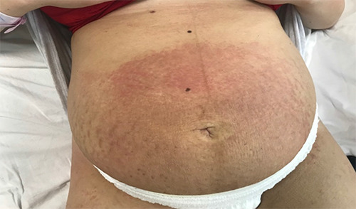
Erythematous papules within abdominal striae, note that periumbilical area is spared.
Figure 2.
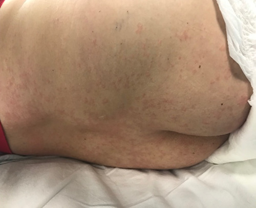
Erythematous papules distributed in the back and buttocks.
Figure 3.
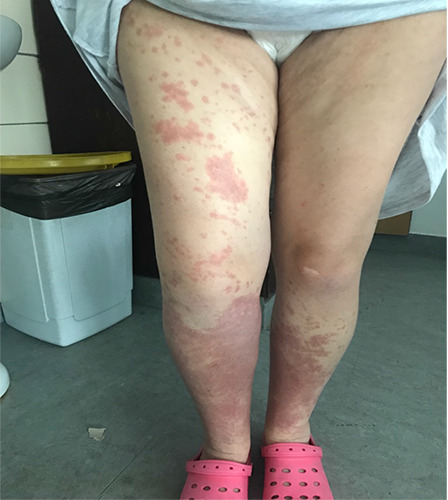
Large red urticated confluent plaques in lower extremities in woman with PEP.
Figure 4.
Large red urticated confluent plaques in lower extremities in woman with PEP.
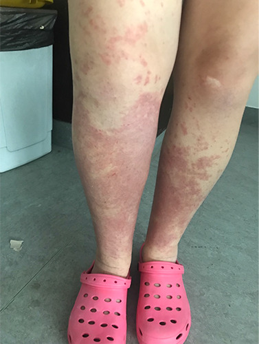
Figure 5.
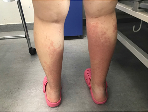
Large red urticated confluent plaques in the posterior compartment of the leg
Figure 6.
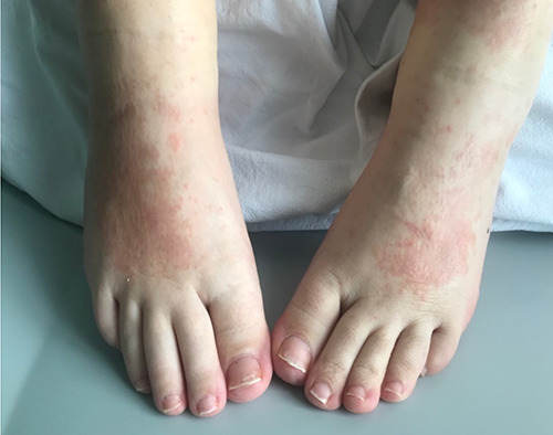
Erythematous papules and plaques are also visible on the dorsum of foot.
Conclusions
In conclusion, a pregnant woman with primary skin lesions and pruritus needs prompt evaluation and diagnosis to differentiate between the benign PEP and other dermatoses which are associated with increased fetal morbidity, namely pemphigoid gestationis and intrahepatic cholestasis of pregnancy.
Funding Statement
Funding: None.
References
- 1.Ambros-Rudolph CM. Dermatoses of pregnancy - clues to diagnosis, fetal risk and therapy. Ann Dermatol 2011;23:265-75. [DOI] [PMC free article] [PubMed] [Google Scholar]
- 2.Massone C, Cerroni L, Heidrun N, et al. Histopathological diagnosis of atopic eruption of pregnancy and polymorphic eruption of pregnancy: a study on 41 cases. Am J Dermatopathol 2014;36:812-21. [DOI] [PubMed] [Google Scholar]
- 3.Lehrhoff S, Pomeranz MK. Specific dermatoses of pregnancy and their treatment. Dermatol Ther 2013;26:274-84. [DOI] [PubMed] [Google Scholar]
- 4.Taylor D, Pappo E, Aronson IK. Polymorphic eruption of pregnancy. Clin Dermatol 2016;34:383-91. [DOI] [PubMed] [Google Scholar]
- 5.Shornick JK. Dermatoses of pregnancy. Semin Cutan Med Surg 1998;17:172-81. [DOI] [PubMed] [Google Scholar]
- 6.Roth MM. Pregnancy dermatoses: diagnosis, management, and controversies. Am J Clin Dermatol 2011;12:25-41. [DOI] [PubMed] [Google Scholar]
- 7.Dominguez-Serrano AJ, Quiroga-Garza A, Jacobo-Baca G, et al. Polymorphic eruption of pregnancy in Mexico. Int J Dermatol 2019;58:259-62. [DOI] [PubMed] [Google Scholar]
- 8.Regnier S, Fermand V, Levy P, et al. A case-control study of polymorphic eruption of pregnancy. J Am Acad Dermatol 2008;58:63-7. [DOI] [PubMed] [Google Scholar]
- 9.Yancey KB, Hall RP, Lawley TJ. Pruritic urticarial papules and plaques of pregnancy. Clinical experience in twenty-five patients. J Am Acad Dermatol 1984;10:473-80. [DOI] [PubMed] [Google Scholar]
- 10.Rudolph CM, Al-Fares S, Vaughan-Jones SA, et al. Polymorphic eruption of pregnancy: clinicopathology and potential trigger factors in 181 patients. Br J Dermatol 2006;154:54-60. [DOI] [PubMed] [Google Scholar]
- 11.Kanj RV, Gerber D, Frey MK, et al. Anaplastic large cell lymphoma in pregnancy. A case report. J Reprod Med 2015;60:265-8. [PubMed] [Google Scholar]
- 12.Scheinfeld N. Pruritic urticarial papules and plaques of pregnancy wholly abated with one week twice daily application of fluticasone propionate lotion: a case report and review of the literature. Dermatol Online J 2008;14:4. [PubMed] [Google Scholar]
- 13.Kroumpouzos G, Cohen LM. Specific dermatoses of pregnancy: an evidencebased systematic review. Am J Obstet Gynecol 2003;188:1083-92. [DOI] [PubMed] [Google Scholar]
- 14.Charles-Holmes R. Polymorphic eruption of pregnancy. Semin Dermatol 1989;8:18-22. [PubMed] [Google Scholar]
- 15.Ramot Y, Ingber A. Dermatoses of pregnancy. Curr Dermatol Rep 2012;1:222-6. [Google Scholar]
- 16.Huilaja L, Mäkikallio K, Tasanen K. Gestational pemphigoid. Orphanet J Rare Dis 2014;9:136. [DOI] [PMC free article] [PubMed] [Google Scholar]
- 17.Trivedi MK, Vaughn AR, Murase JE. Pustular psoriasis of pregnancy: current perspectives. Int J Womens Health 2018;10:109-15. [DOI] [PMC free article] [PubMed] [Google Scholar]
- 18.Hillman SC, Stokes-Lampard H, Kilby MD. Intrahepatic cholestasis of pregnancy. BMJ 2016;353:i1236. [DOI] [PubMed] [Google Scholar]