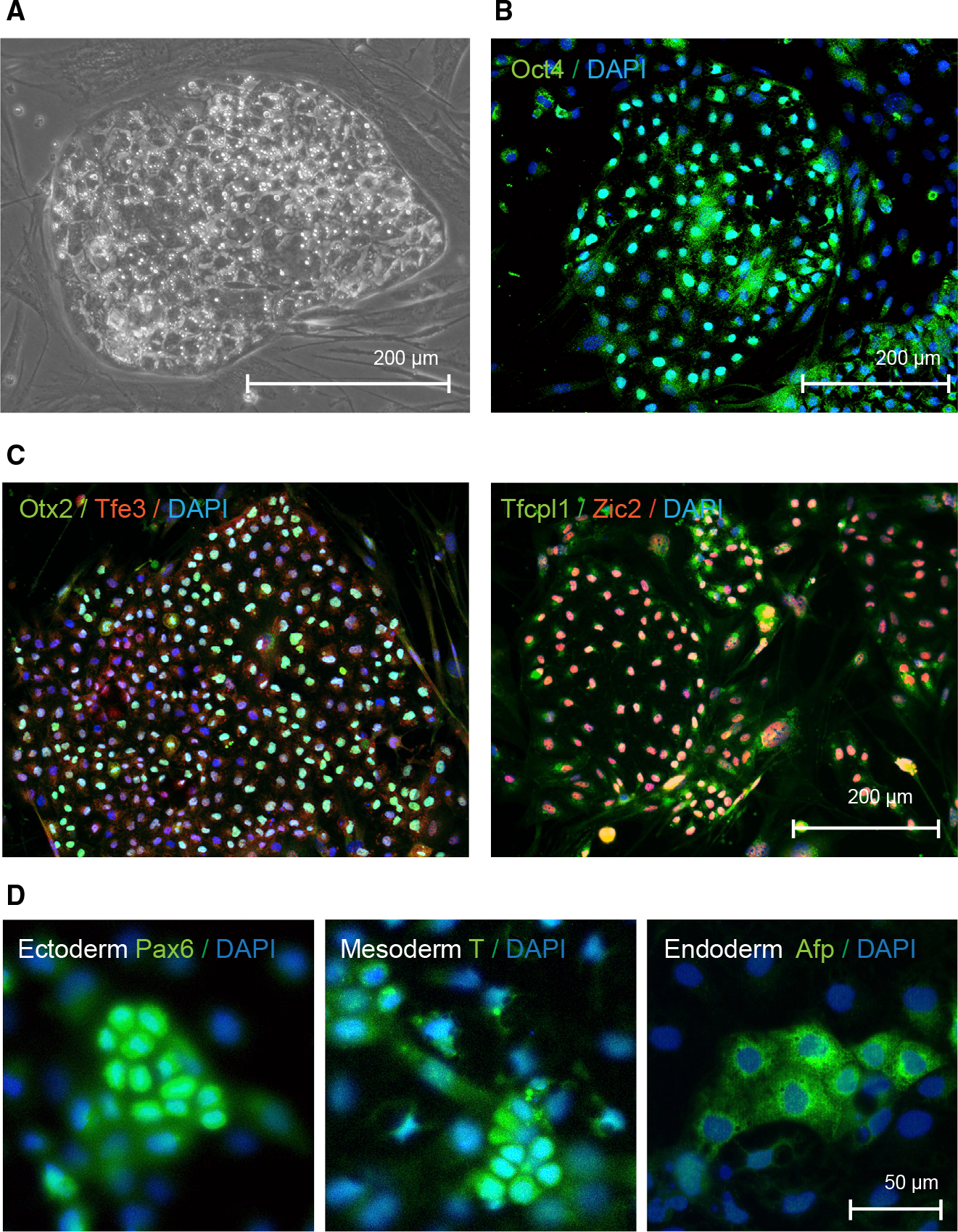
Figure 4. Characterization of induced pluripotent stem cells derived Myotis myotis uropatagium fibroblasts.

(A) Phase contrast image of Myotis myotis iPS cells.
(B) Microscopic image of Myotis myotis iPS cells after immunostaining to detect pluripotency marker Oct4.
(C) Immunofluorescence images of Myotis myotis pluripotent stem cells after staining of markers of naive (Tfe3 and Tfcp2l1) or primed pluripotency (Zic2 and Otx2)
(D) Microscopic images of Myotis myotis iPS cells that underwent differentiation and immunostaining to detect Pax6, brachyury (T) and Afp as markers for ectoderm, mesoderm, and endoderm, respectively.
