Abstract
Background
Inflammation plays an important role in the occurrence, development, and metastasis of tumors. However, the prognostic role of the neutrophil-to-lymphocyte ratio (NLR) in patients with luminal A breast cancer has rarely been reported in the literature. The purpose of this study was to investigate the relationship between preoperative peripheral blood NLR and the survival rate of patients with luminal A breast cancer.
Methods
Data from 226 eligible patients with luminal A breast cancer at the Chongqing University Cancer Hospital between 2011 and 2016 were obtained. The cut-off value for NLR for predicting overall survival (OS) rate was obtained from the receiver operating characteristic (ROC) curve. The baseline characteristics of 2 groups were compared using the Chi-square test or Fisher’s exact test, and OS was estimated using the Kaplan-Meier method. Cox analysis was performed to determine the correlation between clinicopathological parameters and prognosis.
Results
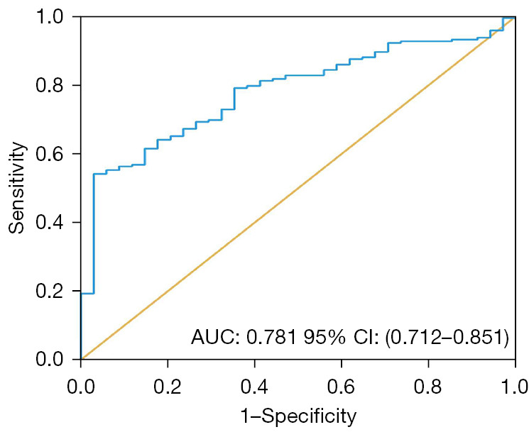
ROC curve analysis showed that the cutoff value for NLR to predict OS was 2.0. Kaplan-Meier analysis revealed that the OS of patients with a NLR <2.0 was significantly longer than that of patients with a higher NLR >2 (P<0.0001). The area under the curve (AUC) for NLR to predict OS was 0.781 [95% confidence interval (CI): 0.712–0.851], sensitivity was 54.17%, and specificity was 97.06%. In univariate Cox regression analysis, NLR, tumor (T) stage (T3–T4 vs. T1–T2), and histological grade (II–III vs. I) were all significantly associated with OS. In multivariate Cox regression analysis, NLR and histology grade (II–III vs. I) were independent prognostic factors for OS.
Conclusions
The results suggested that higher preoperative NLR was associated with worse prognosis in luminal A breast cancer.
Keywords: Breast cancer, luminal A, neutrophil-to-lymphocyte ratio (NLR), prognosis
Highlight box.
Key findings
• NLR had a negative effect on the prognosis of luminal A breast cancer. This finding provides a potential noninvasive prognostic biomarker for luminal A breast cancer patients.
What is known and what is new?
• NLR, as an indicator of systemic inflammation, has been studied in the prognosis of many cancers.
• The relationship between NLR and luminal A breast cancer has been rarely reported. We evaluated the prognostic value of NLR in luminal A breast cancer in this study.
What is the implication, and what should change now?
• In future studies, attention should be given to the tumor development processes and immune heterogeneity of breast carcinoma. Whether anti-inflammatory therapy can improve the sensitivity of breast cancer patients to chemotherapeutic drugs or improve the prognosis of breast cancer patients is a topic worthy of further study.
Introduction
In 2020, breast cancer represented 12% of all human malignancies, surpassing lung cancer, and becoming the most common malignancy worldwide (1,2). Among all types of breast cancer, Luminal A type had relatively less aggressive behavior. It has the best prognosis among all subtypes, and the 5-year overall survival (OS) probability can reach 95.1% (3). However, the axillary lymph node metastasis rate of patients with luminal A breast cancer is still 26.3%, and the local recurrence rate of some patients can reach 29% (4). Thus, the prognostic factors associated with luminal A breast cancer require further investigation.
The immune system is known to have a significant impact on tumor occurrence and progression. Several studies have indicated that the neutrophil-to-lymphocyte ratio (NLR) is a useful biomarker to predict the survival outcome of breast cancer (5-7). A meta-analysis of 39 trials, which included 17,079 breast cancer patients, showed that increased NLR before treatment was correlated with poorer OS (8). Two different preclinical studies of breast cancer have revealed that neutrophil recruitment and accumulation could lead to an increase in metastatic ability and a decrease in the survival rate (9,10). Some studies have shown that women with high lymphocyte concentrations in breast cancer have better survival outcomes (11,12). Thus, the effect of NLR on survival is worthy of further exploration. We therefore performed this study to evaluate the prognostic value of NLR for OS of patients with luminal A breast cancer. We present the following article in accordance with the STARD reporting checklist (available at https://gs.amegroups.com/article/view/10.21037/gs-23-80/rc).
Methods
Patient selection
This retrospective cohort study involved consecutive patients diagnosed with luminal A breast cancer who received surgical intervention at Chongqing University Cancer Hospital between January 2011 and December 2016. The inclusion criteria and exclusion criteria for the study were: (I) patients with complete clinicopathological data were included; (II) patients with hematological diseases, acute and chronic infections, kidney diseases, and other diseases or factors that affect blood indexes were excluded; and (III) patients with luminal A breast cancer were included according to immunohistochemistry (IHC) and fluorescent in situ hybridization (FISH).
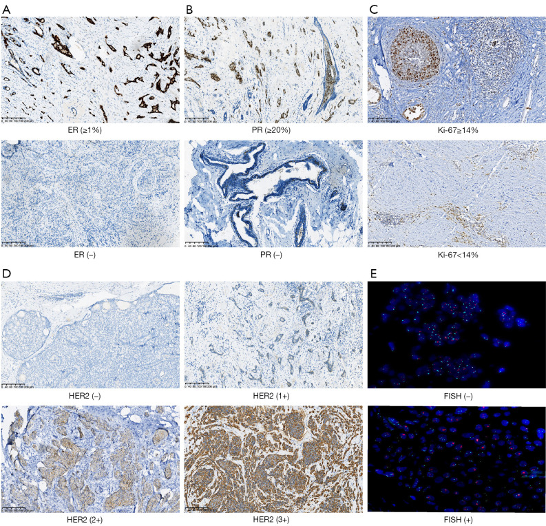
Because other molecular tests such as microarray and PAM50 are not commonly used in clinical examinations, in this study, molecular typing was determined by the results of IHC and FISH. The St Gallen International Expert Consensus 2013 defined luminal A breast cancer with estrogen receptor (ER) ≥1% (breast cancer samples with 1–100% tumor nuclei positive), progesterone receptor (PR) ≥20% (≥20% of tumor nuclei positive), Ki-67 <14% (<14% of tumor nuclei positive), and human epidermal growth factor receptor 2 (HER2) negative. IHC or FISH was used to determine the status of HER2 (13). IHC and FISH were scored as follows: (I) IHC score of 0 or 1 was classified as HER2-negative tumors; (II) patients with an IHC score of 2 were tested by FISH. If the result of the FISH test was HER2 amplification, it was considered HER2-positive; otherwise, if no amplification was found, it was HER2-negative; and (III) IHC3+ staining cases were regarded as HER2-positive (Figure 1). The specific criteria for pathological findings were as follows: IHC0 was defined as invasive breast cancer with incomplete and weak membrane staining ≤10% of tumor cells or unstained. IHC1+ was defined as invasive breast cancer with incomplete membrane staining that was faint/barely perceptible and in >10% of tumor cells. IHC2+ was defined as invasive breast cancer with weak to moderate complete membrane staining observed in >10% of tumor cells; these patients required FISH to determine HER2 status. The amplification of the HER2 gene was defined when the HER2/chromosome enumeration probe 17 (CEP17) ≥2.0 and the average HER2 copy number was ≥4.0 signals per cell. The HER2 status was defined as negative when the HER2/CEP17 was <4.0 signals per cell. When the HER2/CEP17 was <2.0 and the average HER2 copy number was ≥6.0 signals per cell, the HER2 status was defined as HER2 amplification. The HER2 status was defined as negative when the HER2/CEP17 <2.0 and the average HER2 copy number ≥4.0 and <6.0 signals per cell or the average HER2 copy number <4.0 signals per cell. IHC3+ indicated that more than 10% of invasive cancer cells showed intense, complete, and circumferential cell membrane staining (14).
Figure 1.
Representative images. (A) IHC results of ER positive ≥1% and ER-negative breast cancer. (B) IHC findings showing PR ≥20% and PR-negative samples. (C) The IHC findings showing Ki-67 ≥14% and Ki-67 <14%. (D) IHC results of HER2(−), HER2(1+), HER2(2+), and HER2(3+) samples. (E) Patients with an IHC score of 2 tested by FISH. If the FISH test showed HER2 amplification, it was considered HER2-positive, otherwise, it was HER2-negative. ER, estrogen receptor; PR, progesterone receptor; HER2, human epidermal growth factor receptor 2; FISH, fluorescent in situ hybridization; IHC, immunohistochemistry.
To ensure the precision of the results in this study, we asked 2 pathologists to read the pathology slice. One of the experienced pathologists observed the staining with an optical microscope and identified the proportion of positive expression according to the guidelines, and the other then reviewed the results. When the results were inconsistent or difficult to determine, the 2 pathologists decided after discussion. The study was conducted in accordance with the Declaration of Helsinki (as revised in 2013) and was approved by the medical ethics committee of the Chongqing University Cancer Hospital (No. CZLS2023014-A). The ethics committee waived the necessity of informed consent due to the retrospective analysis of routine data. Patient records/information were anonymized and deidentified prior to analysis.
Detection method
The results of a complete blood count test were obtained within 1 week of surgery. In this study, we used the results of the test conducted closest to the date of the operation as the final result. All subjects fasted for 12 h before 2.0 mL of fasting peripheral venous blood was collected in the early morning and placed in an ethylenediaminetetraacetic acid (EDTA) routine blood tube (anticoagulant tube). The sample was mixed before examination and the complete blood count test performed within 1 h. Routine blood examination was performed with the Sysmex XN9000 blood cell analyzer. We collected blood indices, including white blood cell (WBC), red blood cell (RBC), platelet count (PLT), absolute neutrophil count (ANC), absolute lymphocyte count (ALC), and absolute monocyte count (AMC). NLR was defined as the ratio between ANC and ALC.
The determination of sample size and the collection of clinical data
In order to ensure the statistical power of the sample size, we used G*Power to calculate the effective sample size needed for our study, and the final total sample size was set at 220 (15). Medical records were obtained from the hospital’s medical system, and clinical pathological data were collected, including medical history, age, number of axillary lymph node metastases, vascular tumor thrombus; tumor (T), lymph node (N), and metastasis (M) stage; ER, PR, and HER2 status; and histology grade. We temporarily recorded the missing data as unknown if clinical data such as histology grade and T/N stage were missing.
Follow-up
In this study, a combination of electronic medical records, outpatient follow-up, and telephone follow-up was used. The last follow-up was on 23 February 2022. The period from the time of pathological diagnosis to the time of death or the end of follow-up was defined as the survival time.
Statistical analysis
Data were processed using GraphPad Prism 8.0.1 (GraphPad Software Inc., San Diego, CA, USA) and SPSS version 25.0 (IBM Corp., Armonk, NY, USA). Receiver operating characteristic (ROC) curves were used to determine the appropriate cut-off value to balance the specificity and sensitivity of the observation index. A larger area under curve (AUC) value showed that NLR was more likely to make an accurate prediction for OS. Therefore, based on the data at the end of the follow-up, we drew the ROC curve and the maximum value of the Youden index was used to determine the cutoff value of NLR, and patients were divided into a low-NLR group and high-NLR group accordingly. The Chi-square test and the Fisher exact test were used to compare the clinicopathological characteristics of the 2 groups of patients. Survival curves were drawn using the Kaplan-Meier method and the logarithmic rank test was used to compare the differences in survival curves between these 2 groups. The Cox proportional hazards model was used to calculate the hazard ratio (HR) and the 95% confidence interval (95% CI). Results with a P value <0.05 were considered statistically significant.
Results
Characteristics of the included patients
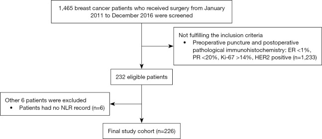
After preliminary screening of the medical records using the hospital database platform, 1,465 breast cancer patients who underwent surgery at the Chongqing University Cancer Hospital were further screened. Among them, 1,233 patients were excluded because they did not meet the molecular typing conditions above, and 6 patients were excluded because they did not have complete preoperative neutrophil and lymphocyte examination results. Finally, a total of 226 patients met the criteria for analysis. According to the G*Power calculation, the sample size included in our study was effective (15). The patient screening flow chart is shown in Figure 2.
Figure 2.
Flow chart of patient screening. ER, estrogen receptor; PR, progesterone receptor; HER2, human epidermal growth factor receptor 2; NLR, neutrophil-to-lymphocyte ratio.
Determination of the cut-off value for NLR
The ROC curve for NLR is shown in Figure 3. As the AUC for NLR to predict OS was 0.781 (95% CI: 0.712–0.851), NLR was considered an accurate marker for predicting OS in patients with luminal A breast cancer. The critical value corresponding to the maximum of the Youden index was the best. In this study, according to the maximum value of the Youden index (0.512), the cut-off value of NLR to predict OS was 2.0, sensitivity was 54.17%, and specificity was 97.06%. The included patients could be divided into high and low groups according to the cut-off value and included 121 and 105 patients, respectively.
Figure 3.

ROC curve of NLR for predicting overall survival. AUC, area under the curve; CI, confidence interval; ROC, receiver operating characteristic; NLR, neutrophil-to-lymphocyte ratio.
Comparison of clinicopathological features in patients with luminal A in the low- and high-NLR groups
We described the clinicopathological and treatment characteristics of the entire population according to NLR group (<2.0 vs. ≥2.0). At the time of this analysis (23 February 2022), a total of 34 patients had died, including 33 from the high group and 1 in the low group. We used the Chi-square test to examine the basic clinical characteristics of patients with high and low NLR (Table 1) and found that the basic clinical characteristics of the 2 groups were similar (P>0.05). Regarding the clinical variables at the time of diagnosis, cases with NLR ≥2.0 were older and had a higher clinical staging of lymph nodes and tumor size, although without statistical significance.
Table 1. Comparison of baseline characteristics between high- and low-NLR groups in luminal A breast cancer.
| Characteristics | High NLR (n=121), No. (%) | Low NLR (n=105), No. (%) | P value |
|---|---|---|---|
| Age at diagnosis | 0.876 | ||
| ≤50 years | 76 (62.8) | 67 (63.8) | |
| >50 years | 45 (37.2) | 38 (36.2) | |
| T stage | 0.386 | ||
| T1 | 44 (36.4) | 46 (43.8) | |
| T2 | 60 (49.6) | 49 (46.7) | |
| T3 | 4 (3.3) | 5 (4.8) | |
| T4 | 12 (9.9) | 5 (4.8) | |
| Unknown | 1 (0.8) | 0 (0.0) | |
| N stage | 0.267 | ||
| N0 | 76 (62.8) | 63 (60.0) | |
| N1 | 17 (14.0) | 25 (23.8) | |
| N2 | 16 (13.3) | 11 (10.5) | |
| N3 | 11 (9.1) | 6 (5.7) | |
| Unknown | 1 (0.8) | 0 (0.0) | |
| Neoadjuvant | 0.815 | ||
| Yes | 21 (17.4) | 17 (16.2) | |
| No | 100 (82.6) | 88 (83.8) | |
| Vascular tumor thrombus | 0.232 | ||
| Yes | 19 (15.7) | 23 (21.9) | |
| No | 102 (84.3) | 82 (78.1) | |
| G-CSF | 0.407 | ||
| Yes | 17 (14.0) | 19 (18.1) | |
| No | 104 (86.0) | 86 (81.9) | |
| Histology grade | 0.407 | ||
| I | 16 (13.2) | 10 (9.5) | |
| II | 75 (62.0) | 65 (61.9) | |
| III | 3 (2.5) | 7 (6.7) | |
| Unknown | 27 (22.3) | 23 (21.9) | |
| PR status | 0.163 | ||
| 20–39 | 15 (12.4) | 7 (6.7) | |
| 40–59 | 6 (5.0) | 11 (10.5) | |
| 60–79 | 23 (19.0) | 15 (14.3) | |
| ≥80 | 77 (63.6) | 72 (68.6) | |
| ER status | 0.695 | ||
| ≤20 | 6 (5.0) | 6 (5.7) | |
| 21–40 | 3 (2.5) | 4 (3.8) | |
| 41–60 | 12 (9.9) | 6 (5.7) | |
| 61–80 | 47 (38.8) | 37 (35.2) | |
| ≥81 | 53 (43.8) | 52 (49.5) | |
NLR, neutrophil-tolymphocyte ratio; G-CSF, granulocyte colony stimulating factor; PR, progesterone receptor; ER, estrogen receptor.
The relationship between NLR and prognosis in patients with luminal A breast cancer
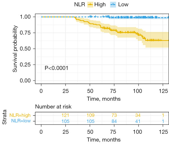
The survival curves of the 2 groups of patients are shown in Figure 4. The Kaplan-Meier analysis showed significant differences in OS between patients with NLR <2.0 and NLR ≥2.0 (HR =33.26; 95% CI: 16.98–65.15; P<0.0001). The OS probability of the low-NLR group was better. Univariate Cox regression analysis showed that T stage (T3–T4 vs. T1–T2), histological grade (II–III vs. I), and NLR affected the OS of patients with luminal A breast cancer (Table 2), and the HRs were 3.004 (95% CI: 1.065–8.475; P=0.0376), 1.676 (95% CI: 1.007–2.791; P=0.047), and 33.26 (95% CI: 16.98–65.15; P<0.0001), respectively. The above variables were incorporated into the multivariate Cox proportional regression analysis, which found that NLR and histology grade (II–III vs. I) were independent risk factors that affected the OS of patients with luminal A breast cancer, and the HRs were 36.003 (95% CI: 4.912–263.92; P<0.0001) and 1.924 (95% CI: 1.14–3.25; P=0.014), respectively (Table 3).
Figure 4.

Survival curve of patients with different NLR levels. NLR, neutrophil-to-lymphocyte ratio.
Table 2. Univariable Cox regression analysis for overall survival in luminal A breast cancer patients.
| Variables | Luminal A breast cancer patients (n=226) | ||
|---|---|---|---|
| HR | 95% CI | P value | |
| Age at diagnosis (≥50 vs. <50 years) | 1.252 | 0.926–1.693 | 0.144 |
| Neoadjuvant (yes vs. no) | 1.192 | 0.509–2.790 | 0.686 |
| Vascular tumor thrombus (yes vs. no) | 1.794 | 0.746–4.313 | 0.192 |
| G-CSF (yes vs. no) | 1.107 | 0.465–2.638 | 0.818 |
| T stage (T3–T4 vs. T1–T2) | 3.004 | 1.065–8.475 | 0.038 |
| N stage (N1–N3 vs. N0) | 1.178 | 0.855–1.623 | 0.315 |
| Histology grade (II–III vs. I) | 1.676 | 1.007–2.791 | 0.047 |
| ER status | 0.999 | 0.983–1.014 | 0.848 |
| PR status | 0.996 | 0.981–1.012 | 0.642 |
| NLR (≥2 vs. <2) | 33.26 | 16.98–65.15 | <0.0001 |
HR, hazard ratio; CI, confidence interval; G-CSF, granulocyte colony stimulating factor; ER, estrogen receptor; PR, progesterone receptor; NLR, neutrophil-to-lymphocyte ratio.
Table 3. Multivariable Cox regression analysis for overall survival in luminal A breast cancer patients.
| Variables | Luminal A breast cancer patients (n=226) | ||
|---|---|---|---|
| HR | 95% CI | P value | |
| T stage (T3–T4 vs. T1–T2) | 1.098 | 0.769–1.566 | 0.608 |
| Histology grade (II–III vs. I) | 1.924 | 1.140–3.250 | 0.014 |
| NLR (≥2 vs. <2) | 36.003 | 4.912–263.920 | <0.0001 |
HR, hazard ratio; CI, confidence interval; NLR, neutrophil-to-lymphocyte ratio.
Discussion
The high heterogeneity of breast cancer has resulted in varying clinical treatment options and clinical outcomes. Based on the molecular classification, breast cancer is divided mainly into 4 types, of which the luminal subtype A is considered to have the best prognosis among all breast cancer types (16). Nevertheless, a fraction of patients with luminal A have a poor prognosis, and the cause of this phenomenon is unclear (17). In this study, we found that NLR and histology grade (II–III vs. I) were independent risk factors for patients with luminal A breast cancer.
The histological grade reflects the morphology of cancer cells (ductal formation), the nuclear morphology of cancer cells (nuclear atypia), and the proliferation ability of cancer cells (nuclear mitosis number), and it can predict prognosis. As in previous reports, our study found that the higher the histological grade, the worse the prognosis of breast cancer (17-20). A study by the Nottingham team, which included 2,219 cases of breast cancer eligible for surgical intervention and with long-term follow-up data, showed that the classification had an important influence on the outcome of breast cancer and played a complementary role for defining the tumor lymph node (LN) stage (17). There is convincing evidence that tumor behavior can be accurately predicted by histology grade, especially in the early stage of small tumors (17-19). The effect of histological classification on the biological behavior of breast cancer was consistent with the clinical results we observed in this study and was closely related to prognosis.
Tumor size often impacts breast cancer prognosis. Previous study indicated that large tumor size adversely affected prognosis in non-luminal A breast cancer (21) However, in luminal A breast cancer, many studies have failed to find association between tumor size and prognosis, regardless of lymph node positivity (22-25). Similarly, in the current study the association between tumor size and prognosis was not found. Lymph node metastasis is another important factor that influences the progression of breast cancer. Some studies reported that lymph node metastasis had adverse effect on the prognosis of patients with luminal A subtype (26,27). While, in the current study, lymph node metastasis status had no independent prognostic value in multifactorial analysis. The contradictory results urge future study with large sample size and a long follow-up period.
Immune status and inflammatory response had a non-negligible effect on the tumors. As a combination index of ANC and ALC, NLR reflects the balance between neutrophil-associated tumor inflammation and lymphocyte-dependent antitumor immune response to some extent. Related studies have shown that NLR can be used to predict the prognosis of colorectal cancer, gastric cancer, lung cancer, and other tumors (28-30), and it has been receiving increasing attention in the field of oncology. Previous studies have evaluated the prognostic role of NLR in different molecular subtypes of breast cancer. Bozkurt et al. (31) and Qiu et al. (32) studied 85 and 406 patients, respectively, with triple-negative breast cancer stages I to III. They obtained similar results and showed that a higher NLR was independently correlated with a worse OS and disease-free survival (DFS), with a cutoff value of 2.0 and 2.85, respectively. Tiainen et al. (33). found that the outcome of HER2 positive patients with high NLR was poor if their adjuvant treatment did not include trastuzumab. However, there have been few reports on the prognostic value of NLR in luminal A breast cancer. The results of our study suggested that NLR may have predictive significance for the prognosis of patients with luminal A subtype breast cancer. In a previous study, elevated NLR was associated with advanced age, larger tumors, and stage of the disease (5). However, in the present study, there was no relationship between NLR and clinicopathological factors. Therefore, the mechanism of how NLR affects survival is worthy of further study to provide a deeper understanding of the biological process and the heterogeneity of immunity in breast carcinoma and to improve the prognosis and treatment strategies of patients.
Biologically, an imbalance in the ratio of neutrophils to lymphocytes has potential effects on tumor progression and prognosis. There is scientific evidence that neutrophils are related to tumor-promoting activities in vivo, such as enhancing angiogenesis, thereby promoting the metastatic ability of tumor cells. Conversely, lymphocytes play an immune monitoring role in cancer (34-36). However, there are few reports on the specific mechanism. In studies evaluating circulating tumor cells (CTCs) in which poor prognosis was mediated by neutrophils, a Gene Ontology (GO) analysis was performed to define the enriched activities involving the 337 target genes. The results showed that CTCs were related to the neutrophil-mediated immune response, especially in the initiation and degranulation of the neutrophil. In addition, this study found that tumor necrosis factor α (TNF-α) signaling via nuclear factor-κB (NF-κB) and hypoxia promote neutrophil activation, particularly neutrophil degranulation, which augments CTC levels. In addition, TNF-α signaling is associated with the activation of neutrophil extracellular traps (NETs) in the inflammatory response. The connection between CTCs and NETs is related to a decrease in cell killing capacity, prompting immune escape and a worse prognosis (37).
One study revealed that neutrophils release their dense chromatin and produce NETS, enormous DNA net-like structures, outside the cell (38). Another study first reported cancer cells in cancer thrombus-delivered granulocyte colony stimulating factor (G-CSF) that could raise neutrophils levels to frame NETs (39). Furthermore, the NET structure in cancer can capture CTCs, permitting relocation and intrusion and leading to a poor prognosis (40). Other basic research supports the above results. In the above study, 2 cell lines were used: MDA-MB-231 cells, which show a high production of G-CSF, and MCF-7, which rarely secrete G-CSF. The neutrophils cocultured with MDA-MB-231 cells formed extensive NETs which increased cell migration. Although neutrophils cocultured with MCF-7 cells formed few NETs and neutrophils, which were not activated, they had a negative influence on the migration of cell (41). The same study also found that in a dominant high-risk luminal A breast cancer subtype, the high expression of G-CSF was accompanied by neutrophil aggregation, which may explain our findings in patients with luminal A breast cancer. Neutrophils have also been considered a therapeutic target in some experimental therapies (42). Cancer cells stimulate the expression of interleukin (IL-17) from gamma-delta T (gdT) cells, leading to a cascade effect of systemic immune activation, with gdT cells and neutrophils jointly driving the metastasis of breast carcinoma (43). Thus, to improve prognosis for these patients, in future studies, greater attention should be given to the tumor development processes and immune heterogeneity of breast carcinoma. It has been reported that inflammation can promote angiogenesis and metastasis of malignant cells and alter the response to hormones and chemotherapeutic agents (34). Therefore, whether anti-inflammatory therapy can improve the sensitivity of breast cancer patients to chemotherapeutic drugs or improve the prognosis of breast cancer patients is a topic worthy of further study.
There were some limitations to our study. First, although we strictly enforced the inclusion and exclusion criteria, the peripheral blood cell count may still have been affected by potential confounding factors such as smoking and drinking. Second, this study was performed at a single center, based only on retrospective data collection and analysis of a small number of patients. Although our sample size was sufficient according to the G*Power calculation, our findings may be more persuasive if verified by larger clinical multicenter research in the future. Third, we were unable to evaluate the response to treatment because this was a retrospective study with a long duration. Therefore, in the future, we will explore the relationship between the treatment response of patients with NLR and luminal A breast cancer. Fourth, in our study, the critical cutoff value of the NLR was determined using ROC curve analysis. However, the ROC curve only considers “death” as the prognostic outcome and survival time is not taken into account, so prognosis could not be accurately judged. Additional studies are needed to explore the best critical value to evaluate prognosis. Finally, the use of G-CSF in some patients might have affected the number of neutrophils and influenced our results.
Conclusions
The results of our study were consistent with the results of previous studies and further demonstrated the relationship between a high NLR with poor outcomes in patients with luminal A breast cancer. However, further basic research is needed to demonstrate the mechanism of how NLR affects the prognosis of patients with luminal A breast cancer.
Supplementary
The article’s supplementary files as
Acknowledgments
We thank all the patients who contributed to this study.
Funding: This work was supported by the Science and Technology Research Program of Chongqing Municipal Education Commission (No. KJZD-K202000104), Chongqing Science and Health Joint Medical Research Project (No. 2021MSXM085), Talent Program of Chongqing (No. CQYC20200303137), and Chongqing Municipal Health and Health Commission (No. 2019NLTS005).
Ethical Statement: The authors are accountable for all aspects of the work in ensuring that questions related to the accuracy or integrity of any part of the work are appropriately investigated and resolved. The study was conducted in accordance with the Declaration of Helsinki (as revised in 2013) and was approved by the medical ethics committee of the Chongqing University Cancer Hospital (No. CZLS2023014-A). The ethics committee waived the necessity of informed consent due to the retrospective analysis of routine data.
Reporting Checklist: The authors have completed the STARD reporting checklist. Available at https://gs.amegroups.com/article/view/10.21037/gs-23-80/rc
Data Sharing Statement: Available at https://gs.amegroups.com/article/view/10.21037/gs-23-80/dss
Conflicts of Interest: All authors have completed the ICMJE uniform disclosure form (available at https://gs.amegroups.com/article/view/10.21037/gs-23-80/coif). The authors have no conflicts of interest to declare.
(English Language Editor: A. Muylwyk)
References
- 1.Sung H, Ferlay J, Siegel RL, et al. Global Cancer Statistics 2020: GLOBOCAN Estimates of Incidence and Mortality Worldwide for 36 Cancers in 185 Countries. CA Cancer J Clin 2021;71:209-49. 10.3322/caac.21660 [DOI] [PubMed] [Google Scholar]
- 2.Liu H, Qiu C, Wang B, et al. Evaluating DNA Methylation, Gene Expression, Somatic Mutation, and Their Combinations in Inferring Tumor Tissue-of-Origin. Front Cell Dev Biol 2021;9:619330. 10.3389/fcell.2021.619330 [DOI] [PMC free article] [PubMed] [Google Scholar]
- 3.Hennigs A, Riedel F, Gondos A, et al. Prognosis of breast cancer molecular subtypes in routine clinical care: A large prospective cohort study. BMC Cancer 2016;16:734. 10.1186/s12885-016-2766-3 [DOI] [PMC free article] [PubMed] [Google Scholar]
- 4.Lale Atahan I, Yildiz F, Ozyigit G, et al. Percent positive axillary lymph node metastasis predicts survival in patients with non-metastatic breast cancer. Acta Oncol 2008;47:232-8. 10.1080/02841860701678761 [DOI] [PubMed] [Google Scholar]
- 5.Guthrie GJ, Charles KA, Roxburgh CS, et al. The systemic inflammation-based neutrophil-lymphocyte ratio: experience in patients with cancer. Crit Rev Oncol Hematol 2013;88:218-30. 10.1016/j.critrevonc.2013.03.010 [DOI] [PubMed] [Google Scholar]
- 6.Koh CH, Bhoo-Pathy N, Ng KL, et al. Utility of pre-treatment neutrophil-lymphocyte ratio and platelet-lymphocyte ratio as prognostic factors in breast cancer. Br J Cancer 2015;113:150-8. 10.1038/bjc.2015.183 [DOI] [PMC free article] [PubMed] [Google Scholar]
- 7.Ethier JL, Desautels D, Templeton A, et al. Prognostic role of neutrophil-to-lymphocyte ratio in breast cancer: a systematic review and meta-analysis. Breast Cancer Res 2017;19:2. 10.1186/s13058-016-0794-1 [DOI] [PMC free article] [PubMed] [Google Scholar]
- 8.Guo W, Lu X, Liu Q, et al. Prognostic value of neutrophil-to-lymphocyte ratio and platelet-to-lymphocyte ratio for breast cancer patients: An updated meta-analysis of 17079 individuals. Cancer Med 2019;8:4135-48. 10.1002/cam4.2281 [DOI] [PMC free article] [PubMed] [Google Scholar]
- 9.Kowanetz M, Wu X, Lee J, et al. Granulocyte-colony stimulating factor promotes lung metastasis through mobilization of Ly6G+Ly6C+ granulocytes. Proc Natl Acad Sci U S A 2010;107:21248-55. 10.1073/pnas.1015855107 [DOI] [PMC free article] [PubMed] [Google Scholar]
- 10.Yan HH, Pickup M, Pang Y, et al. Gr-1+CD11b+ myeloid cells tip the balance of immune protection to tumor promotion in the premetastatic lung. Cancer Res 2010;70:6139-49. 10.1158/0008-5472.CAN-10-0706 [DOI] [PMC free article] [PubMed] [Google Scholar]
- 11.Dieci MV, Criscitiello C, Goubar A, et al. Prognostic value of tumor-infiltrating lymphocytes on residual disease after primary chemotherapy for triple-negative breast cancer: a retrospective multicenter study. Ann Oncol 2014;25:611-8. Erratum in: Ann Oncol 2015;26:1518. 10.1093/annonc/mdt556 [DOI] [PMC free article] [PubMed] [Google Scholar]
- 12.Luen SJ, Salgado R, Dieci MV, et al. Prognostic implications of residual disease tumor-infiltrating lymphocytes and residual cancer burden in triple-negative breast cancer patients after neoadjuvant chemotherapy. Ann Oncol 2019;30:236-42. 10.1093/annonc/mdy547 [DOI] [PubMed] [Google Scholar]
- 13.Goldhirsch A, Winer EP, Coates AS, et al. Personalizing the treatment of women with early breast cancer: highlights of the St Gallen International Expert Consensus on the Primary Therapy of Early Breast Cancer 2013. Ann Oncol 2013;24:2206-23. 10.1093/annonc/mdt303 [DOI] [PMC free article] [PubMed] [Google Scholar]
- 14.Wolff AC, Hammond MEH, Hicks DG, et al. Recommen-dations for Human Epidermal Growth Factor Receptor 2 Testing in Breast Cancer: American Society of Clinical Oncology/College of American Pathologists Clinical Practice Guideline Update. J Clin Oncol 2013;31:3997-4013. 10.1200/JCO.2013.50.9984 [DOI] [PubMed] [Google Scholar]
- 15.Faul F, Erdfelder E, Lang AG, et al. G*Power 3: a flexible statistical power analysis program for the social, behavioral, and biomedical sciences. Behav Res Methods 2007;39:175-91. 10.3758/bf03193146 [DOI] [PubMed] [Google Scholar]
- 16.Kim MJ, Ro JY, Ahn SH, et al. Clinicopathologic significance of the basal-like subtype of breast cancer: a comparison with hormone receptor and Her2/neu-overexpressing phenotypes. Hum Pathol 2006;37:1217-26. 10.1016/j.humpath.2006.04.015 [DOI] [PubMed] [Google Scholar]
- 17.Rakha EA, El-Sayed ME, Lee AH, et al. Prognostic significance of Nottingham histologic grade in invasive breast carcinoma. J Clin Oncol 2008;26:3153-8. 10.1200/JCO.2007.15.5986 [DOI] [PubMed] [Google Scholar]
- 18.Galea MH, Blamey RW, Elston CE, et al. The Nottingham Prognostic Index in primary breast cancer. Breast Cancer Res Treat 1992;22:207-19. 10.1007/BF01840834 [DOI] [PubMed] [Google Scholar]
- 19.Frkovic-Grazio S, Bracko M. Long term prognostic value of Nottingham histological grade and its components in early (pT1N0M0) breast carcinoma. J Clin Pathol 2002;55:88-92. 10.1136/jcp.55.2.88 [DOI] [PMC free article] [PubMed] [Google Scholar]
- 20.Uchinaka E, Amisaki M, Morimoto M, et al. Utility and Limitation of Preoperative Neutrophil Lymphocyte Ratio as a Prognostic Factor in Hepatocellular Carcinoma. Yonago Acta Med 2018;61:197-203. 10.33160/yam.2018.12.002 [DOI] [PMC free article] [PubMed] [Google Scholar]
- 21.Kustic D, Lovasic F, Belac-Lovasic I, et al. Impact of HER2 receptor status on axillary nodal burden in patients with non-luminal A invasive ductal breast carcinoma. Rev Med Chil 2019;147:557-67. 10.4067/S0034-98872019000500557 [DOI] [PubMed] [Google Scholar]
- 22.Uchida N, Suda T, Ishiguro K. Effect of chemotherapy for luminal a breast cancer. Yonago Acta Med 2013;56:51-6. [PMC free article] [PubMed] [Google Scholar]
- 23.Alramadhan M, Ryu JM, Rayzah M, et al. Goserelin plus tamoxifen compared to chemotherapy followed by tamoxifen in premenopausal patients with early stage-, lymph node-negative breast cancer of luminal A subtype. Breast 2016;30:111-7. 10.1016/j.breast.2016.08.011 [DOI] [PubMed] [Google Scholar]
- 24.Herr D, Wischnewsky M, Joukhadar R, et al. Does chemotherapy improve survival in patients with nodal positive luminal A breast cancer? A retrospective Multicenter Study. PLoS One 2019;14:e0218434. 10.1371/journal.pone.0218434 [DOI] [PMC free article] [PubMed] [Google Scholar]
- 25.Li Y, Ma L. Efficacy of chemotherapy for lymph node-positive luminal A subtype breast cancer patients: an updated meta-analysis. World J Surg Oncol 2020;18:316. 10.1186/s12957-020-02089-y [DOI] [PMC free article] [PubMed] [Google Scholar]
- 26.Min SK, Lee SK, Woo J, et al. Relation Between Tumor Size and Lymph Node Metastasis According to Subtypes of Breast Cancer. J Breast Cancer 2021;24:75-84. 10.4048/jbc.2021.24.e4 [DOI] [PMC free article] [PubMed] [Google Scholar]
- 27.Chas M, Boivin L, Arbion F, et al. Clinicopathologic predictors of lymph node metastasis in breast cancer patients according to molecular subtype. J Gynecol Obstet Hum Reprod 2018;47:9-15. 10.1016/j.jogoh.2017.10.008 [DOI] [PubMed] [Google Scholar]
- 28.Okui M, Yamamichi T, Asakawa A, et al. Prognostic significance of neutrophil-lymphocyte ratios in large cell neuroendocrine carcinoma. Gen Thorac Cardiovasc Surg 2017;65:633-9. 10.1007/s11748-017-0804-y [DOI] [PubMed] [Google Scholar]
- 29.Zhang Y, Lu JJ, Du YP, et al. Prognostic value of neutrophil-to-lymphocyte ratio and platelet-to-lymphocyte ratio in gastric cancer. Medicine (Baltimore) 2018;97:e0144. 10.1097/MD.0000000000010144 [DOI] [PMC free article] [PubMed] [Google Scholar]
- 30.Malietzis G, Giacometti M, Kennedy RH, et al. The emerging role of neutrophil to lymphocyte ratio in determining colorectal cancer treatment outcomes: a systematic review and meta-analysis. Ann Surg Oncol 2014;21:3938-46. 10.1245/s10434-014-3815-2 [DOI] [PubMed] [Google Scholar]
- 31.Bozkurt O, Karaca H, Berk V, et al. Predicting the role of the pretreatment neutrophil to lymphocyte ratio in the survival of early triple-negative breast cancer patients. J BUON 2015;20:1432-9. [PubMed] [Google Scholar]
- 32.Qiu X, Song Y, Cui Y, et al. Increased neutrophil-lymphocyte ratio independently predicts poor survival in non-metastatic triple-negative breast cancer patients. IUBMB Life 2018;70:529-35. 10.1002/iub.1745 [DOI] [PubMed] [Google Scholar]
- 33.Tiainen S, Rilla K, Hämäläinen K, et al. The prognostic and predictive role of the neutrophil-to-lymphocyte ratio and the monocyte-to-lymphocyte ratio in early breast cancer, especially in the HER2+ subtype. Breast Cancer Res Treat 2021;185:63-72. 10.1007/s10549-020-05925-7 [DOI] [PMC free article] [PubMed] [Google Scholar]
- 34.Coussens LM, Werb Z. Inflammation and cancer. Nature 2002;420:860-7. 10.1038/nature01322 [DOI] [PMC free article] [PubMed] [Google Scholar]
- 35.Shankaran V, Ikeda H, Bruce AT, et al. IFNgamma and lymphocytes prevent primary tumour development and shape tumour immunogenicity. Nature 2001;410:1107-11. 10.1038/35074122 [DOI] [PubMed] [Google Scholar]
- 36.De Larco JE, Wuertz BR, Furcht LT. The potential role of neutrophils in promoting the metastatic phenotype of tumors releasing interleukin-8. Clin Cancer Res 2004;10:4895-900. 10.1158/1078-0432.CCR-03-0760 [DOI] [PubMed] [Google Scholar]
- 37.Zhang W, Qin T, Yang Z, et al. Telomerase-positive circulating tumor cells are associated with poor prognosis via a neutrophil-mediated inflammatory immune environment in glioma. BMC Med 2021;19:277. 10.1186/s12916-021-02138-7 [DOI] [PMC free article] [PubMed] [Google Scholar]
- 38.Brinkmann V, Reichard U, Goosmann C, et al. Neutrophil extracellular traps kill bacteria. Science 2004;303:1532-5. 10.1126/science.1092385 [DOI] [PubMed] [Google Scholar]
- 39.Shang B, Guo L, Shen R, et al. Prognostic Significance of NLR About NETosis and Lymphocytes Perturbations in Localized Renal Cell Carcinoma With Tumor Thrombus. Front Oncol 2021;11:771545. 10.3389/fonc.2021.771545 [DOI] [PMC free article] [PubMed] [Google Scholar]
- 40.Cools-Lartigue J, Spicer J, McDonald B, et al. Neutrophil extracellular traps sequester circulating tumor cells and promote metastasis. J Clin Invest 2013;123:3446-58. 10.1172/JCI67484 [DOI] [PMC free article] [PubMed] [Google Scholar]
- 41.Guo L, Chen G, Zhang W, et al. A high-risk luminal A dominant breast cancer subtype with increased mobility. Breast Cancer Res Treat 2019;175:459-72. 10.1007/s10549-019-05135-w [DOI] [PMC free article] [PubMed] [Google Scholar]
- 42.Jaillon S, Ponzetta A, Di Mitri D, et al. Neutrophil diversity and plasticity in tumour progression and therapy. Nat Rev Cancer 2020;20:485-503. 10.1038/s41568-020-0281-y [DOI] [PubMed] [Google Scholar]
- 43.Coffelt SB, Kersten K, Doornebal CW, et al. IL-17-producing γδ T cells and neutrophils conspire to promote breast cancer metastasis. Nature 2015;522:345-8. 10.1038/nature14282 [DOI] [PMC free article] [PubMed] [Google Scholar]
Associated Data
This section collects any data citations, data availability statements, or supplementary materials included in this article.
Supplementary Materials
The article’s supplementary files as


