An 8-year-old boy presented with a 2-week history of fever and the onset of a scarlatinaform rash. He was found to have mild bulbar conjunctivitis, a “strawberry” tongue and palpable lymph nodes (neck and groin). These clinical features could be caused by Scarlet fever, but they were also in accord with the diagnostic criteria for Kawasaki disease, as outlined in recent reviews of this acute systemic vasculitis of unknown cause.1,2
The boy was treated with a 5-day course of amoxicillin (250 mg 3 times a day). Throat and blood cultures were negative for group A streptococcus. The boy also received gamma globulin (2 g/kg) intravenously followed by high-dose anti-inflammatory therapy with ASA (90 mg/kg daily) for 2 weeks to reduce inflammation and decrease the risk of coronary artery aneurysms, which are the main complications of Kawasaki disease. Baseline and 2 follow-up echocardiograms (at 2 and 10 weeks after presentation) showed no evidence of coronary artery aneurysms. The boy's condition gradually improved during the 2 weeks of anti-inflammatory therapy.
A week after completing the ASA therapy, the skin on the tips of the boy's fingers and toes began to peel. This periungual desquamation is a common late sign observed in Kawasaki disease and other severe systemic diseases, including Scarlet fever. Usually the desquamation does not affect the nails and resolves spontaneously over 1 to 2 weeks.
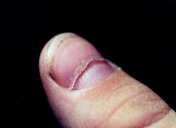
The periungual desquamation had started to heal when, 1 week later, all of the boy's fingernails and toenails spontaneously separated from the matrix (onychomadesis) (Fig. 1). Despite the generalized nature and severity of this nailbed damage, the proximal nails subsequently grew in normally, with minimal evidence of residual scarring.

Figure F1. Photo by: Courtesy: Allen R. Ciastko
We could find only one other report of such nail shedding secondary to Kawasaki disease.3 Milder variations of nail damage associated with Kawasaki disease have previously been reported in the form of Beau's lines (transverse grooves in the nails),4,5 pincer nails (transverse curling of the nail along its longitudinal axis)6 and leukonychia partialis (abnormally white proximal portion of nail).7 These nail abnormalities are nonspecific and, in the context of Kawasaki disease or other systemic triggers, generally resolve spontaneously within 1 to 2 months.
Allen R. Ciastko Pediatrician Kamloops, BC
References
- 1.Han RK, Sinclair B, Newman A, Silverman ED, Taylor GW, Walsh P, et al. Recognition and management of Kawasaki disease. CMAJ 2000;162(6):807-12. Available: www.cmaj.ca/cgi/content/full/162/6/807 [PMC free article] [PubMed]
- 2.Rowley AH, Shulman ST. Kawasaki syndrome. Pediatr Clin North Am 1999;46:313-29. [DOI] [PubMed]
- 3.Pilapil VR, Quizon DF. Nail shedding in Kawasaki syndrome [letter]. Am J Dis Child 1990; 144: 142-3. [DOI] [PubMed]
- 4.Bures FA. Beau's lines in mucocutaneous lymph node syndrome [letter]. Am J Dis Child 1981; 135: 383. [DOI] [PubMed]
- 5.Lindsley CB. Nail-bed lines in Kawasaki disease [letter]. Am J Dis Child 1992;146:659-60. [DOI] [PubMed]
- 6.Vanderhooft SL, Vanderhooft JE. Pincer nail deformity after Kawasaki's disease. J Am Acad Dermatol 1999;41:341-2. [DOI] [PubMed]
- 7.Iosub S, Gromisch DS. Leukonychia partialis in Kawasaki disease. J Infect Dis 1984;150:617-8. [DOI] [PMC free article] [PubMed]


