



Abstract
Due to protection of extracellular polymeric substances, the therapeutic efficiency of conventional antimicrobial agents is often impeded by their poor infiltration and accumulation in biofilm. Herein, one type of surface charge adaptable nitric oxide (NO) nanogenerator was developed for biofilm permeation, retention and eradication. This nanogenerator (PDG@Au–NO/PBAM) is composed of a core-shell structure: thermo-sensitive NO donor conjugated AuNPs on cationic poly(dopamine-co-glucosamine) nanoparticle (PDG@Au–NO) served as core, and anionic phenylboronic acid-acryloylmorpholine (PBAM) copolymer was employed as a shell. The NO nanogenerator featured long circulation and good biocompatibility. Once the nanogenerator reached acidic biofilm, its surface charge would be switched to positive after shell dissociation and cationic core exposure, which was conducive for the nanogenerator to infiltrate and accumulate in the depth of biofilm. In addition, the nanogenerator could sustainably generate NO to disturb the integrity of biofilm at physiological temperature, then generate hyperthermia and explosive NO release upon NIR irradiation to efficiently eradicate drug-resistant bacteria biofilm. Such rational design offers a promising approach for developing nanosystems against biofilm-associated infections.
Keywords: Antibacterial, Charge reversal, Photothermal therapy, Gasotransmitter, Biofilm microenvironment
Graphical abstract
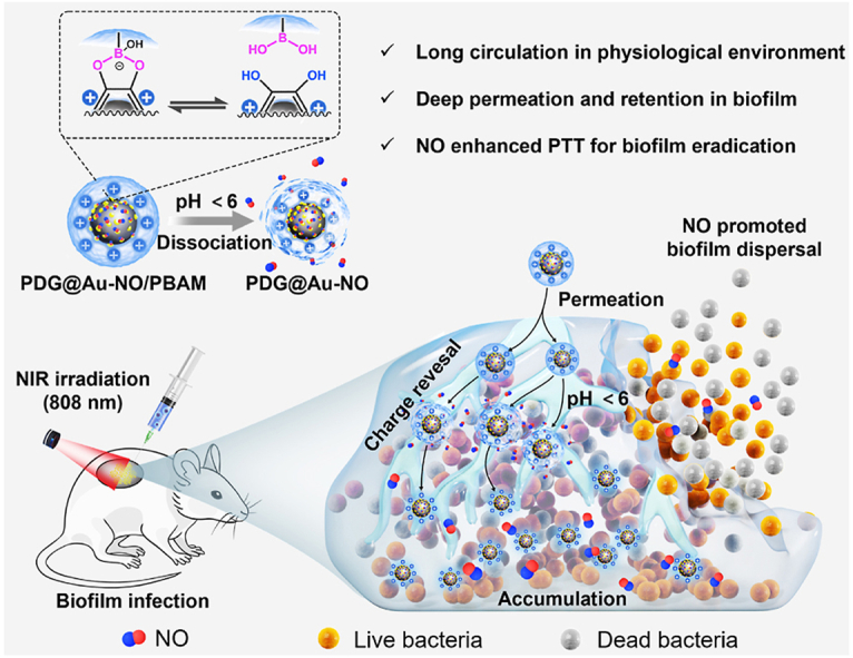
Highlights
-
•
The NO nanogenerator was designed for NO enhanced photothermal therapy to combat bacterial biofilm-associated infections.
-
•
The nanogenerator was negative charged in physiological condition but switched to positive in biofilm microenvironment.
-
•
The nanogenerator sustainably generated NO at physiological temperature and accelerated NO release under NIR irradiation.
1. Introduction
Infections induced by pathogenic bacteria are persistently imperiling human health throughout history [1]. The rapid emerging bacterial resistance arising from the intemperate and inappropriate use of antibiotics since mid-20th century is now entangling the world in a public health crisis [2,3]. In addition, bacteria always strive to settle and proliferate in territory suitable for themselves and form multicellular communities termed biofilm [4]. Bacteria that live in the biofilm are encapsulated by self-produced extracellular polymeric substances (EPS) which provides bacterial inhabitant with a fortress that not only prevents bacteria from being aggressed by host immune response but also defends permeation of antibacterial drugs [5,6]. Therefore, biofilm formation further exacerbates the expansion of bacteria resistance and instigates grievous and intractable infections [7,8]. The United States National Institutes of Health disclosed that biofilm is to be blamed for more than 60% of all nosocomial infections [6]. Due to the lack of valid treatments, biofilm-associated infections (BAI) have caused huge suffering and financial burden to patients.
As the conventional antibiotics shriveled in battling with BAI, alternative strategies attracted abundant research interest in the past decades, such as non-antibiotic chemotherapy by using cationic antimicrobial peptides and macromolecules [9], phototherapy by generating hyperthermia or reactive oxygen species [10,11], immunotherapy [12], and gas therapy [13,14], etc. Among these novel antibacterial technologies, light-driven phototherapy is a promising approach due to its spatial-temporal precision, low-invasive manipulation, broad-spectrum biocidal activities and scarce emergence of bacterial resistance [15,16]. Photothermal therapy (PTT) utilizing nanosized photothermal agents could convert photo energy to hyperthermia for physically denaturation of proteins, lipids and nucleic acids, which ultimately cause bacterial death [17,18]. However, conventional photothermal agents are usually hard to completely eradicate biofilm due to the low infiltration and accumulation in biofilm [17,19]. To obtain aspired curative efficiency, PTT usually requires prolonged hyperthermia under strong laser density, which will inevitably accompany by undesired harm to nearby tissue [19,20]. Hence, the integration of PTT with other therapeutic techniques is favored to promote anti-biofilm efficacy and diminish adverse effects simultaneously [11].
Nitric oxide (NO) as an important gasotransmitter that regulates multiple biofunctions in human body has also been engaged as a bactericidal and anti-biofilm agent recently [[21], [22], [23]]. The antibacterial abilities of NO are dose-dependent, which could either inactivate bacteria at relatively high concentrations (typically above 10−6 M) through nitrosative and oxidative stress to induce DNA damage, lipid peroxidation and membrane destruction [24,25], or enable depletion of exopolysaccharide in EPS and dispersion of biofilm at lower levels (10−12–10−9 M) by down-regulating biofilm-related second messengers [26,27]. Recently, several attempts have been exploited to combine NO therapy and PTT for the purpose of enhancing anti-biofilm efficacy. For example, photothermal agent (mesoporous polydopamine) conjugated with cationic NO donor (L-Arg) was fabricated by Yuan et al., which achieved enhanced eradication of Gram-positive bacteria biofilm [20]. Then, a kind of anionic NO-releasing photothermal graphene nanosheet was prepared for synergistically eradication of Gram-negative bacteria biofilm [28]. Recently, our group has developed a thermo-sensitive NO donor conjugated gold nanocage, which effectively eradicated methicillin-resistant Staphylococcus aureus (MRSA) biofilm both in vitro and in vivo [29]. Although such integrations of NO and PTT have yielded preliminary prospects in combating BAI, these reported nanosystems still have some restrictions in permeating the deep interior of biofilm [[30], [31], [32]].
The thick and dense EPS matrix is a protective barrier that prevents antimicrobial agents from diffusing into the interior of biofilm [5]. The surface of biofilm usually exhibits apparent negative charge due to the presence of anionic EPS components such as polysaccharides, proteins, nucleic acids, and lipids [5,33]. While the interior of biofilm could be more acidic owing to bacteria undergoing anaerobic glycolysis in biofilm [34]. Cationic materials are prone to target negatively charged biofilms due to their electrostatic affinity [35]. However, they always suffer from non-specific protein adsorption and are easily recognized then cleared by immune system [36]. Besides, anionic materials usually are more stable in the body and have longer circulation time, but the repulsion makes them tricky to enter and interact with the negatively charged biofilms [36]. Therefore, it is a great challenge for single-charged materials to achieve favorable permeation and accumulation in the deep interior of biofilm. In such case, surface charge adaptive materials that could maintain negative charged in normal physiological environment while switch to positive upon reaching biofilm for deep penetration and accumulation are desired [37,38].
In this work, we present a surface charge adaptable NO nanogenerator which could penetrate and accumulate in biofilm for NO enhanced PTT to efficiently eradicate BAI. As depicted in Fig. 1a, gold nanoparticles (AuNPs) decorated poly(dopamine-co-glucosamine) (PDG) hybrid nanoparticle which possessed positive surface charge was prepared as photothermal core (PDG@Au), thiol-functionalized thermo-sensitive NO donor was subsequently loaded via Au–S conjugation, and a stealth shell was further constructed through the grafting of phenylboronic acid and acryloylmorpholine (PBAM) copolymer. Such PDG@Au–NO/PBAM nanogenerator exhibits negative charged surface in normal physiological condition whereas exposes positively charged surface by the cleavage of pH-sensitive boronate ester bonds once it reaches the acidic microenvironment of biofilm. This surface charge adaptability was a prominent innovation in the PDG@Au–NO/PBAM nanogenerator, which ensured its sustainability and good biocompatibility, as well as infiltration and accumulation into the acidic deep interior of biofilm. In addition, after the PDG@Au-NO/PBAM nanogenerator infiltrating into the acidic deep interior of biofilm, it could slowly release NO to actively disrupt the protecting EPS matrix, thus making the biofilm bacteria more susceptible. And upon NIR irradiation, the PDG@Au–NO/PBAM nanogenerator could accelerate the NO release and cause local hyperthermia, achieving enhanced eradication of biofilm (Fig. 1b).
Fig. 1.

Schematic illustrations of surface charge adaptable NO nanogenerator for BAI treatment. a) Design and preparation of surface charge adaptable NO nanogenerator. b) Schematic illustration of NO nanogenerator with surface charge adaptability for NO enhanced PTT to eradicate BAI.
2. Results and discussion
2.1. Synthesis and characterization of PDG
Nano-sized polydopamine (PDA) nanoparticles in virtue of their good biocompatibility, high photothermal conversion efficiency, and exceptional tailorable surface properties have attracted increasing attention in the field of photothermal therapy [39]. At physiological pH, PDA normally exhibits negative charge due to the deprotonation of catechol groups on the surface, which is favorable for circulation in the body [40]. However, the electrostatic repulsion between PDA and negatively charged EPS matrix makes it difficult for PDA to infiltrate and accumulate into biofilm under physiological condition. Therefore, we expect to endow PDA nanoparticles with cationic charge to promote its affinity with biofilm. Versatile PDA nanomaterials could be constructed by the copolymerization of dopamine (DA) monomer with multi-type functional molecules during the self-oxidative polymerization process [[41], [42], [43]]. Herein, a precursor of cationic glucosamine, N-acetyl-d-glucosamine (NG), was chosen to copolymerized with DA to prepare poly(dopamine-co-N-acetyl-d-glucosamine) (PDNG) nanoparticles. Subsequently, the obtained PDNG nanoparticles were further deacetylated to fabricate cationic PDG nanoparticles (Fig. 2a), which were expected to promote adhesion and affinity with the negatively charged bacterial cell membrane and biofilm.
Fig. 2.

Synthesis and characterization of PDG. a) Schematic illustration for preparation of PDG. b) SEM and TEM images of PDA, PDNG and PDG. c) XPS O1s and d) N1s binding energy spectra of PDA and PDG nanoparticles. e) MALDI-TOF MS spectra of PDA and PDG nanoparticles. f) The non-covalent interaction of glucosamine moiety with DA monomer.
The molar ratios of DA:NG varied by 1:0, 1:1 and 1:2.5, the as prepared nanoparticles were denoted as PDA, PDNG1 and PDNG2.5, respectively. After deacetylation, the obtained cationic PDG nanoparticles with different feed ratios were named as PDG1 and PDG2.5, respectively. The scanning electron microscopy (SEM) and transmission electron microscope (TEM) were employed to observe the morphology of the prepared nanoparticles. As shown in Fig. 2b and Fig. S1, PDA, PDNG and PDG all showed similar uniform spherical shape with diameter around 150 nm, and no obvious difference was observed in morphology before and after deacetylation, suggesting that the glucosamine modification scarcely affected the intrinsic spherical morphology of PDA. Furthermore, hydrodynamic diameter and zeta potential of the aforementioned nanoparticles were determined using dynamic light scattering (DLS). As shown in Fig. S2a, the diameter of PDA was measured as 157 ± 3 nm. Compared with PDA, the diameter of PDNG showed a slight increase as the monomer molar ratio increased. And after deacetylation, the size of PDG1 and PDG2.5 was slightly reduced to around 145 nm. As presented in Fig. S2b, the zeta potential of PDA was determined as −5 mV at pH 7.4, and both PDNG1 and PDNG2.5 were detected as −11 mV. While after deacetylation, the zeta potentials of PDG1 and PDG2.5 were inverted positive to +21 mV and +25 mV, which were attributed to protonation of abundant amine groups of glucosamine on PDG surface. These above results suggested that the cationic glucosamine moiety was successfully doped in PDA nanoparticles. The PDG1 and PDG2.5 exhibited similar morphology. However, the zeta potential distribution results presented in Fig. S2b showed that PDG1 was positively charged with a narrow distribution. However, PDG2.5 also showed a certain distribution in the range of negative charge, which might be ascribed to that the acetyl groups in PDNG2.5 were not completely hydrolyzed in the deacetylation process. Thus, PDG1 was chosen to prepare PDG@Au–NO/PBAM nanogenerator in the subsequent experiments.
In addition, X-ray photoelectron spectroscopy (XPS) was also carried out. Compared with PDA, a new peak at 530.3 eV attributed to C–O–C species from the glucosamine segment was observed in the O1s spectra of PDG (Fig. 2c). The proportion of –NH2 (398.2 eV) increased in the N1s spectra may be ascribed to the abundant amine groups of glucosamine in PDG (Fig. 2d). These results further indicated the successful preparation of PDG. Matrix-assisted laser desorption/ionization-time of flight mass spectrometry (MALDI-TOF MS) was used to investigate the structure of PDG. As shown in Fig. 2e, the peaks of m/z 171 and m/z 269 found in PDA spectra were attributed to indoledione and dihydroxyindole/tris(hydroxymethyl)methyl aminomethane (Tris, from the buffer solution) complex, respectively [40,44]. For PDG, new MS fragments around m/z 180 and m/z 301 were detected which could be attributed to protonated glucosamine and glucosamine/Tris complex. This result unambiguously confirmed the existence of glucosamine segments in PDG nanoparticles and suggested that the glucosamine moiety potentially interacted with DA monomer via weak non-covalent interaction such as hydrogen bonding or van der Waals attraction rather than covalently conjugation (Fig. 2f) [45]. Furthermore, we explored whether it was possible to prepare PDG through a one-step strategy by direct oxidative copolymerization of DA with glucosamine or its polymeric derivative (chitosan oligosaccharide) to improve the reaction efficiency. However, the morphology study of those resulting nanomaterials by SEM exhibited obvious aggregation, which might be ascribed to the crosslinking of amino groups in glucosamine with quinone groups in PDA through Michael addition and/or Schiff base reaction (Fig. S3). The above result justified that this two-step acetyl-protection/deprotection strategy is reasonable and reliable to prepare cationic poly(dopamine-co-glucosamine) nanoparticles.
2.2. Synthesis and characterization of PDG@Au–NO/PBAM nanogenerator
A thiolated N-nitrosamines NO donor (cupferron) has been constructed in our previous work, which could be conveniently tethered to gold surface via Au–S conjugation for thermo-responsive NO release [29]. It was reported that AuNPs with the size below 20 nm exhibit reduced uptake by the liver and could be easily excreted from the body [46]. Therefore, we aimed to generate AuNPs on the surface of PDG in situ to construct PDG@Au hybrid that could load thiolated cupferron (TCup). The chloroauric acid (HAuCl4) with a series of concentrations (0.125, 0.25, 0.75, 1.25 mM) was in-situ reduced to AuNPs utilizing the reducibility of the catechol groups on PDG (Fig. 1a). SEM and TEM observation confirmed that many AuNPs distributed on the surface of PDG (Fig. 3a and Fig. S4). The sizes of AuNPs on PDG@Au0.125 and PDG@Au0.25 were both below 20 nm, whereas the quantity of AuNPs on PDG@Au0.125 was less than that on PDG@Au0.25. And unfortunately, obvious aggregation and oversized AuNPs were found on both PDG@Au0.75 and PDG@Au1.25. Meanwhile, inductively coupled plasma source mass spectrometer (ICP-MS) manifested that PDG@Au0.25 had the highest Au content among all four kinds of nanocomposites (Fig. S5). Thus, PDG@Au0.25 was selected for the loading of NO donors. The PDG@Au–NO was prepared by mixed PDG@Au with TCup in the dark. The photothermal performance of PDG@Au nanocomposite and thermo-induced NO release property of resulted PDG@Au–NO will be discussed later in this work.
Fig. 3.

Synthesis and characterization of PDG@Au–NO/PBAM nanogenerator. a) SEM and TEM images of PDG@Au nanocomposite. b) The average hydrodynamic sizes of PDG@Au and PDG@Au/PBAM at pH 7.4 and 5.5. c) The zeta potential of PDG@Au/PBAM at pH 7.4 and 5.5. d) UV–Vis spectra of PDG@Au and PDG@Au/PBAM. e) The heating-cooling curve of PDG@Au with varying concentrations under NIR light irradiation (808 nm 1.0 W cm−2). f) The on/off generation of NO from PDG@Au–NO/PBAM under NIR light irradiation (808 nm, 1 W cm−2). g) The relatively long-term NO generation profiles of PDG@Au–NO and PDG@Au–NO/PBAM.
Thereafter, a pH-responsive stealthy shell was constructed for PDG@Au–NO to temporarily mask its positive surface charge (Fig. 1a). Hydrophilic acryloylmorpholine (AM) has been typically employed as blocks to construct stealth surface for nanomaterials, which could resist protein non-specific adsorption, reduce immune recognition, improve their biocompatibility and circulation time in the body [36]. Morpholine was negatively charged in the physiological environment due to oxygen's negative inductive effect as well as nitrogen's relatively lower basicity [47]. Phenylboronic acids could specifically bind to diols (such as catechol groups) to form pH-sensitive boronate ester bonds [48,49]. Herein, N-3-acrylamidophenylboronic acid and AM were polymerized together to produce a hydrophilic PBAM copolymer. The successful synthesis of PBAM was confirmed by proton nuclear magnetic resonance (1H NMR in Fig. S6). The designed NO nanogenerator (PDG@Au–NO/PBAM) was prepared by encapsulating PDG@Au–NO with PBAM copolymer through the formation of boronate ester bonds between boronic acid groups on the copolymer and catechol moieties on PDG. SEM (Fig. S7) and TEM (Fig. S8) observation confirmed that after decoration of PBAM copolymer the nanocomposites still maintained the original spherical structure.
2.3. pH-sensitive surface charge adaptability
The PDG@Au/PBAM nanocomposite without loading of NO was employed to verify the pH-sensitive surface charge switchability. The size distribution and zeta potential of PDG@Au and PDG@Au/PBAM were measured by DLS at pH 7.4 and 5.5. As shown in Fig. 3b, the hydrodynamic size of PDG@Au/PBAM (287 nm ± 5 nm) was found to be much larger than that of PDG@Au (220 nm ± 6 nm) at physiological pH, which confirmed the successful decoration of PBAM on the nanocomposites. However, the hydrodynamic size of PDG@Au/PBAM reduced to approximately 230 nm at pH 5.5 which was similar to that of PDG@Au suggesting that PBAM copolymer dissociated. Furthermore, this dissociation of PBAM copolymer under acidic condition was also manifested by change of zeta potential result. As presented in Fig. 3c, the zeta potential of PDG@Au/PBAM was negative (−28 mV) at physiological pH, whereas switched to positive (+19 mV) when the pH dropped from 7.4 to 5.5. This result was attributed to the dissociation of stealthy polymeric coating by the cleavage of the boronate ester bonds under acidic condition and the exposure of cationic PDG@Au core [50]. The reduced particle size and switched zeta potential implied the pH-sensitive surface charge adaptability of PDG@Au/PBAM. In addition, the UV–Vis adsorption spectra of PDG@Au and PDG@Au/PBAM were determined. As presented in Fig. 3d, no obvious UV–Vis absorption spectral change between PDG@Au and PDG@Au/PBAM was observed, verifying that the stealthy shell structure barely obstructed light absorption property of PDG@Au.
2.4. Thermo-responsive NO generation
Firstly, the photothermal conversion performance of PDG@Au was investigated. The temperature changes of PDG@Au suspension in phosphate buffer solution (PBS) with varying concentrations under NIR irradiation (808 nm 1.0 W cm−2) were depicted in Fig. 3e. The temperature of PDG@Au suspension was positively correlated with its concentration and laser irradiation time. Especially, the temperature of PDG@Au suspension at concentration of 150 μg mL−1 could increase to 54 °C after 10 min of NIR irradiation. When the concentration of PDG@Au suspension increased to 300 μg mL−1, the maximum temperature could reach approximately 70 °C after 10 min irradiation suggesting excellent photothermal conversion efficiency of PDG@Au. It has been reported that temperature above 50 °C could cause bacteria death though inducing physical destruction of bacteria cells [51]. The higher temperature could definitely enhance the photothermal antimicrobial efficiency, but concurrently generate higher toxicity to surrounding healthy tissue [17]. Thus, 150 μg mL−1 was chosen as the appropriate concentration of PDG@Au for subsequent PTT evaluation.
Then, NO generation capability of our nanogenerator was determined by Griess assay [52]. As shown in Fig. 3f, the amount of NO generated by PDG@Au–NO/PBAM increased rapidly upon NIR irradiation, demonstrating the effective photothermal activation of TCup. When the NIR was turned off, the NO generation amount was retarded and the release profile was almost horizontal. The nanogenerator could rapidly release NO once irradiated with NIR light. These results evidently confirmed the NIR-responsive NO generation capability of the PDG@Au–NO/PBAM. In addition, from the relatively long-term NO generation profile presented in Fig. 3g, the PDG@Au–NO/PBAM could slowly generate NO for more than 48 h at physiological temperature. And without polymeric coating decoration, PDG@Au–NO could generate higher amounts of NO. The maximum NO release amount of PDG@Au–NO and PDG@Au–NO/PBAM at physiological temperature were 4.82 μM g−1 and 3.40 μM g−1, respectively. When exposed to 10 min NIR irradiation, the burst release of NO was achieved, and the maximum NO release amount of PDG@Au–NO and PDG@Au–NO/PBAM could reach 8.67 μM g−1 and 6.76 μM g−1, respectively. Notably, the NO release amount in PDG@Au–NO/PBAM group was lower than that in PDG@Au–NO group either irradiated with NIR light or not. This lagged NO release profile of PDG@Au–NO/PBAM may owe to the shielding effect of its polymeric shell coating. Above results indicated that the nanogenerator could slowly generate relatively low dosage NO at physiological temperature, and burst release of high dosage NO under NIR irradiation.
2.5. Hemocompatibility and cytocompatibility evaluation
To evaluate the potential toxicity of the prepared PDG@Au–NO/PBAM nanogenerator, hemolysis and cytotoxicity assays were performed in vitro. First, PDG@Au–NO/PBAM nanogenerator and its precursors were incubated with rabbit red blood cells to assess their hemocompatibility. As presented in Fig. 4a, all the tested samples maintained a low hemolytic level below 7% in the concentration range from 37.5 to 600 μg mL−1. The PDG@Au–NO nanocomposite displayed the most obvious concentration-dependent performance and showed the highest hemolysis rates of 5.5% and 6.2% at concentration of 300 and 600 μg mL−1, respectively. In sharp contrast, the hemolysis rates of PDG@Au–NO/PBAM kept below 1% even at the concentration of 300 and 600 μg mL−1, which was substantially lower than that of PDG@Au–NO. This phenomenon was attributed to the physical barrier formed by the morpholine containing PBAM copolymer shell, which improved the compatibility of PDG@Au–NO/PBAM nanogenerator with erythrocytes [53].
Fig. 4.

In vitro hemocompatibility towards rabbit red blood cells and cytocompatibility towards mouse embryonic fibroblast NIH3T3 cells. a) Hemolysis rates of different nanocomposites and 0.1% of Triton X-100 (positive control). b) Cell viability of NIH3T3 fibroblasts at different concentrations of nanocomposites.
Moreover, the in vitro cytotoxicity of PDG@Au–NO/PBAM nanogenerator with mouse embryonic fibroblast cells (NIH3T3) was investigated by alamarBlue assays. As depicted in Fig. 4b, at concentration range of 37.5–150 μg mL−1, the cell viability in PDG@Au–NO/PBAM group was higher than that in PDG@Au/PBAM group with or without 10 min NIR irradiation, indicating that the NO generated by the PDG@Au–NO/PBAM nanogenerator in this concentration range could promote cell survival and proliferation through protection against apoptosis [54]. However, as the concentration rose to 300 and 600 μg mL−1, the cell viability of PDG@Au–NO/PBAM group was relatively lower than PDG@Au/PBAM group, which might be ascribed to the higher concentration of generated NO caused cell death through pro-apoptotic responses and cell cycle arrest [54]. Meanwhile, without NIR irradiation, PDG@Au/PBAM and PDG@Au–NO/PBAM at concentration of 150 μg mL−1 exhibited cell viabilities of 91% ± 11% and 95% ± 7%, respectively, indicating acceptable cytocompatibility. Once irradiated by NIR light the cell viability of PDG@Au/PBAM + NIR group reduced to 46% ± 1%, suggesting apparent toxicity. But its apparent cytotoxicity was significantly counteracted by NO, the PDG@Au–NO/PBAM + NIR group maintaining 70% ± 8% of cell viability. Collectively, these hemolysis and cell viability results confirmed the desired biocompatibility of the prepared PDG@Au–NO/PBAM nanogenerator with concentration of 37.5 μg mL−1 to 150 μg mL−1. However, PDG@Au–NO/PBAM with concentration of 300 and 600 μg mL−1 was toxic to fibroblast cells. Thus, 150 μg mL−1 of PDG@Au–NO/PBAM nanogenerator and its precursors were chosen in subsequent bactericidal experiments.
2.6. In vitro antibacterial effect against planktonic bacteria
Two typical drug-resistant bacteria, MRSA (Gram-positive) and tetracycline-resistant Escherichia coli (TREC, Gram-negative) were chosen to evaluate the in vitro NO enhanced PTT bactericidal efficacy of NO nanogenerator. The PDG@Au–NO/PBAM nanogenerator and its precursors at 150 μg mL−1 were incubated with planktonic MRSA or TREC (108 CFU mL−1) for 4 h at 37 °C and further irradiated with or without NIR for 10 min, then the corresponding bactericidal rates were assessed by colony forming unit (CFU) counting. As shown in Fig. 5a, without NIR irradiation, PDG and PDG@Au already exhibited a certain degree of antibacterial effect against MRSA, with log reductions of 1.26 ± 0.20 (96.30 ± 1.48% killing) and of 1.27 ± 0.23 (96.37 ± 1.60% killing), respectively. However, after loading of NO donors, the antibacterial rates of PDG@Au–NO and PDG@Au–NO/PBAM were not improved but rather slightly decreased, with log reductions of 0.89 ± 0.34 (90.54 ± 5.34% killing) and of 0.95 ± 0.15 (92.74 ± 2.48% killing), respectively. These results should be ascribed to that PDG@Au–NO and PDG@Au–NO/PBAM could only produce low level NO in the absence of NIR irradiation, which had limited bacterial-killing effect [13]. Unlike gram-positive bacteria, PDG and PDG@Au exhibited little inherent bactericidal effect against TREC in the absence of NIR irradiation, with log reductions of 0.49 ± 0.30 (64.74 ± 18.84% killing) and of 0.48 ± 0.36 (62.11 ± 25.26% killing) (Fig. 5b). Similarly, the antibacterial activities of PDG@Au–NO and PDG@Au–NO/PBAM against TREC without irradiation also were not enhanced, with log reductions of 0.62 ± 0.52 (67.58 ± 23.63% killing) and of 0.27 ± 0.14 (47.37 ± 17.96% killing), respectively. This inherent bactericidal efficacy of PDG and PDG@Au against planktonic MRSA should be ascribed to their cationic surface charge that is capable of disrupting the integrity of anionic cell membrane of bacteria and leading to apoptosis [55]. In addition, the Gram-positive bacterial cell wall is composed of thick peptidoglycan. The carbohydrate-carbohydrate interaction between the peptidoglycan layer existing in MRSA surface and glucosamine decorated on PDG further enhanced the disruption [56]. On the contrary, PDG and PDG@Au showed lower killing efficiency against TREC because the Gram-negative bacterial cell wall has a single thin peptidoglycan layer and its peptidoglycan layer is surrounded by another outer membrane [57].
Fig. 5.

In vitro antibacterial activity of PDG@Au–NO/PBAM nanogenerator. The log reduction of a) MRSA and b) TREC after treated by different nanocomposites with or without 10 min NIR light irradiation (808 nm, 1.0 W cm−2). c) Photographs of surviving bacterial colonies on agar plates under different treatments. d) SEM morphologies of MRSA and TREC under different treatments. *p < 0.05, **p < 0.01, ***p < 0.001, ****p < 0.0001, and not significant (NS).
In sharp contrast, once the systems were irradiated by NIR for 10 min, the bactericidal rates of PDG@Au–NO/PBAM nanogenerator and its precursors increased significantly (Fig. 5a). In detail, the bacterial log reductions of PDG + NIR and PDG@Au + NIR against MRSA increased to 3.14 ± 0.08 (>99.9% killing) and 2.73 ± 0.18 (>99% killing), respectively. This enhanced bactericidal activity was attributed to local hyperthermia generated by PDG and PDG@Au under NIR irradiation. Notably, PDG@Au–NO + NIR and PDG@Au–NO/PBAM + NIR groups exhibited further enhanced antimicrobial efficacy for MRSA, with log reductions of 4.67 ± 0.36 (>99.99% killing) and of 4.22 ± 0.06 (>99.99% killing), respectively. Likewise, 10 min NIR irradiation also greatly amplified the antimicrobial effect of the NO nanogenerator against TREC (Fig. 5b). The log reductions of TREC under treatments of PDG + NIR, PDG@Au + NIR, PDG@Au–NO + NIR and PDG@Au–NO/PBAM + NIR were 1.65 ± 0.06 (>98% killing), 2.02 ± 0.50 (>99% killing), 3.81 ± 0.21 and 3.97 ± 0.06 (>99.9% killing), respectively. Their antibacterial performance could also be clearly observed from survival MRSA and TREC colonies on agar plates, where the colonies in PDG@Au–NO + NIR and PDG@Au–NO/PBAM + NIR group were greatly reduced (Fig. 5c). These results demonstrated the excellent NO enhanced PTT bactericidal effect of PDG@Au–NO + NIR and PDG@Au–NO/PBAM + NIR against planktonic drug-resistant bacteria.
The morphologies of MRSA and TREC before and after treatments of different nanocomposites were observed by SEM. As presented in Fig. 5d, all MRSA and TREC cells exhibited smooth surface and intact cell membranes in control group. Contrastively, without NIR irradiation, minor impact on bacterial cell membrane integrity was observed in PDG@Au, PDG@Au–NO and PDG@Au–NO/PBAM treated groups. However, after treated by PDG@Au together with 10 min NIR irradiation, it resulted in obvious impact on bacterial cell morphologies, where both MRSA and TREC exhibited collapse of membranes. Prominently, after treatment with PDG@Au–NO + NIR and PDG@Au–NO/PBAM + NIR, both two bacteria displayed severe cell membranes damage. Specifically, most MRSA cells were seriously shriveled and collapsed, and TREC cells were also seriously shrank and destructed (as indicated by the red arrows). In addition, abundant nanocomposites attached were observed on the bacterial surface, especially for MRSA. It indicated that our prepared nanocomposites had higher affinity with Gram-positive MRSA and therefore, their killing abilities for MRSA were much more potent than that of TREC. This trend was consistent with the aforementioned antimicrobial testing.
2.7. In vitro anti-biofilm effect
After verifying the excellent antibacterial properties against planktonic bacteria, the anti-biofilm effect of NO nanogenerator was explored. The PDG@Au–NO/PBAM NO nanogenerator (150 μg mL−1) was firstly co-incubated with MRSA biofilm at 37 °C for different times and then irradiated with an 808 nm laser at a power density of 1.0 W cm−2 for 10 min to explore the effect of co-incubation time on anti-biofilm efficiency. As shown in Fig. S9, when the co-incubation time of PDG@Au–NO/PBAM nanogenerator and MRSA biofilm increased from 0 to 4 h, the survival rate of MRSA in biofilm significantly reduced. Then with co-incubation time continued increasing, the number of survival bacteria in biofilm was not significantly decreased. This result indicated that the PDG@Au–NO/PBAM nanogenerator generated enough NO to disperse biofilm when it co-incubated with MRSA biofilm at 37 °C for 4 h. Therefore, in the anti-biofilm experiment, the MRSA and TREC biofilms were firstly co-incubated with PDG@Au–NO/PBAM nanogenerator and its different precursors at 37 °C for 4 h.
The PDG@Au–NO and PDG@Au–NO/PBAM with concentrations from 0 to 600 μg mL−1 were incubated with MRSA or TREC biofilms at 37 °C for 4 h, respectively, then the residual biofilms mass in different treatment groups were stained by crystal violet staining assay. The amounts of extracellular polysaccharides existed in biofilm could be stained purple with the crystal violet dye [58]. As shown in Fig. S10, the biofilm dissipation effect was positively correlated with the concentration of PDG@Au–NO and PDG@Au–NO/PBAM. With the concentration of PDG@Au–NO and PDG@Au–NO/PBAM increasing the biofilm dissipation rate gradually rose, indicating that the biofilm dissipation capacity of the NO nanogenerator was NO concentration-dependent. Then 150 μg mL−1 of PDG@Au–NO/PBAM nanogenerator and its precursors were co-incubated with MRSA or TREC biofilms to investigate the biofilm dissipation performance. As shown in Fig. 6a–b, MRSA and TREC biofilms in PDG, PDG@Au and PDG@Au/PBAM groups were relatively intact in comparison with PBS-treated control groups, indicating almost no biofilm dispersal effect. However, the biofilms biomass in PDG@Au–NO and PDG@Au–NO/PBAM treated groups were significantly decreased. According to the afore-determined NO release profiles, these two nanogenerators continuously release low level NO at 37 °C, which could effectively promote biofilm dispersal. In addition, it is worth noting that PDG@Au–NO/PBAM group was better than PDG@Au–NO group for the eradication of both MRSA and TREC biofilms. The corresponding quantitative result also confirmed that the biofilm dispersal rates of PDG@Au–NO/PBAM (67.7% ± 5.4% for MRSA and 70.3% ± 5.1% for TREC) were higher than those of PDG@Au–NO (46.5% ± 7.6% for MRSA and 65.0% ± 8.5% for TREC). This result could be ascribed to PDG@Au–NO/PBAM nanogenerator with surface charge adaptability could permeate into deep interior of biofilm and generate NO to dissipate the biofilm.
Fig. 6.

In vitro biofilm dispersal ability of PDG@Au–NO/PBAM nanogenerator. Quantitative calculation of biofilm biomass of a) MRSA and b) TREC by the crystal violet staining assay. c) SEM images of MRSA and TREC biofilms before and after treatment by different nanocomposites. d) Schematic illustrations of biofilm dispersal by PDG@Au–NO/PBAM nanogenerator. *p < 0.05, **p < 0.01, ***p < 0.001, ****p < 0.0001, and not significant (NS).
The PDG@Au–NO/PBAM nanogenerator induced infiltration and disruption of MRSA and TREC biofilms were further observed by SEM. As shown in Fig. 6c, biofilms with densely stacked bacteria were successfully formed (control). After being treated with 150 μg mL−1 of PDG, PDG@Au and PDG@Au/PBAM, there were massive nanocomposites stuck on the surface of the biofilm bacteria (marked with red circles and arrows). And PDG@Au/PBAM nanocomposites were even distributed in the crevices and water channels of biofilm. Although PDG@Au/PBAM was negatively charged, its PBAM shell would dissociate in response to the acidic biofilm environment allowing the exposure of positively charged PDG@Au core to interact with negatively charged biofilm through electrostatic affinity (Fig. 6d (i)). The dissociation of this stealthy polymeric coating and charge conversion under acidic condition had been confirmed by the afore-discussed DLS and zeta potential analysis (Fig. 3b–c). After further loading of NO, only a few bacteria were left and the integrity of biofilms was considerably disrupted when treated by PDG@Au–NO/PBAM nanogenerator. Such sharp contrast should be ascribed to the biofilms dispersal ability of NO which is capable of down-regulating the exopolysaccharide production second messengers such as cyclic-di-guanosine monophosphate (c-di-GMP), thus breaking the integrity of EPS matrix (Fig. 6d (ii)) [31]. The SEM observation was consistent with the crystal violet staining study that verified PDG@Au–NO/PBAM nanogenerator holds significant biofilm dispersal effect.
After the validation of its biofilm dispersal ability without NIR irradiation, the bactericidal efficacy of PDG@Au–NO/PBAM nanogenerator based on NO enhanced PTT was further studied. After co-incubation of PDG@Au–NO/PBAM nanogenerator and its precursors with biofilms for 4 h plus 10 min NIR irradiation (808 nm 1.0 W cm−2), the survival bacteria in biofilm were counted by colony counting. Different from the bactericidal results against planktonic bacteria, PDG and PDG@Au showed negligible killing abilities against bacteria in two biofilms without NIR irradiation (Fig. 7a–b). The PDG@Au–NO and PDG@Au–NO/PBAM without the NIR irradiation groups also exhibited neglectable bactericidal efficiency for two biofilms. Considering crystal violet and colony counting results together, it could be deduced that low level NO generation lead to dispersal of biofilm rather than death of bacteria [13]. However, the bactericidal abilities of PDG + NIR and PDG@Au + NIR for MRSA biofilm were greatly improved, with log reduction of 3.38 ± 0.13 and 3.73 ± 0.18 (>99.9% killing), respectively (Fig. 7a). Such results clearly indicated the local hyperthermia generated by PDG and PDG@Au in the presence of NIR irradiation enabled to cause abundant death of bacteria in biofilm. Moreover, the addition of NO further promoted the bactericidal efficacy, and the viable MRSA under PDG@Au–NO + NIR treatment was reduced by 5.70 ± 0.15 logarithms (>99.999% killing). More interestingly, PDG@Au–NO/PBAM + NIR showed the strongest antibacterial efficiency for MRSA biofilm, with log reduction of 6.61 ± 0.42 (>99.9999% killing), which was superior to any of its precursors. Such outstanding biofilm eradication effect could be attributed to the surface charge adaptive shell of the PDG@Au–NO/PBAM nanogenerator promoted infiltration into biofilm, and then the locally released NO dissociated the EPS matrix and enhanced PTT damage to bacteria. For TREC biofilm, PDG@Au–NO/PBAM nanogenerator and its precursors exhibited similar trend (Fig. 7b). Among all tested groups, PDG@Au–NO/PBAM + NIR also showed the most potent bactericidal ability against TREC biofilm, with log reduction of 2.20 ± 0.29 (>99% killing).
Fig. 7.

In vitro anti-biofilm ability of PDG@Au–NO/PBAM nanogenerator. The log reduction of MRSA biofilm a) and TREC biofilm b) after treated by different nanocomposites with or without 10 min NIR light irradiation (808 nm, 1.0 W cm−2). c) 3D confocal images of fluorescent stained MRSA and TREC biofilms under different treatments. *p < 0.05, **p < 0.01, ***p < 0.001, ****p < 0.0001, and not significant (NS).
In addition, confocal laser scanning microscopy (CLSM) was carried out to further investigate the anti-biofilm effect of NO nanogenerator. As presented in Fig. 7c, when MRSA and TREC biofilms were treated by PBS, PDG@Au/PBAM and PDG@Au–NO/PBAM in the absence of NIR irradiation, bright green fluorescence was observed, suggesting most bacteria were alive. But the biofilm density and thickness (shown in Fig. S11) in PDG@Au–NO/PBAM treated groups decreased significantly compared with that in PBS and PDG@Au/PBAM groups, implying that low level NO generation effectively disrupted the biofilm. This phenomenon agreed with crystal violet and SEM observations, confirming PDG@Au–NO/PBAM could effectively dissipate biofilm biomass in the absence of NIR irradiation. In PDG@Au/PBAM + NIR group without NO released group, the red fluorescence signals of dead bacteria increased, but green fluorescence signals of survival bacteria were still obviously observed. And the biofilm thickness in PDG@Au/PBAM + NIR treatment group kept similar to that in PBS group (Fig. S11). This result suggested 10 min NIR irradiation alone was not strong enough to thoroughly eradicate biofilm. Conspicuously, after PDG@Au–NO/PBAM + NIR treatment, almost all the MRSA and the majority of TREC were stained red, and the biofilms density and thickness observably decreased, demonstrating the potent biofilm eradication efficacy of NO enhanced PTT.
2.8. In vivo anti-biofilm activity
Encouraged by the outstanding in vitro anti-biofilm efficiency of PDG@Au–NO/PBAM nanogenerator towards MRSA, its in vivo anti-biofilm performance was further investigated. In clinical, biofilm infections on implantable medical devices was high-risk and obstinate. Therefore, we performed a subcutaneous implant-associated biofilm infection model to investigate in vivo anti-biofilm performance of PDG@Au–NO/PBAM nanogenerator (Fig. 8a). The polydimethylsiloxane (PDMS) slices with MRSA biofilm were subcutaneously implanted into the back of the mice for 24 h to establish local infection, and slices without biofilm were also implanted to set as the negative control. Then PBS, PDG@Au–NO/PBAM nanogenerator and its different precursors as well as vancomycin were correspondingly administrated by subcutaneous injection. Subsequent 10 min of 808 nm NIR irradiation (1.0 W cm−2) was carried out 4 h after injection. As shown in Fig. 8b, 3 days after administration, obvious suppuration was observed near the implanted site in PBS, PBS + NIR, PDG@Au/PBAM and PDG@Au–NO/PBAM groups, implying severe infection. Comparatively, the suppuration alleviated slightly in PDG@Au/PBAM + NIR and vancomycin treated groups, proving that PTT or antibiotic therapy alone showed a certain degree of therapeutic effect. Strikingly, after being treated by the PDG@Au–NO/PBAM + NIR, the suppuration was significantly diminished, which kept a similar phenomenon with the negative uninfected control group.
Fig. 8.

In vitro anti-biofilm efficiency of PDG@Au–NO/PBAM nanogenerator in a subcutaneous implant-associated biofilm infection model. a) Schematic illustration of subcutaneous implant-associated biofilm infection model. b) Representative photographs of the implanted site every 12 h. Quantitative evaluation of bacterial counts on the c) implanted PDMS and d) tissue after different treatments. e) H&E staining of the vicinity tissues on the implanted site. f) The pro-inflammatory cytokines (IL-6 and TNF-α) analysis by immunohistochemistry. *p < 0.05, **p < 0.01, ***p < 0.001, ****p < 0.0001 and not significant (NS).
Moreover, the implants and their surrounding tissue were collected at day 3 post administration, and their infection states were quantitatively assessed. As shown in Fig. 8c–d, abundant survival bacteria existed in PBS (log10 (CFU mL−1) 6.89 ± 1.18 on implants and 7.00 ± 0.45 in tissue) and PBS + NIR treated wounds (7.10 ± 0.55 on implants and 6.55 ± 0.44 in tissue), which were much higher than non-infected negative control group (1.92 ± 0.45 on implants and 3.07 ± 0.40 on tissues), affirmed the successful generation of infection. The administration of PDG@Au/PBAM did not obviously reduce the number of colonies on implants or tissue, where the survival bacteria numbers (log10 (CFU mL−1) 6.12 ± 0.51 on implants and 6.18 ± 0.72 on tissues) showed no significant difference in comparison with PBS treated group. However, with the irradiation of NIR, the antibacterial efficacy of PDG@Au/PBAM + NIR was greatly enhanced, the survival bacteria number reduced to the log10 (CFU mL−1) 3.63 ± 0.64 (implant) and 4.89 ± 0.57 (tissue). With the loading of NO, the bacteria number on the implants was drastically reduced to log10 (CFU mL−1) 3.10 ± 0.99 under PDG@Au–NO/PBAM treatment without NIR, but large number of survival bacteria were also counted in the surrounding tissue (6.84 ± 0.30) which is close to the infected control group. Such result should attribute to that low concentration NO released from PDG@Au–NO/PBAM could only disperse the implant surface attached biofilm, but was unable to cause death of bacteria in the absence of NIR irradiation, resulting in abundant survival bacteria existing in surrounding tissue. However, when supplemented with NIR irradiation, the number of bacteria on the tissues was obviously reduced to log10 (CFU mL−1) 4.27 ± 0.76. Specifically, the bactericidal rates of PDG@Au–NO/PBAM + NIR group on implants and surrounding tissues were 99.99 ± 0.01% and 99.18 ± 0.08%, respectively. Conversely, for the vancomycin (one last-line antibiotic) treated group, although the counts of bacteria in surrounding tissue were greatly reduced to the level of non-infected negative control group (log10 (CFU mL−1) 3.30 ± 0.43), its survival bacteria number on implants (4.27 ± 0.36) was 13.8 times higher than PDG@Au–NO/PBAM + NIR group (3.13 ± 0.52). Such result indicates that vancomycin could only kill planktonic bacteria in the biofilm surrounding tissue, but was inefficient to kill biofilm encapsulated bacteria owing to inefficient biofilms dispersion of antibiotics. Overall, these results illustrated the PDG@Au–NO/PBAM + NIR exhibited a potent biofilm eradication effect in vivo through NO enhanced PTT.
Furthermore, the histological analysis of surrounding tissue was performed by Hematoxylin and Eosin (H&E) staining. As shown in Fig. 8e, compared with non-infected negative control group, the surrounding tissue in PBS and PBS + NIR treated groups displayed pronounced infiltration of neutrophils (purple spots), indicating an obvious inflammation state under bacterial infection [17]. Comparatively, although the number of neutrophils decreased in PDG@Au/PBAM, PDG@Au/PBAM + NIR and PDG@Au–NO/PBAM treated groups, they are still higher than that in the non-infected negative control group, indicating slightly alleviated inflammation. In addition, vancomycin treated group displayed relatively high number of neutrophils although its survival bacterial number already dropped to a level close to the negative control. It might ascribe to the ineffectiveness of vancomycin in dealing with implant attached biofilms, and the abundant survival bacteria on the implant still cause relatively high inflammation state in surrounding tissue [59]. Among all bacteria infected groups, the NO enhanced PTT by PDG@Au–NO/PBAM + NIR exhibited minimal inflammatory infiltrates similar to the non-infected negative control group. To further demonstrate the inflammation conditions after different treatments, the expression of pro-inflammatory cytokines (IL-6 and TNF-α) was analyzed by immunohistochemistry. As shown in Fig. 8f, the amount of pro-inflammatory cytokines (dyed brown) kept at a high level for the surrounding tissue of MRSA infected implant in PBS ± NIR, PDG@Au/PBAM ± NIR, PDG@Au–NO/PBAM, and vancomycin treatment groups. However, the MRSA-infected implant surrounding tissue in PDG@Au–NO/PBAM + NIR treated group showed a low expression of IL-6 and TNF-α as the non-infected negative control. The expression results of IL-6 and TNF-α were consistent with the H&E staining study, which verified PDG@Au–NO/PBAM nanogenerator under NIR light irradiation exhibited excellent therapeutic effect against subcutaneous implant-associated MRSA biofilm infection. This remarkable anti-biofilm performance of PDG@Au–NO/PBAM nanogenerator should be attributed to its self-activating cascade processes involving long circulation in physiological environment, charge adaptability enabled deep permeation and accumulation in biofilm, and NO enhanced PTT treatment under NIR irradiation. The rationally designed nanogenerator could cross the protective EPS barrier of biofilm that conventional antibiotics cannot overcome.
3. Conclusions
In summary, one type of surface charge adaptable NO nanogenerator that could simultaneously generate NO for enhanced PTT was successfully fabricated. This NO nanogenerator exhibited negative charged in physiological environment which ensured its in vivo sustainability and biocompatibility. Whereas the surface charge of PDG@Au–NO/PBAM nanogenerator could be switched to positive in acidic biofilm microenvironment on account of the cleavage of boronate ester bonds between the PDG@Au–NO core and PBAM shell, which promoted deep permeation and accumulation of the nanogenerator into biofilm. Afterwards, the decorticated PDG@Au–NO could continuously and spontaneously generated NO at physiological temperature to dissipate biofilm and make the bacteria in a more sensitive state. Upon NIR irradiation, the locally generated hyperthermia and burst release of NO cooperatively exacerbated collapse of biofilm and the death of bacteria. Such PDG@Au–NO/PBAM nanogenerator exhibited potent biofilm eradication efficacy towards drug-resistant bacteria MRSA both in vitro and in vivo, even surpassed vancomycin, one of the last-line antibiotics, in a subcutaneous implant-associated biofilm infection model. Moreover, this NO enhanced PTT strategy not only enabled to efficiently kill drug-resistant bacteria but was not susceptible to inducing bacterial resistance due to its physical destruction and multiple cellular processes action mode rather than specific cellular targets. Overall, the proposed NO enhanced PTT strategy validated by PDG@Au–NO/PBAM nanogenerator sheds light on the design of nanotherapeutics to overcome the barriers of biofilm and achieve thorough eradication of drug-resistant bacteria biofilm.
4. Experimental section
4.1. Preparation of PDG
The DA (0.22 g) and Tris (0.24 g) were first added into 200 mL of deionized water and dissolved under mechanical stirring for 10 min. Then, NG (0 g, 0.26 g or 0.52 g) was added to the above system and stirred for additional 1 h to obtain PDA, PDNG1, PDNG2.5 nanoparticles. After that, these nanoparticles were purified by centrifugation and washing with deionized water for 3 times. Subsequently, 4 mL of PDNG (1.5 mg mL−1) suspension and 8 mL of hydrochloric acid were added into a flask, and then the system was condensed and refluxed under magnetic stirring at 85 °C for 1 h to deacetylate. The resulting PDG nanoparticles were purified after repetitive centrifugation and washing with deionized water.
4.2. Preparation of NO nanogenerator
Firstly, the PDG@Au hybrid nanocomposite was prepared though in-situ reduction of Au3+ to AuNPs on PDG nanoparticle surface. Briefly, the PDG nanoparticle (0.1 mg mL−1) and HAuCl4 (concentration varied from 0.125 to 1.25 mM) were added in 20 mL deionized water with magnetically stirring. After 1 h of stirring, the PDG@Au hybrid nanocomposite was purified by centrifugation and washed twice with deionized water. The thermo-sensitive NO donor TCup was prepared according to our previous report [29]. The 0.5 mL of TCup (20 mg mL−1 in PBS) and 19.5 mL of PDG@Au (0.1 mg mL−1 in PBS) were mixed into a 40 mL glass vial, and the reaction mixture was stirred in the dark in an ice bath for 72 h. The resulting PDG@Au–NO was purified by centrifugation and washing with PBS. Finally, 20 mL of PDG@Au–NO (0.1 mg mL−1) and PBAM copolymer (0.7 μg mL−1) in PBS (pH 9.5) was stirred magnetically for 8 h in an ice-salt bath in the dark to prepare PDG@Au–NO/PBAM nanogenerator. The obtained PDG@Au–NO/PBAM NO nanogenerator was purified by centrifugation and washed twice with deionized water. Additionally, PDG/PBAM and PDG@Au/PBAM were prepared by directly mixing PBAM copolymer with PDG and PDG@Au, respectively. Similarly, the mixed solution was stirred magnetically for 8 h in an ice bath, and the obtained product was purified by centrifugation and washed with deionized water.
4.3. Evaluation of photothermal performance
200 μL of PDG@Au dispersions with different concentrations (0, 37.5, 75, 150, 300 μg mL−1 in PBS) were irradiated with an 808 nm laser at a power density of 1.0 W cm−2 for 10 min. The corresponding temperature changes of the dispersions were recorded by the infrared thermal imager for a certain time interval to plot the heating-cooling curve.
4.4. Measurement of NO generation
The NO release from PDG@Au–NO and PDG@Au–NO/PBAM nanogenerator was tested by Griess assay. NO in aqueous solution was easily transferred to nitrite, which could react with Griess agent and produce prink azo compounds that showed high absorbance at 540 nm. Firstly, the thermo-responsive controlled NO generation capability of PDG@Au–NO/PBAM was investigated through alternate NIR irradiation/dark of 2.5 min for cycling 4 times. Then accumulated NO release from PDG@Au–NO and PDG@Au–NO/PBAM were determined by incubation at 37 °C for 1–72 h with or without 10 min of NIR irradiation. After that, the PDG@Au–NO and PDG@Au–NO/PBAM dispersions were quickly centrifuged, and 50 μL of supernatant was mixed with 50 μL of Griess agent I and 50 μL of Griess agent II in the dark for 20 min for detecting the absorbance at 540 nm using a microplate reader.
4.5. In vivo hemocompatibility and cytocompatibility assays
Rabbit red blood cells (RBCs) were collected from fresh rabbit blood. The collected RBCs were diluted with Tris buffer to obtain a 5% (v/v) of RBCs suspension. Then, 50 μL of PDG, PDG@Au, PDG@Au–NO and PDG@Au–NO/PBAM in Tris buffer with different concentrations (75, 150, 300, 600, 1200 μg mL−1) were added into 96-well plate, respectively, and incubated with 50 μL of RBCs suspension for 4 h at 37 °C. Normal saline (NS, 0.9% NaCl) solution and Triton X-100 (0.1% in NS) were selected as negative control and positive control, respectively. The plate was centrifuged at 1000 rpm for 10 min. After that, 80 μL of the supernatant were carefully transferred to another 96-well plate to measure the absorbance at 540 nm by a microplate reader. The hemolysis rate was calculated as:
where A was the absorbance value at 540 nm.
The mouse fibroblasts (NIH3T3) were used to evaluate cytotoxicity of the PDG@Au–NO/PBAM NO nanogenerator. The NIH3T3 cells were seeded to a 96-well plate (approximate 5000 cells each well, 100 μL) and cultured in high-glucose Dulbecco's modified Eagle's medium (DMEM) supplemented with 10% newborn calf serum and 1% penicillin-streptomycin solution. The cells were then cultured at 37 °C with 5% CO2 in an incubator for 12 h. Subsequently, PDG@Au/PBAM or PDG@Au–NO/PBAM with various concentrations (0, 37.5, 75, 150, 300, 600 μg mL−1) in DMEM were co-incubated with NIH3T3 cells for additional 24 h. After that, the cell viability was investigated using the alamarBlue assay.
4.6. Antibacterial assay
Gram-positive MRSA (ATCC BAA-40) and Gram-positive TREC (ATCC ER2738) were employed to evaluate the antibacterial and anti-biofilm efficacy of PDG@Au–NO/PBAM NO nanogenerator. The bacteria were cultured in LB medium to the mid-log growth phase and then washed with PBS three times for further use. 100 μL of MRSA or TREC suspension (108 CFU mL−1) was incubated with PBS, PDG, PDG@Au, PDG@Au–NO and PDG@Au–NO/PBAM (100 μL, 150 μg mL−1 in PBS), respectively. After incubation at 37 °C for 4 h, and further with or without NIR irradiation (1 W cm−2) for 10 min, the suspensions were 10-fold diluted to a series of concentration and spread on agar plates. Then, the plates were incubated at 37 °C for 18 h to count bacterial colonies number.
4.7. Anti-biofilm assay
100 μL of MRSA or TREC (107 CFU mL−1) in TSB were added into a 96-well plate and cultured at 37 °C for 24 h to form biofilm. Then the TSB medium was carefully removed and the biofilms were washed gently with PBS for 3 times. To investigate whether the biofilm dissipation capacity of the NO nanogenerator was NO concentration-dependent, 100 μL of PDG@Au–NO or PDG@Au–NO/PBAM with concentrations from 0 to 600 μg mL−1 were incubated with MRSA or TREC biofilms at 37 °C for 4 h, respectively, for crystal violet staining analysis. The medium was gently removed and 100 μL of methanol was added to each well for 30 min. After removing methanol and air-drying, residual biofilms were stained by crystal violet (1% w/v) for 15 min. Then, the stained biofilms were gently washed with PBS 3 times and dissolved by acetic acid (100 μL, 30%) to measure the absorbance value at 590 nm by a microplate reader. For biofilm dissipation experiment, the MRSA or TREC biofilm was incubated with 100 μL of PBS, PDG, PDG@Au, PDG@Au/PBAM, PDG@Au–NO and PDG@Au–NO/PBAM (150 μg mL−1) at 37 °C for 4 h and analyzed by crystal violet staining. Thereafter, to quantitatively evaluate the anti-biofilm effect, MRSA or TREC biofilm was co-cultured with 100 μL of PBS, PDG, PDG@Au, PDG@Au–NO and PDG@Au–NO/PBAM (150 μg mL−1), respectively. After incubation at 37 °C for 4 h, the NIR groups were irradiated with or without NIR irradiation (1 W cm−2) for 10 min. Finally, the plate was ultrasonicated for 1 min and diluted to a series of concentrations to count bacterial colony numbers. In addition, confocal laser scanning microscopy was used to observe the anti-biofilm ability by LIVE/DEAD staining assay. In brief, the biofilms were first established by adding 2 mL of TREC or MRSA suspension (107 CFU mL−1 in TSB) into the confocal dishes and cultured at 37 °C for 24 h. The biofilms were washed with PBS for 3 times and then treated by PDG@Au/PBAM and PDG@Au–NO/PBAM for 4 h and further with or without NIR irradiation (1 W cm−2, 10 min). After that, the supernatant was aspirated and the treated biofilms were washed with PBS for 3 times and stained by SYTO9 and PI dyes solution for 20 min for CLSM observation. The biofilm thickness in different treatment groups was also quantified via 3D CLSM images, according to the literature [60,61]. The biofilm thickness of each sample was recorded, and each sample was evaluated in three fields.
4.8. Morphology observation of bacteria and biofilm
After being treated by PBS, PDG@Au–NO/PBAM nanogenerator, and its different precursors (150 μg mL−1) with or without NIR irradiation, the morphologies of MRSA and TREC and their biofilm were observed by SEM. In brief, the treated bacteria and biofilm were fixed with 4% glutaraldehyde at 4 °C overnight followed by washing with PBS for 3 times and dehydrated with different concentrations of ethanol (20%, 40%, 60%, 80%, 90%, and 100%) each lasting for 30 min. At last, the samples were air-dried and sprayed with gold (10 mA, 90 s) for SEM observation.
4.9. In vivo subcutaneous implant-associated biofilm infection model
All the animal experiments were carried out by following the Animal Ethics Procedures and Guidelines of the People's Republic of China and approved by the Northwestern Polytechnic University Institutional Animal Care and Use Committee. In vivo subcutaneous implant-associated biofilm infection model was constructed using Kunming mice (7-week-old, female, 18–25 g). Briefly, PDMS slices (0.3 × 0.3 cm) were immersed in 4 mL of MRSA suspension (107 CFU mL−1 in TSB) and cultured for 24 h to form biofilm. The PDMS slices with biofilm were then surgically implanted into the back of the mice. After implantation for 24 h, implant-associated MRSA biofilm infection model was established. The slices without biofilm were also implanted to set as negative control. Then, PBS, PDG@Au/PBAM and PDG@Au–NO/PBAM nanogenerator (150 μg mL−1), vancomycin (0.2 mg mL−1) and were injected into the infection site at a dose of 375 μg kg−1. After 4 h, NIR groups were irradiated with NIR (1 W cm−2) for 10 min. The digital photographs of implant site were recorded by a camera. After 3 days of administration, the PDMS slices and implant surrounding tissues were collected for CFU counting. Meanwhile, surrounding tissues were also fixed with 4% paraformaldehyde for H&E staining and immunohistochemical (IL-6 and TNF-α) staining.
Ethics approval and consent to participate
All experimental procedures were conducted in accordance with institutional guidelines for the care and use of laboratory animals and protocols, which were approved by the Northwestern Polytechnical University Institutional Animal Care and Use Committee (code: NPU-202001038).
CRediT authorship contribution statement
Huifang Ma: Methodology, Investigation, Formal analysis, Data curation, Visualization, Writing – original draft. Yizhang Tang: Methodology, Investigation, Formal analysis, Data curation, Writing – original draft. Fan Rong: Methodology, Formal analysis, Investigation. Kun Wang: Investigation, Formal analysis. Tengjiao Wang: Conceptualization, Methodology, Supervision, Funding acquisition, Investigation, Project administration, Writing – review & editing. Peng Li: Conceptualization, Methodology, Supervision, Funding acquisition, Project administration, Resources, Writing – review & editing.
Declaration of competing interest
The authors declare that they have no known competing financial interests or personal relationships that could have appeared to influence the work reported in this paper.
Acknowledgements
The authors acknowledge the financial support from the National Natural Science Foundation of China (52073230 and 52003224), the Shaanxi Provincial Science Fund for Distinguished Young Scholars (2023-JC-JQ-32) and the Natural Science Basic Research Program of Shaanxi Province (2020GXLH-Z-013 and 2019JQ-157).
Footnotes
Peer review under responsibility of KeAi Communications Co., Ltd.
Supplementary data to this article can be found online at https://doi.org/10.1016/j.bioactmat.2023.03.022.
Contributor Information
Tengjiao Wang, Email: iamtjwang@nwpu.edu.cn.
Peng Li, Email: iampli@nwpu.edu.cn.
Appendix A. Supplementary data
The following is the Supplementary data to this article:
References
- 1.Kalelkar P., Riddick M., García A. Biomaterial-based antimicrobial therapies for the treatment of bacterial infections. Nat. Rev. Mater. 2022;7:39–54. doi: 10.1038/s41578-021-00362-4. [DOI] [PMC free article] [PubMed] [Google Scholar]
- 2.Makabenta J., Nabawy A., Li C.-H., Schmidt-Malan S., Patel R., Rotello V. Nanomaterial-based therapeutics for antibiotic-resistant bacterial infections. Nat. Rev. Microbiol. 2021;19:23–36. doi: 10.1038/s41579-020-0420-1. [DOI] [PMC free article] [PubMed] [Google Scholar]
- 3.Willyard C. The drug-resistant bacteria that pose the greatest health threats. Nature. 2017;543 doi: 10.1038/nature.2017.21550. 15-15. [DOI] [PubMed] [Google Scholar]
- 4.Flemming H.-C., Wingender J., Szewzyk U., Steinberg P., Rice S., Kjelleberg S. Biofilms: an emergent form of bacterial life. Nat. Rev. Microbiol. 2016;14:563–575. doi: 10.1038/nrmicro.2016.94. [DOI] [PubMed] [Google Scholar]
- 5.Flemming H.-C., Wingender J. The biofilm matrix. Nat. Rev. Microbiol. 2010;8:623–633. doi: 10.1038/nrmicro2415. [DOI] [PubMed] [Google Scholar]
- 6.Rumbaugh K.P., Sauer K. Biofilm dispersion. Nat. Rev. Microbiol. 2020;18:571–586. doi: 10.1038/s41579-020-0385-0. [DOI] [PMC free article] [PubMed] [Google Scholar]
- 7.Bi Y., Xia G., Shi C., Wan J., Liu L., Chen Y., Wu Y., Zhang W., Zhou M., He H., Liu R. Therapeutic strategies against bacterial biofilms. Fundam. Res. 2021;1:193–212. [Google Scholar]
- 8.Stewart P.S., William Costerton J. Antibiotic resistance of bacteria in biofilms. Lancet. 2001;358:135–138. doi: 10.1016/s0140-6736(01)05321-1. [DOI] [PubMed] [Google Scholar]
- 9.Yu L., Li K., Zhang J., Jin H., Saleem A., Song Q., Jia Q., Li P. Antimicrobial peptides and macromolecules for combating microbial infections: from agents to interfaces. ACS Appl. Bio Mater. 2022;5:366–393. doi: 10.1021/acsabm.1c01132. [DOI] [PubMed] [Google Scholar]
- 10.Jia Q., Song Q., Li P., Huang W. Rejuvenated photodynamic therapy for bacterial infections. Adv. Healthc. Mater. 2019;8 doi: 10.1002/adhm.201900608. [DOI] [PubMed] [Google Scholar]
- 11.Huo J., Jia Q., Huang H., Zhang J., Li P., Dong X., Huang W. Emerging photothermal-derived multimodal synergistic therapy in combating bacterial infections. Chem. Soc. Rev. 2021;50:8762–8789. doi: 10.1039/d1cs00074h. [DOI] [PubMed] [Google Scholar]
- 12.Pang X., Liu X., Cheng Y., Zhang C., Ren E., Liu C., Zhang Y., Zhu J., Chen X., Liu G. Sono-immunotherapeutic nanocapturer to combat multidrug-resistant bacterial infections. Adv. Mater. 2019;31 doi: 10.1002/adma.201902530. [DOI] [PubMed] [Google Scholar]
- 13.Rong F., Tang Y., Wang T., Feng T., Song J., Li P., Huang W. Nitric oxide-releasing polymeric materials for antimicrobial applications: a review. Antioxidants. 2019;8:556. doi: 10.3390/antiox8110556. [DOI] [PMC free article] [PubMed] [Google Scholar]
- 14.Wang T.-Y., Zhu X.-Y., Wu F.-G. Antibacterial gas therapy: strategies, advances, and prospects. Bioact. Mater. 2023;23:129–155. doi: 10.1016/j.bioactmat.2022.10.008. [DOI] [PMC free article] [PubMed] [Google Scholar]
- 15.Luo Y., Li B., Liu X., Zheng Y., Wang E., Li Z., Cui Z., Liang Y., Zhu S., Wu S. Simultaneously enhancing the photocatalytic and photothermal effect of NH2-MIL-125-GO-Pt ternary heterojunction for rapid therapy of bacteria-infected wounds. Bioact. Mater. 2022;18:421–432. doi: 10.1016/j.bioactmat.2022.03.035. [DOI] [PMC free article] [PubMed] [Google Scholar]
- 16.Tian Y., Li Y., Liu J., Lin Y., Jiao J., Chen B., Wang W., Wu S., Li C. Photothermal therapy with regulated Nrf2/NF-κB signaling pathway for treating bacteria-induced periodontitis. Bioact. Mater. 2022;9:428–445. doi: 10.1016/j.bioactmat.2021.07.033. [DOI] [PMC free article] [PubMed] [Google Scholar]
- 17.Li J., Liu X., Tan L., Cui Z., Yang X., Liang Y., Li Z., Zhu S., Zheng Y., Yeung K., Wang X., Wu S. Zinc-doped Prussian blue enhances photothermal clearance of Staphylococcus aureus and promotes tissue repair in infected wounds. Nat. Commun. 2019;10:4490. doi: 10.1038/s41467-019-12429-6. [DOI] [PMC free article] [PubMed] [Google Scholar]
- 18.Zhu W., Mei J., Zhang X., Zhou J., Xu D., Su Z., Fang S., Wang J., Zhang X., Zhu C. Photothermal nanozyme-based microneedle patch against refractory bacterial biofilm infection via iron-actuated Janus ion therapy. Adv. Mater. 2022;34 doi: 10.1002/adma.202207961. [DOI] [PubMed] [Google Scholar]
- 19.Li M., Li L., Su K., Liu X., Zhang T., Liang Y., Jing D., Yang X., Zheng D., Cui Z., Li Z., Zhu S., Yeung K.W.K., Zheng Y., Wang X., Wu S. Highly effective and noninvasive near-infrared eradication of a staphylococcus aureus biofilm on implants by a photoresponsive coating within 20 min. Adv. Sci. 2019;6 doi: 10.1002/advs.201900599. [DOI] [PMC free article] [PubMed] [Google Scholar]
- 20.Yuan Z., Lin C., He Y., Tao B., Chen M., Zhang J., Liu P., Cai K. Near-Infrared Light-triggered nitric-oxide-enhanced photodynamic therapy and low-temperature photothermal therapy for biofilm elimination. ACS Nano. 2020;14:3546–3562. doi: 10.1021/acsnano.9b09871. [DOI] [PubMed] [Google Scholar]
- 21.Gao L., Cheng J., Shen Z., Zhang G., Liu S., Hu J. Orchestrating nitric oxide and carbon monoxide signaling molecules for synergistic treatment of MRSA infections. Angew. Chem. Int. Ed. 2022;61 doi: 10.1002/anie.202112782. [DOI] [PubMed] [Google Scholar]
- 22.Shen Z., Zheng S., Xiao S., Shen R., Liu S., Hu J. Red-light-mediated photoredox catalysis enables self-reporting nitric oxide release for efficient antibacterial treatment. Angew. Chem. Int. Ed. 2021;60:20452–20460. doi: 10.1002/anie.202107155. [DOI] [PubMed] [Google Scholar]
- 23.Duan Y., Zhang M., Shen Z., Zhang M., Zheng B., Cheng S., Hu J. Photoresponsive vesicles enabling sequential release of nitric oxide (NO) and gentamicin for efficient biofilm eradication. Macromol. Rapid Commun. 2021;42 doi: 10.1002/marc.202000759. [DOI] [PubMed] [Google Scholar]
- 24.Yang L., Feura E., Ahonen M., Schoenfisch M. Nitric oxide–releasing macromolecular scaffolds for antibacterial applications. Adv. Healthc. Mater. 2018;7 doi: 10.1002/adhm.201800155. [DOI] [PMC free article] [PubMed] [Google Scholar]
- 25.Sadrearhami Z., Thuy-Khanh N., Namivandi-Zangeneh R., Jung K., Wong E., Boyer C. Recent advances in nitric oxide delivery for antimicrobial applications using polymer-based systems. J. Mater. Chem. B. 2018;6:2945–2959. doi: 10.1039/c8tb00299a. [DOI] [PubMed] [Google Scholar]
- 26.Howlin R., Cathie K., Hall-Stoodley L., Cornelius V., Duignan C., Allan R., Fernandez B., Barraud N., Bruce K., Jefferies J., Kelso M., Kjelleberg S., Rice S., Rogers G., Pink S., Smith C., Sukhtankar P., Salib R., Legg J., Carroll M., Daniels T., Feelisch M., Stoodley P., Clarke S., Connett G., Faust S., Webb J. Low-dose nitric oxide as targeted anti-biofilm adjunctive therapy to treat chronic Pseudomonas aeruginosa infection in cystic fibrosis. Mol. Ther. 2017;25:2104–2116. doi: 10.1016/j.ymthe.2017.06.021. [DOI] [PMC free article] [PubMed] [Google Scholar]
- 27.Arora D., Hossain S., Xu Y., Boon E.M. Nitric oxide regulation of bacterial biofilms. Biochemistry. 2015;54:3717–3728. doi: 10.1021/bi501476n. [DOI] [PubMed] [Google Scholar]
- 28.Zhao B., Wang H., Dong W., Cheng S., Li H., Tan J., Zhou J., He W., Li L., Zhang J., Luo G., Qian W. A multifunctional platform with single-NIR-laser-triggered photothermal and NO release for synergistic therapy against multidrug-resistant Gram-negative bacteria and their biofilms. J. Nanobiotechnol. 2020;18:59. doi: 10.1186/s12951-020-00614-5. [DOI] [PMC free article] [PubMed] [Google Scholar]
- 29.Tang Y., Wang T., Feng J., Rong F., Wang K., Li P., Huang W. Photoactivatable nitric oxide-releasing gold nanocages for enhanced hyperthermia treatment of biofilm-associated infections. ACS Appl. Mater. Interfaces. 2021;13:50668–50681. doi: 10.1021/acsami.1c12483. [DOI] [PubMed] [Google Scholar]
- 30.Su L., Li Y., Liu Y., An Y., Shi L. Recent advances and future prospects on adaptive biomaterials for antimicrobial applications, Macromol. Bioscience. 2019;19 doi: 10.1002/mabi.201900289. [DOI] [PubMed] [Google Scholar]
- 31.Qi M., Ren X., Li W., Sun Y., Sun X., Li C., Yu S., Xu L., Zhou Y., Song S., Dong B., Wang L. NIR responsive nitric oxide nanogenerator for enhanced biofilm eradication and inflammation immunotherapy against periodontal diseases. Nano Today. 2022;43 [Google Scholar]
- 32.Gong C., Guan W., Liu X., Zheng Y., Li Z., Zhang Y., Zhu S., Jiang H., Cui Z., Wu S. Biomimetic bacteriophage-like particles formed from probiotic extracts and NO donors for eradicating multidrug-resistant Staphylococcus aureus. Adv. Mater. 2022;34 doi: 10.1002/adma.202206134. [DOI] [PubMed] [Google Scholar]
- 33.Fulaz S., Vitale S., Quinn L., Casey E. Nanoparticle–biofilm interactions: the role of the EPS matrix. Trends Microbiol. 2019;27:915–926. doi: 10.1016/j.tim.2019.07.004. [DOI] [PubMed] [Google Scholar]
- 34.Zhang F., Li Q., Zhu J., Liu X., Ding J., Sun J., Liu Y., Jiang T. Surface-charge-switch triggered self assembly of vancomycin modified carbon nanodots for enhanced photothermal eradication of vancomycin-resistant Enterococci biofilms. Colloids Surf., B. 2023;224 doi: 10.1016/j.colsurfb.2023.113207. [DOI] [PubMed] [Google Scholar]
- 35.Forier K., Raemdonck K., De Smedt S.C., Demeester J., Coenye T., Braeckmans K. Lipid and polymer nanoparticles for drug delivery to bacterial biofilms. J. Contr. Release. 2014;190:607–623. doi: 10.1016/j.jconrel.2014.03.055. [DOI] [PubMed] [Google Scholar]
- 36.Liu Y., Shi L., Su L., van der Mei H.C., Jutte P.C., Ren Y., Busscher H.J. Nanotechnology-based antimicrobials and delivery systems for biofilm-infection control. Chem. Soc. Rev. 2019;48:428–446. doi: 10.1039/c7cs00807d. [DOI] [PubMed] [Google Scholar]
- 37.Hu D., Deng Y., Jia F., Jin Q., Ji J. Surface charge switchable supramolecular nanocarriers for nitric oxide synergistic photodynamic eradication of biofilms. ACS Nano. 2020;14:347–359. doi: 10.1021/acsnano.9b05493. [DOI] [PubMed] [Google Scholar]
- 38.Wang C., Zhao W., Cao B., Wang Z., Zhou Q., Lu S., Lu L., Zhan M., Hu X. Biofilm-responsive polymeric nanoparticles with self-adaptive deep penetration for in vivo photothermal treatment of implant infection. Chem. Mater. 2020;32:7725–7738. [Google Scholar]
- 39.Yang P., Zhu F., Zhang Z., Cheng Y., Wang Z., Li Y. Stimuli-responsive polydopamine-based smart materials. Chem. Soc. Rev. 2021;50:8319–8343. doi: 10.1039/d1cs00374g. [DOI] [PubMed] [Google Scholar]
- 40.Liebscher J., Mrówczyński R., Scheidt H.A., Filip C., Hădade N.D., Turcu R., Bende A., Beck S. Structure of polydopamine: a never-ending story? Langmuir. 2013;29:10539–10548. doi: 10.1021/la4020288. [DOI] [PubMed] [Google Scholar]
- 41.Zou Y., Chen X., Yang P., Liang G., Yang Y., Gu Z., Li Y. Regulating the absorption spectrum of polydopamine. Sci. Adv. 2020;6 doi: 10.1126/sciadv.abb4696. [DOI] [PMC free article] [PubMed] [Google Scholar]
- 42.Yang P., Gu Z., Zhu F., Li Y. Structural and functional tailoring of melanin-like polydopamine radical scavengers. CCS Chem. 2020;2:128–138. [Google Scholar]
- 43.Jin Z., Yang L., Shi S., Wang T., Duan G., Liu X., Li Y. Flexible polydopamine bioelectronics. Adv. Funct. Mater. 2021;31 [Google Scholar]
- 44.Liu Y., Ai K., Lu L. Polydopamine and its derivative materials: synthesis and promising applications in energy, environmental, and biomedical fields. Chem. Rev. 2014;114:5057–5115. doi: 10.1021/cr400407a. [DOI] [PubMed] [Google Scholar]
- 45.Fan H., Yu X., Liu Y., Shi Z., Liu H., Nie Z., Wu D., Jin Z. Folic acid–polydopamine nanofibers show enhanced ordered-stacking via π–π interactions. Soft Matter. 2015;11:4621–4629. doi: 10.1039/c5sm00732a. [DOI] [PubMed] [Google Scholar]
- 46.Zhang G., Yang Z., Lu W., Zhang R., Huang Q., Tian M., Li L., Liang D., Li C. Influence of anchoring ligands and particle size on the colloidal stability and in vivo biodistribution of polyethylene glycol-coated gold nanoparticles in tumor-xenografted mice. Biomaterials. 2009;30:1928–1936. doi: 10.1016/j.biomaterials.2008.12.038. [DOI] [PMC free article] [PubMed] [Google Scholar]
- 47.Tzara A., Xanthopoulos D., Kourounakis A. Morpholine as a scaffold in medicinal chemistry: an update on synthetic strategies. ChemMedChem. 2020;15:392–403. doi: 10.1002/cmdc.201900682. [DOI] [PubMed] [Google Scholar]
- 48.Zeng Y., Zhu C., Tao L. Stimuli-responsive multifunctional phenylboronic acid polymers via multicomponent reactions: from synthesis to application. Macromol. Rapid Commun. 2021;42 doi: 10.1002/marc.202100022. [DOI] [PubMed] [Google Scholar]
- 49.Zhou X., Gao X., Song F., Wang C., Chu F., Wu S. A sensing approach for dopamine determination by boronic acid-functionalized molecularly imprinted graphene quantum dots composite. Appl. Surf. Sci. 2017;423:810–816. [Google Scholar]
- 50.Anzai J.-i. Recent progress in electrochemical biosensors based on phenylboronic acid and derivatives. Mater. Sci. Eng. C. 2016;67:737–746. doi: 10.1016/j.msec.2016.05.079. [DOI] [PubMed] [Google Scholar]
- 51.Hu D., Li H., Wang B., Ye Z., Lei W., Jia F., Jin Q., Ren K., Ji J. Surface-adaptive gold nanoparticles with effective adherence and enhanced photothermal ablation of methicillin-resistant staphylococcus aureus biofilm. ACS Nano. 2017;11:9330–9339. doi: 10.1021/acsnano.7b04731. [DOI] [PubMed] [Google Scholar]
- 52.Miranda K., Espey M., Wink D. A rapid, simple spectrophotometric method for simultaneous detection of nitrate and nitrite. Nitric Oxide. 2001;5:62–71. doi: 10.1006/niox.2000.0319. [DOI] [PubMed] [Google Scholar]
- 53.Gorman M., Chim Y., Hart A., Riehle M., Urquhart A. Poly(N-acryloylmorpholine): a simple hydrogel system for temporal and spatial control over cell adhesion. J. Biomed. Mater. Res. 2014;102:1809–1815. doi: 10.1002/jbm.a.34853. [DOI] [PubMed] [Google Scholar]
- 54.Coneski P., Schoenfisch M. Nitric oxide release: Part III. Measurement and reporting. Chem. Soc. Rev. 2012;41:3753–3758. doi: 10.1039/c2cs15271a. [DOI] [PMC free article] [PubMed] [Google Scholar]
- 55.Hancock R., Sahl H.-G. Antimicrobial and host-defense peptides as new anti-infective therapeutic strategies. Nat. Biotechnol. 2006;24:1551–1557. doi: 10.1038/nbt1267. [DOI] [PubMed] [Google Scholar]
- 56.Bucior I., Scheuring S., Engel A., Burger M. Carbohydrate–carbohydrate interaction provides adhesion force and specificity for cellular recognition. J. Cell Biol. 2004;165:529–537. doi: 10.1083/jcb.200309005. [DOI] [PMC free article] [PubMed] [Google Scholar]
- 57.Banahene N., Kavunja H.W., Swarts B.M. Chemical reporters for bacterial glycans: development and applications. Chem. Rev. 2022;122:3336–3413. doi: 10.1021/acs.chemrev.1c00729. [DOI] [PMC free article] [PubMed] [Google Scholar]
- 58.Doll K., Jongsthaphongpun K., Stumpp N., Winkel A., Stiesch M. Quantifying implant-associated biofilms: comparison of microscopic, microbiologic and biochemical methods. J. Microbiol. Methods. 2016;130:61–68. doi: 10.1016/j.mimet.2016.07.016. [DOI] [PubMed] [Google Scholar]
- 59.Xiu W., Shan J., Yang K., Xiao H., Yuwen L., Wang L. Recent development of nanomedicine for the treatment of bacterial biofilm infections. View. 2021;2 [Google Scholar]
- 60.He Y., Na R., Niu X., Xiao B., Yang H. Lactobacillus rhamnosus and Lactobacillus casei affect various stages of gardnerella species biofilm formation. Front. Cell. Infect. Microbiol. 2021;11 doi: 10.3389/fcimb.2021.568178. [DOI] [PMC free article] [PubMed] [Google Scholar]
- 61.Beaudoin T., Yau Y., Stapleton P., Gong Y., Wang P., Guttman D., Waters V. Staphylococcus aureus interaction with Pseudomonas aeruginosa biofilm enhances tobramycin resistance. npj Biofilms Microbiol. 2017;3:25. doi: 10.1038/s41522-017-0035-0. [DOI] [PMC free article] [PubMed] [Google Scholar]
Associated Data
This section collects any data citations, data availability statements, or supplementary materials included in this article.