

Abstract
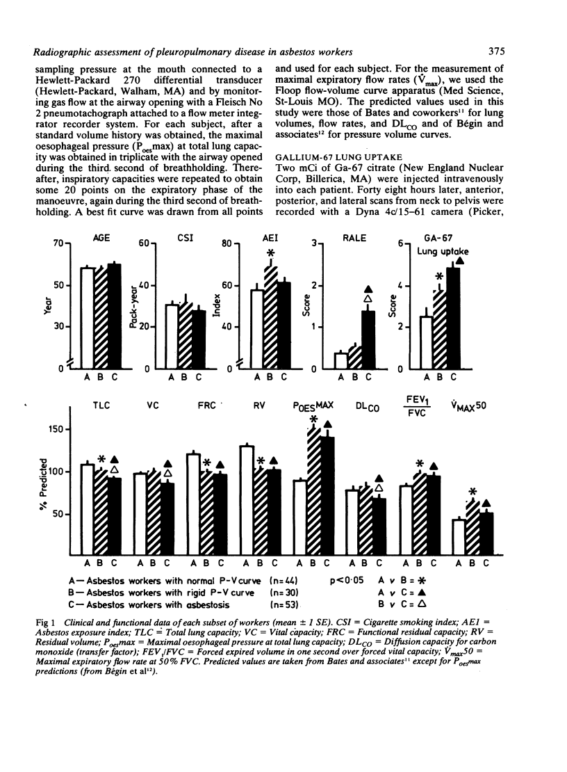
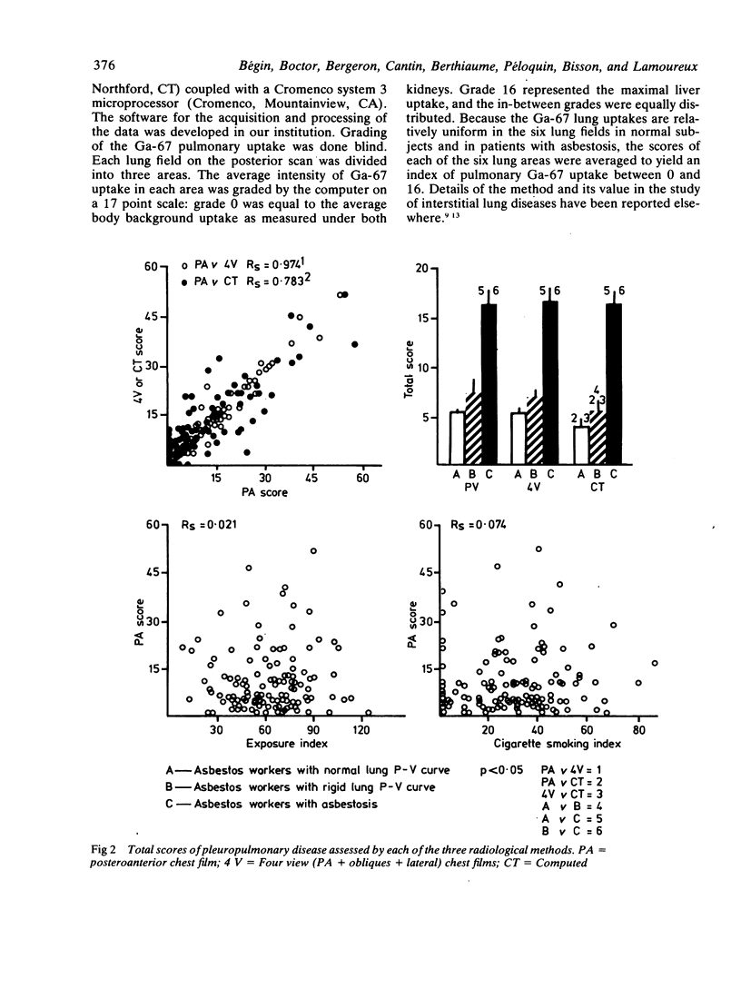
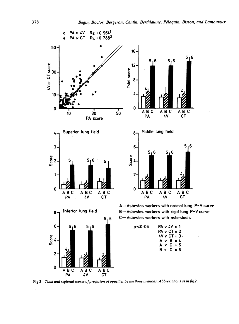
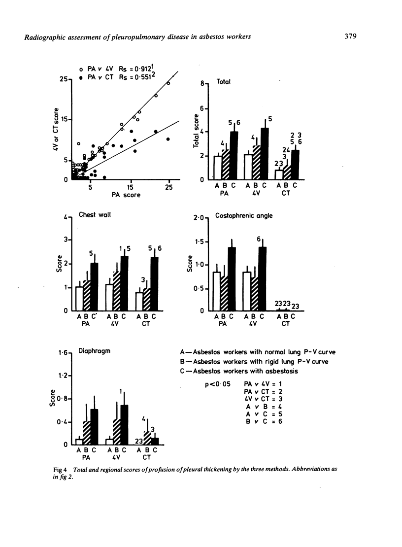
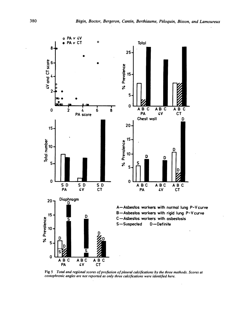
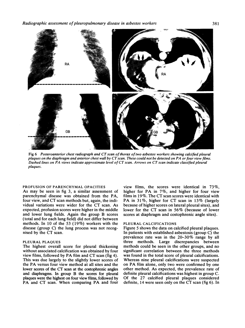
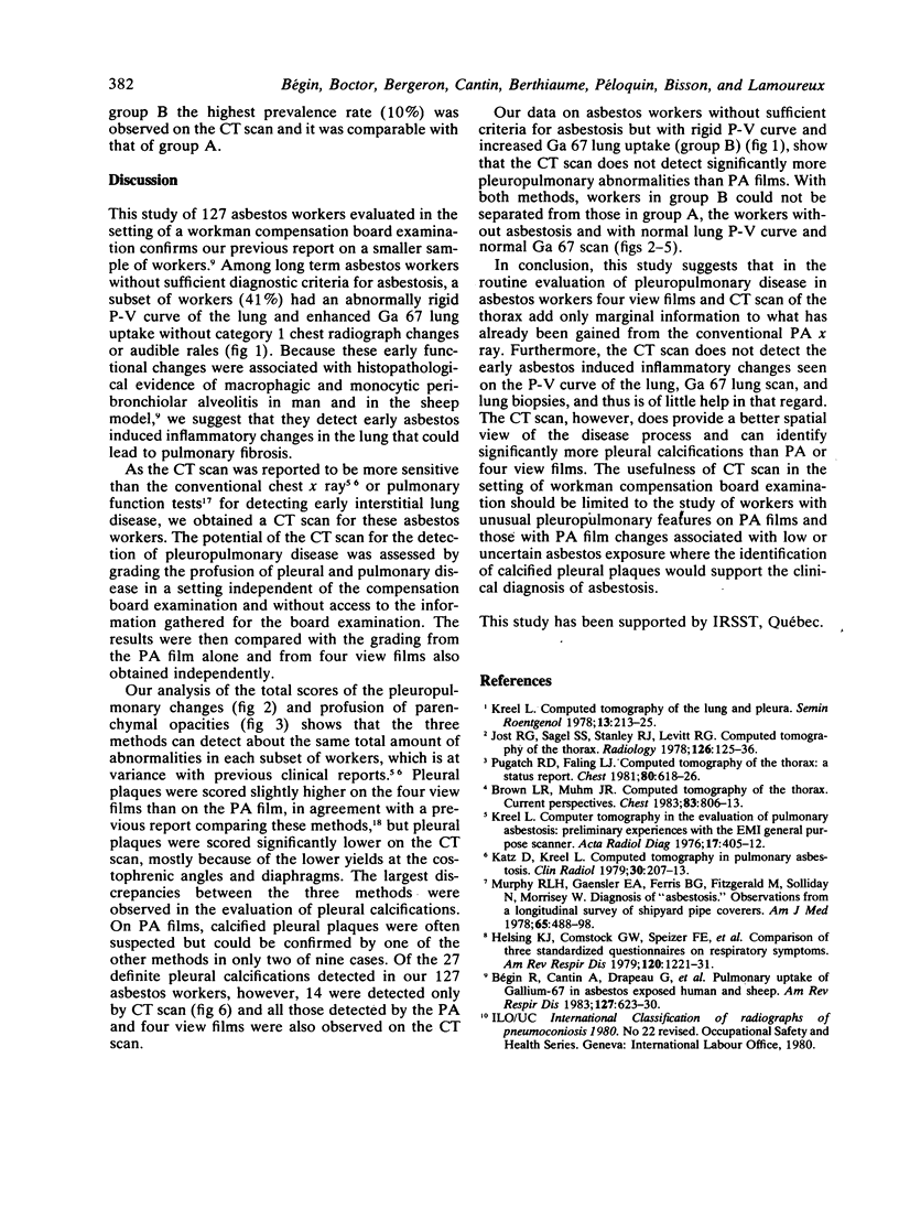
To study the clinical usefulness of computed tomography (CT) scanning of the thorax in asbestos related pleuropulmonary disease, 127 long term asbestos workers of the mines and mills in the Eastern Townships of Québec were examined. The CT scan was compared with the standard posteroanterior (PA) chest film and the four view films using the ILO grading system for profusion of disease. Six lung areas and six pleural sites were studied. On the basis of the usual diagnostic criteria, 41% of the workers had asbestosis. For profusion of parenchymal disease, there was an excellent correlation (r = 0.96, p less than 0.001) between PA and four view films and the latter did not significantly increase the total profusion score; the CT scan correlated less well with the PA film (r = 0.79, p less than 0.01) and the scatter of the data was larger. In 10 of the 53 (19%) workers with asbestosis the pulmonary lesions were not recognised by CT scan. For profusion of pleural plaques, there was an excellent correlation (p = 0.91, p less than 0.001) between PA and four view films; scores were identical in 73%, higher for PA in 7%, and higher for four view films in 19%. CT scan scores, however, were identical with PA films in 31%, higher for CT scan in 13% (owing to higher scores on lateral pleural sites), and lower for CT scan in 56% (owing to lower scores at diaphragm and costophrenic angle sites). Pleural calcifications were identified in 24 workers for a total of 40 sites; 13 as possible, 31% identified by two modes, and 27 as definite. Of the latter, 14 were seen only on CT scan. In the workers with rigid pressure volume curve of the lung and increased Gallium-67 lung uptake only, CT scan total scores were not significantly higher than in those without these markers of early interstitial lung disease (5 +/- 1 v 4 +/- 1, p less than 0.05). Thus the four view films and CT scan appear to be useful mainly in the assessment of pleural disease. The four view film identifies more sites of pleural plaques and the CT scan more pleural calcified plaques.
Full text
PDF

Images in this article
Selected References
These references are in PubMed. This may not be the complete list of references from this article.
- Begin R., Renzetti A. D., Jr, Bigler A. H., Watanabe S. Flow and age dependence of airway closure and dynamic compliance. J Appl Physiol. 1975 Feb;38(2):199–207. doi: 10.1152/jappl.1975.38.2.199. [DOI] [PubMed] [Google Scholar]
- Bisson G., Drapeau G., Lamoureux G., Cantin A., Rola-Pleszczynski M., Bégin R. Computer-based quantitative analysis of gallium-67 uptake in normal and diseased lungs. Chest. 1983 Nov;84(5):513–517. doi: 10.1378/chest.84.5.513. [DOI] [PubMed] [Google Scholar]
- Brown L. R., Muhm J. R. Computed tomography of the thorax. Current perspectives. Chest. 1983 May;83(5):806–813. doi: 10.1378/chest.83.5.806. [DOI] [PubMed] [Google Scholar]
- Bégin R., Cantin A., Drapeau G., Lamoureux G., Boctor M., Massé S., Rola-Pleszczynski M. Pulmonary uptake of gallium-67 in asbestos-exposed humans and sheep. Am Rev Respir Dis. 1983 May;127(5):623–630. doi: 10.1164/arrd.1983.127.5.623. [DOI] [PubMed] [Google Scholar]
- Fahey P. J., Utell M. J., Mayewski R. J., Wandtke J. D., Hyde R. W. Early diagnosis of bleomycin pulmonary toxicity using bronchoalveolar lavage in dogs. Am Rev Respir Dis. 1982 Jul;126(1):126–130. doi: 10.1164/arrd.1982.126.1.126. [DOI] [PubMed] [Google Scholar]
- Helsing K. J., Comstock G. W., Speizer F. E., Ferris B. G., Lebowitz M. D., Tockman M. S., Burrows B. Comparison of three standardized questionnaires on respiratory symptoms. Am Rev Respir Dis. 1979 Dec;120(6):1221–1231. doi: 10.1164/arrd.1979.120.6.1221. [DOI] [PubMed] [Google Scholar]
- Jost R. G., Sagel S. S., Stanley R. J., Levitt R. G. Computed tomography of the thorax. Radiology. 1978 Jan;126(1):125–136. doi: 10.1148/126.1.125. [DOI] [PubMed] [Google Scholar]
- Katz D., Kreel L. Computed tomography in pulmonary asbestosis. Clin Radiol. 1979 Mar;30(2):207–213. doi: 10.1016/s0009-9260(79)80163-4. [DOI] [PubMed] [Google Scholar]
- Kreel L. Computed tomography of the lung and pleura. Semin Roentgenol. 1978 Jul;13(3):213–225. doi: 10.1016/0037-198x(78)90042-1. [DOI] [PubMed] [Google Scholar]
- Kreel L. Computer tomography in the evaluation of pulmonary asbestosis. Preliminary experiences with the EMI general purpose scanner. Acta Radiol Diagn (Stockh) 1976 Jul;17(4):405–412. doi: 10.1177/028418517601700403. [DOI] [PubMed] [Google Scholar]
- Murphy R. L., Jr, Gaensler E. A., Ferris B. G., Fitzgerald M., Solliday N., Morrisey W. Diagnosis of "asbestosis". Observations from a longitudinal survey of shipyard pipe coverers. Am J Med. 1978 Sep;65(3):488–498. doi: 10.1016/0002-9343(78)90775-1. [DOI] [PubMed] [Google Scholar]
- Pugatch R. D., Faling L. J. Computed tomography of the thorax: a status report. Chest. 1981 Nov;80(5):618–626. doi: 10.1378/chest.80.5.618. [DOI] [PubMed] [Google Scholar]
- Reger R. B., Ames R. G., Merchant J. A., Polakoff P. P., Sargent E. N., Silbiger M., Whittlesey P. The detection of thoracic abnormalities using posterior-anterior (PA) vs PA and oblique roentgenograms. Chest. 1982 Mar;81(3):290–295. doi: 10.1378/chest.81.3.290. [DOI] [PubMed] [Google Scholar]