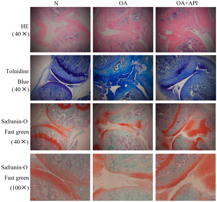
Figure 1.
Apigenin protected articular cartilage in OA mice. Representative photographs of sections of articular cartilage with H&E, toluidine blue, and Safranin-O Fast green staining in the N, OA, and OA + API groups (magnification, ×40, ×100). Compared with the N group, the OA group exhibited serious degradation of the cartilage, obvious proteoglycan loss, and deep cartilage erosion. In contrast, the OA + API group exhibited a smoother cartilage surface and reduced the loss of proteoglycan.

