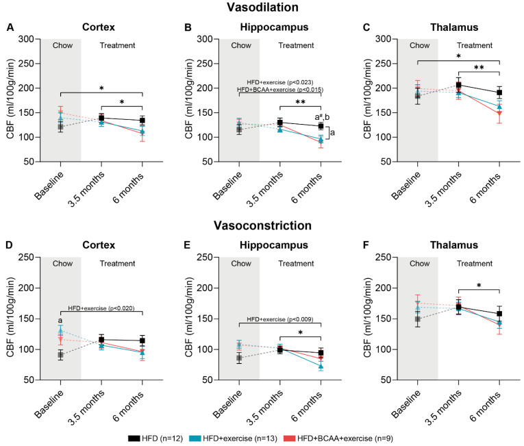
Figure 8.
Measurement of cerebral blood flow under vasodilative and vasoconstrictive conditions. Cerebral blood flow (CBF) was measured by arterial spin labeling at baseline (chow) and after 3.5 and 6 months of treatment with HFD, HFD + exercise, and HFD + BCAA + exercise. At each neuroimaging session, CBF was first assessed under vasodilative conditions in the (A) cortex, (B) hippocampus, and (C) thalamus, and afterwards under vasoconstrictive conditions in the (D) cortex, (E) hippocampus, and (F) thalamus. High-fat diet (HFD), branched-chain amino acids (BCAA), cerebral blood flow (CBF). Data are presented as mean ± SEM. * p < 0.05, ** p < 0.01, # 0.05 < p < 0.08 (trend); a: significant difference HFD vs. HFD + exercise group, b: significant difference HFD vs. HFD + BCAA + exercise group.

