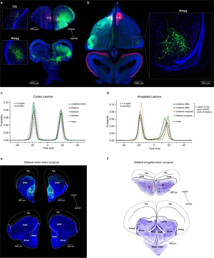
Extended Data Fig. 5. Cortex and amygdala lesions do not prevent dominance switches.
a,b, Identification of contralateral inputs and distribution of GFP-labelled neurons in the forebrain after injection of rAAV2-retro into the Pogona claustrum on one side. a, Transverse sections through the telencephalon, showing very sparse labelling in the contralateral cortex (top), and dense labelling of neurons in the contralateral amygdala. b, Horizontal section through the tel- and mesencephalon, showing contralateral input from the amygdala. Also note the absence of labelled cells in the contralateral claustrum (within stippled line; pink fluorescence: hippocalcin). c, Distributions of peak-correlation lags between left and right claustra for four animals with cortex lesions. d, Same as in c, but for amygdala lesions. Note that we removed the cortical sheet before injecting IBA for excitotoxic lesions, and before removing the amygdala surgically. Additional lesions were made to the adjacent DVR and parts of the striatum. None affected the inter-claustral apparent competition. e, Transverse sections at the level of the claustrum and more caudal telencephalon, showing the recording locations (red circles, top), and the bilateral absence of cortex along the rostro-caudal axis (top and bottom). f, Nissl stains of transverse sections at the level of the claustrum and caudal telencephalon, comparable to e. Indicated are the approximate positions of the bilaterally lesioned amygdala and other brain areas.

