

Abstract
Background
Leukemic presentation of follicular lymphoma (FL) is uncommon, with most cases reported in older adults.
Design
This report describes an unusual case of a young adult diagnosed with leukemic phase of FL. We reviewed the existing literature on this rare presentation of the disease and its potential impact on patient outcomes.
Results
Leukemic phase of FL in young adults can be mistaken for other high‐grade hematologic malignancies. Morphology assessment and ancillary testing, such as flow cytometry and FISH analysis, can assist in achieving an accurate diagnosis of the leukemic phase of FL. Notably, our young patient responded well to therapy, which is different from what is typically observed in older patients who have a poorer prognosis. Further cases are needed to investigate the prognostic impact of the leukemic phase of FL in younger patients.
Keywords: BCL2 rearrangement, follicular lymphoma, leukemic phase, lymphocytosis
Leukemic presentation of follicular lymphoma (FL) is uncommon, with most cases reported in older adults. This case report describes an unusual case of a young adult diagnosed with leukemic phase of FL. We also review the existing literature on this rare manifestation of the disease and its potential impact on patient outcomes. It is important to note that leukemic phase of FL in young adults can be mistaken for other high‐grade hematologic malignancies. The use of morphology assessment and ancillary testing, such as flow cytometry and FISH analysis, can aid in making an accurate diagnosis. However, the prognosis for young patients with leukemic phase of FL may differ from older patients, as the literature suggests a poor prognosis in the latter group.
1. INTRODUCTION
Follicular lymphoma (FL) is the second most common type of non‐Hodgkin lymphoma (NHL) in Western countries, accounting for 35% of cases. 1 It typically affects elderly patients with a median age of diagnosis at 65 years. 1 , 2 The presentation of the disease can vary, from no symptoms to mild or severe symptoms such as lymphadenopathy, fever, excessive night sweats, and unintentional weight loss. 2 , 3 , 4 Bone marrow involvement is present in 70% of cases. The (14;18) translocation, which leads to overexpression of the BCL‐2 protein, is present in around 85% of patients with FL. 5
Leukemic presentation of FL is rare, with most cases reported in patients over 60 years old. 3 , 6 In recent years, the use of rituximab treatment has improved the overall survival rate for patients with follicular lymphoma to around 80%. 7 However, studies have shown that leukemic presentation of follicular lymphoma and high‐risk follicular Lymphoma International Prognostic Index (FLIPI) have been associated with shorter progression‐free survival (PFS). 8 In this case report, we present a young adult patient with an uncommon presentation of follicular lymphoma in a leukemic phase.
2. CASE PRESENTATION
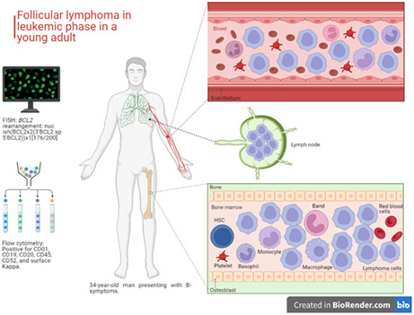
A 34‐year‐old man with a history of asthma presented to the emergency department complaining of fever, drenching night sweats, a 20‐pound weight loss, and fatigue over the past two months. Laboratory test results showed anemia, thrombocytopenia, and marked leukocytosis with lymphocytosis. The peripheral blood smear revealed numerous atypical lymphoid cells, predominantly small to intermediate in size, with bilobed to multilobed nuclei and clumped chromatin. Some cells had butterfly‐shaped nuclei, and most had clover‐like nuclei. (Figure 1A–F) No left‐shift granulocytes or blasts were identified. Flow cytometry revealed an aberrant B‐cell population that was positive for CD10, CD19, CD20, CD45, and CD52 and negative for CD2, CD3, CD4, CD5, CD7, CD8, CD34, and TDT. (Figure 1G) Fluorescence in situ hybridization (FISH) analysis using FISH probes for high‐grade/large B‐cell lymphoma and t(8;14) demonstrated the abnormalities involving the BCL2 probe set (1R1G1F, 88.0%, normal <5.7%) indicative of BCL2 rearrangement, and the t(8;14) probe set (2R3G2A, 85.0%, normal <5.0%) suggestive of duplication of IGH/chromosome 14 or an IGH rearrangement to a locus other than MYC. BCL6 rearrangement and MYC rearrangement/amplification were not detected. The overall findings were consistent with follicular lymphoma (FL) in the leukemic phase.
FIGURE 1.
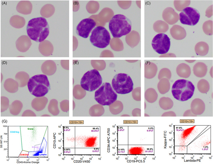
Panels A–F: Peripheral blood smear showed numerous small atypical lymphoid cells with cleaved or lobulated nuclei, clumped chromatin without nucleoli, and scant agranular cytoplasm. Panel G: Flow cytometry analysis on peripheral blood specimen revealed an aberrant B‐cell population (81%) positive for CD10 (98%), CD19, CD20, CD45, and monotypic surface Kappa immunoglobulin light chain expression. 4
On follow‐up examination, the patient was found to have palpable bilateral cervical lymph nodes and splenomegaly. A bone marrow biopsy showed a hypercellular marrow with decreased maturing hematopoiesis and a marked lymphoid infiltrate, consistent with a diagnosis of follicular lymphoma. The patient was classified as high risk for FL based on the FLIPI score of 4 and was started on cyclophosphamide, hydroxydaunorubicin, oncovin, and prednisone (CHOP) chemotherapy. Due to COVID‐19 infection and treatment with Remdesivir, Rituxan was not administered. Despite this, the patient has responded well to therapy.
3. DISCUSSION
Literature review shows that reported cases of leukemic phase FL primarily occur in older patients. 3 , 4 However, in our case, the patient was diagnosed with leukemic phase FL at the age of 34. Leukemic phase follicular lymphoma is characterized by unusual lymphoid cells with “notched” or “buttock” shaped nuclei. Our patient's peripheral blood smear showed abnormal lymphoid cells with nuclei that were “clover‐like” in shape, which is a previously undescribed cytomorphological feature of leukemic phase follicular lymphoma.
Initially, the unusual appearance of the lymphoid cells and the patient's young age made it difficult to diagnose. Further testing with FISH analysis allowed us to confirm the diagnosis and rule out other possibilities, such as mantle cell lymphoma (MCL) in a leukemic phase, chronic lymphocytic leukemia (CLL), and high‐grade B‐cell lymphomas. 9 , 10 Flow cytometry revealed that these lymphoid cells had an immunophenotype characteristic of follicular lymphoma. It is worth noting that lack of CD10 expression cannot exclude the possibility of leukemic phase FL, as reported by Maeshima et al. 11 , 12
Studies have shown that high‐risk FLIPI and leukemic phase are associated with shorter progression‐free survival (PFS) in patients with follicular lymphoma. 8 Furthermore, patients with FL in leukemic phase may show suboptimal response to rituximab‐containing regimens. 13 Our younger patient had a high‐risk FLIPI score and presented with an aggressive form of follicular lymphoma but responded well to CHOP therapy. This outcome is different from what is typically observed in older patients, which raises the possibility that the prognostic impact of the leukemic phase of FL may differ between younger and older patients. Further investigation is warranted to explore this possibility. In addition, this case highlights the need to study the incidence of histologic transformation in patients with the leukemic phase of FL, particularly in younger patients.
In conclusion, when evaluating a young adult patient with leukocytosis and lymphocytosis, the possibility of follicular lymphoma in a leukemic phase should be considered. An accurate diagnosis can be made by evaluating the morphology of the lymphoid cells and utilizing ancillary testing such as flow cytometry and FISH analysis on peripheral blood specimens. In addition, our patient's positive response to chemotherapy might indicate the need for an individualized treatment approach for young patients with leukemic phase of follicular lymphoma.
AUTHOR CONTRIBUTIONS
Daniel Rivera wrote the article. Zhihong Hu diagnosed this case and provided critical review and revisions to the article. Wei Wang and Zubaidah Al‐Jumaili participated in the diagnosis of the patient. All authors reviewed and approved the final version of the article.
CONFLICT OF INTEREST STATEMENT
The authors have no conflicts of interest to report.
ACKNOWLEDGMENT
This case was written under the mentorship of Zhihong Hu, M.D, Ph.D.
Rivera D, Wang W, Al‐Jumaili Z, Hu Z. Leukemic phase follicular lymphoma in a young adult. J Clin Lab Anal. 2023;37:e24869. doi: 10.1002/jcla.24869
DATA AVAILABILITY STATEMENT
Not applicable.
REFERENCES
- 1. Swerdlow SH, Campo E, Pileri SA, et al. The 2016 revision of the World Health Organization classification of lymphoid neoplasms. Blood. 2016;127(20):2375‐2390. [DOI] [PMC free article] [PubMed] [Google Scholar]
- 2. Carbone A, Roulland S, Gloghini A, et al. Follicular lymphoma. Nat Rev Dis Primers. 2019;5(1):83. [DOI] [PubMed] [Google Scholar]
- 3. Medeiros LJ. Ioachim's Lymph Node Pathology. 5th Edition. Philadelphia: Wolters Kluwer Health; 2021:479‐481. [Google Scholar]
- 4. Jacobsen E. Follicular lymphoma: 2023 update on diagnosis and management. Am J Hematol. 2022;97:1638‐1651. [DOI] [PubMed] [Google Scholar]
- 5. Green MR. Chromatin modifying gene mutations in follicular lymphoma. Blood. 2018;131(6):595‐604. [DOI] [PMC free article] [PubMed] [Google Scholar]
- 6. Beltran BE, Castillo JJ, Quiñones P, et al. Follicular lymphoma with leukemic phase at diagnosis: an aggressive disease. report of seven cases and review of the literature. Leuk Res. 2013;37(9):1116‐1119. [DOI] [PMC free article] [PubMed] [Google Scholar]
- 7. Sarkozy C, Maurer MJ, Link BK, et al. Cause of death in follicular lymphoma in the first decade of the rituximab era: a pooled analysis of French and US cohorts. J Clin Oncol. 2019;37(2):144‐152. [DOI] [PMC free article] [PubMed] [Google Scholar]
- 8. Sarkozy C, Baseggio L, Callet‐Bauchu E, et al. Detection of leukemic phase in patients with follicular lymphoma At diagnosis: a rare event associated with poor prognosis. Blood. 2012;120(21):1594. [Google Scholar]
- 9. Nelson B, Variakojis D, Peterson L. Leukemic phase of B‐cell lymphomas mimicking chronic lymphocytic leukemia and variants at presentation. Mod Pathol. 2002;15:1111‐1120. [DOI] [PubMed] [Google Scholar]
- 10. Chekol S, Kimball A, Ning Y, Nanaji N. Transformation of follicular lymphoma to leukemic phase of burkitt lymphoma. American Journal of Clinical Pathology. 2012;138(Issue suppl_1):A222. [Google Scholar]
- 11. Maeshima AM, Taniguchi H, Tanioka K, et al. Clinicopathological characteristics of follicular lymphoma with peripheral blood involvement. Leuk Lymphoma. 2015;56(7):2000‐2004. [DOI] [PubMed] [Google Scholar]
- 12. Al‐Nawakil C, Kosmider O, Stern MH, et al. Leukemic phase of follicular lymphomas: an atypical presentation. Leuk Lymphoma. 2011;52(8):1504‐1508. [DOI] [PubMed] [Google Scholar]
- 13. Kodaira M, Takeuchi K, Nara E, et al. Kiyohiko Hatake; leukemic presentation is predictive indicator for relapse for patients with follicular lymphoma treated with rituximab containing initial therapy. Blood. 2009;114(22):4763. [Google Scholar]
Associated Data
This section collects any data citations, data availability statements, or supplementary materials included in this article.
Data Availability Statement
Not applicable.