Abstract
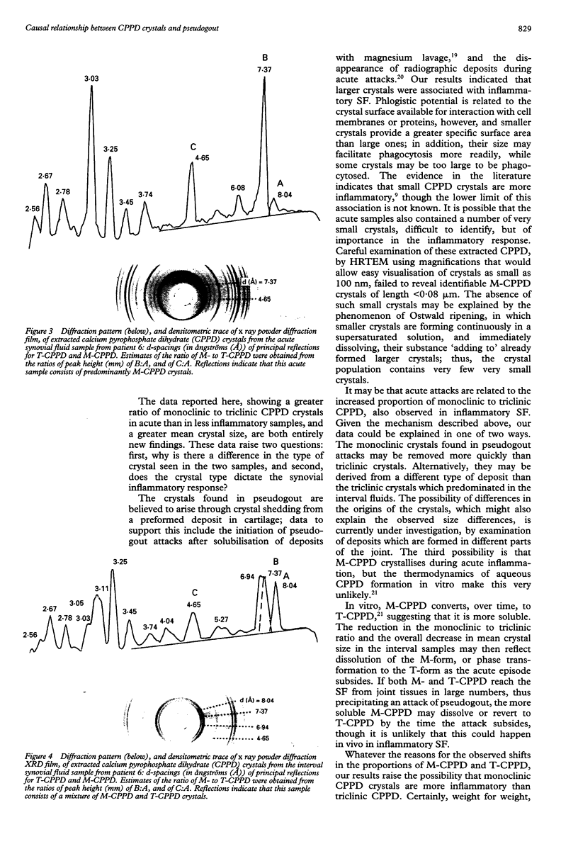
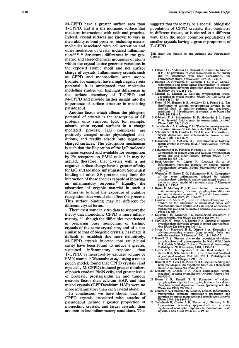
OBJECTIVE--To investigate any relationship between the nature, size, and numbers of synovial fluid (SF) calcium pyrophosphate dihydrate (CPPD) crystals, and attacks of pseudogout. METHODS--Knee SF was aspirated from nine selected patients, first during an attack of pseudogout (acute sample) and again later when the attack had subsided (interval sample). CPPD crystals were extracted, weighed, examined by high resolution transmission electron microscopy (HRTEM), and characterised by size and crystal habit (monoclinic or triclinic). Structural analysis was carried out by x ray powder diffraction (XRD) and the proportions of monoclinic to triclinic CPPD were estimated from densitometric measurements of selected key reflections. RESULTS--The mean crystal size, by HRTEM, indicated that the crystals in the acute sample were larger than those in the interval sample. The ratio of monoclinic to triclinic CPPD, whether estimated from their morphological appearance by HRTEM, or from XRD, was greater in the acute than in the interval sample in all nine patients. The total amount of extracted mineral varied, but in every patient the concentration of CPPD per ml of fluid, and the total mineral per joint, were greater in the acute sample than in the interval sample. CONCLUSION--In this highly selected group of patients, the large numbers of CPPD crystals associated with attacks of pseudogout included a greater proportion of monoclinic crystals, and larger crystals, than those present when inflammation had subsided. A special, phlogistic population of crystals may exist, originating in different joint tissues, or cleared in a different manner, than the more common populations of smaller crystals with a greater proportion of triclinic CPPD, seen in chronic disease.
Full text
PDF
Images in this article
Selected References
These references are in PubMed. This may not be the complete list of references from this article.
- Doherty M., Dieppe P. A. Acute pseudogout: "crystal shedding" or acute crystallization? Arthritis Rheum. 1981 Jul;24(7):954–957. doi: 10.1002/art.1780240716. [DOI] [PubMed] [Google Scholar]
- Felson D. T., Anderson J. J., Naimark A., Kannel W., Meenan R. F. The prevalence of chondrocalcinosis in the elderly and its association with knee osteoarthritis: the Framingham Study. J Rheumatol. 1989 Sep;16(9):1241–1245. [PubMed] [Google Scholar]
- Gibilisco P. A., Schumacher H. R., Jr, Hollander J. L., Soper K. A. Synovial fluid crystals in osteoarthritis. Arthritis Rheum. 1985 May;28(5):511–515. doi: 10.1002/art.1780280507. [DOI] [PubMed] [Google Scholar]
- Gordon T. P., Ahern M. J., Reid C., Roberts-Thomson P. J. Studies on the interaction of rheumatoid factor with monosodium urate crystals and case report of coexistent tophaceous gout and rheumatoid arthritis. Ann Rheum Dis. 1985 Jun;44(6):384–389. doi: 10.1136/ard.44.6.384. [DOI] [PMC free article] [PubMed] [Google Scholar]
- Guerne P. A., Terkeltaub R., Zuraw B., Lotz M. Inflammatory microcrystals stimulate interleukin-6 production and secretion by human monocytes and synoviocytes. Arthritis Rheum. 1989 Nov;32(11):1443–1452. doi: 10.1002/anr.1780321114. [DOI] [PubMed] [Google Scholar]
- Halverson P. B., McCarty D. J. Identification of hydroxyapatite crystals in synovial fluid. Arthritis Rheum. 1979 Apr;22(4):389–395. doi: 10.1002/art.1780220412. [DOI] [PubMed] [Google Scholar]
- Hearn P. R., Russell R. G. Formation of calcium pyrophosphate crystals in vitro: implications for calcium pyrophosphate crystal deposition disease (pseudogout). Ann Rheum Dis. 1980 Jun;39(3):222–227. doi: 10.1136/ard.39.3.222. [DOI] [PMC free article] [PubMed] [Google Scholar]
- KELLGREN J. H., LAWRENCE J. S. Radiological assessment of osteo-arthrosis. Ann Rheum Dis. 1957 Dec;16(4):494–502. doi: 10.1136/ard.16.4.494. [DOI] [PMC free article] [PubMed] [Google Scholar]
- KOHN N. N., HUGHES R. E., McCARTY D. J., Jr, FAIRES J. S. The significance of calcium phosphate crystals in the synovial fluid of arthritic patients: the "pseudogout syndrome". II. Identification of crystals. Ann Intern Med. 1962 May;56:738–745. doi: 10.7326/0003-4819-56-5-738. [DOI] [PubMed] [Google Scholar]
- Kozin F., McCarty D. J. Protein binding to monosodium urate monohydrate, calcium pyrophosphate dihydrate, and silicon dioxide crystals. I. Physical characteristics. J Lab Clin Med. 1977 Jun;89(6):1314–1325. [PubMed] [Google Scholar]
- McGill N. W., Swan A., Dieppe P. A. Survival of calcium pyrophosphate crystals in stored synovial fluids. Ann Rheum Dis. 1991 Dec;50(12):939–941. doi: 10.1136/ard.50.12.939. [DOI] [PMC free article] [PubMed] [Google Scholar]
- Resnick D., Niwayama G., Goergen T. G., Utsinger P. D., Shapiro R. F., Haselwood D. H., Wiesner K. B. Clinical, radiographic and pathologic abnormalities in calcium pyrophosphate dihydrate deposition disease (CPPD): pseudogout. Radiology. 1977 Jan;122(1):1–15. doi: 10.1148/122.1.1. [DOI] [PubMed] [Google Scholar]
- Roch-Arveiller M., Legros R., Chanaud B., Muntaner O., Strzalko S., Thuret A., Willoughby D. A., Giroud J. P. Inflammatory reactions induced by various calcium pyrophosphate crystals. Biomed Pharmacother. 1990;44(9):467–474. doi: 10.1016/0753-3322(90)90207-p. [DOI] [PubMed] [Google Scholar]
- Schumacher H. R., Fishbein P., Phelps P., Tse R., Krauser R. Comparison of sodium urate and calcium pyrophosphate crystal phagocytosis by polymorphonuclear leukocytes. Effects of crystal size and other factors. Arthritis Rheum. 1975 Nov-Dec;18(6 Suppl):783–792. doi: 10.1002/art.1780180723. [DOI] [PubMed] [Google Scholar]
- Terkeltaub R. A., Ginsberg M. H. The inflammatory reaction to crystals. Rheum Dis Clin North Am. 1988 Aug;14(2):353–364. [PubMed] [Google Scholar]
- Terkeltaub R., Curtiss L. K., Tenner A. J., Ginsberg M. H. Lipoproteins containing apoprotein B are a major regulator of neutrophil responses to monosodium urate crystals. J Clin Invest. 1984 Jun;73(6):1719–1730. doi: 10.1172/JCI111380. [DOI] [PMC free article] [PubMed] [Google Scholar]
- Watanabe W., Baker D. G., Schumacher H. R., Jr Comparison of the acute inflammation induced by calcium pyrophosphate dihydrate, apatite and mixed crystals in the rat air pouch model of a synovial space. J Rheumatol. 1992 Sep;19(9):1453–1457. [PubMed] [Google Scholar]


