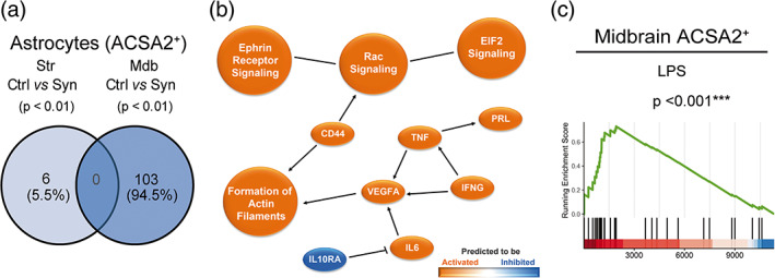
FIGURE 3.

Transcriptomic analysis of astrocytes purified from the striatum and the midbrain of αSyn overexpressing mice. Animals were sacrificed at 2 weeks after AAV9 administration in the SNpc. The striatum and the midbrain were dissected out and a cell suspension was prepared from these regions. Astrocytes were separated based on ACSA2 expression for RNA sequencing. (a) Venn diagram showing overlap of differentially expressed genes (p < .01) between ACSA2+ cells from control and αSyn mice of the two regions(n = 3 animals/group). (b) Graphical summary of the pathways, upstream regulators and biological functions predicted to be altered in ACSA2+ cells from the midbrain of αSyn mice. (c) GSEA plot of the LPS reactive astrocyte signature enriched in midbrain astrocytes of αSyn mice.
