ABSTRACT
This study describes the case of a health professional infected first by influenza virus A(H3N2) and then by severe acute respiratory syndrome coronavirus 2 (SARS-CoV-2) 11 days later. Respiratory samples and clinical data were collected from the patient and from close contacts. RNA was extracted from samples and reverse transcription–quantitative polymerase chain reaction (RT-qPCR) was used to investigate the viruses. The patient presented with two different illness events: the first was characterized by fever, chest and body pain, prostration and tiredness, which ceased on the ninth day; RT-qPCR was positive only for influenza virus A(H3N2). Eleven days after onset of the first symptoms, the patient presented with sore throat, nasal congestion, coryza, nasal itching, sneezing and coughing, and a second RT-qPCR test was positive only for SARS-CoV-2; in the second event, symptoms lasted for 11 days. SARS-CoV-2 sequencing identified the Omicron BA.1 lineage. Of the patient’s contacts, one was coinfected with influenza A(H3N2) and SARS-CoV-2 lineage BA.1.15 and the other two were infected only with SARS-CoV-2, one also with Omicron BA.1.15 and the other with BA.1.1. Our findings reinforce the importance of testing for different viruses in cases of suspected respiratory viral infection during routine epidemiological surveillance because common clinical manifestations of COVID-19 mimic those of other viruses, such as influenza.
Keywords: Influenza A virus, H3N2 subtype; COVID-19; SARS-CoV-2; respiratory tract infections
RESUMEN
Este estudio describe el caso de un profesional de la salud que contrajo la infección primero por el virus de la gripe A (H3N2) y a continuación por el coronavirus 2 del síndrome respiratorio agudo grave (SARS-CoV-2) 11 días después. Se recogieron muestras respiratorias y datos clínicos del paciente y sus contactos cercanos. Se extrajo ARN de muestras y se utilizó la reacción en cadena de la polimerasa cuantitativa con transcripción inversa (RT-qPCR, por su sigla en inglés) para investigar los virus. El paciente presentó dos procesos infecciosos distintos: el primero se caracterizó por fiebre, dolor corporal y torácico, postración y cansancio, que cesó en el noveno día. La prueba mediante RT-qPCR solo fue positiva en el virus de la gripe A (H3N2). Once días después del inicio de los primeros síntomas, el paciente manifestó dolor de garganta, congestión nasal, catarro, picazón nasal, estornudos y tos. Una segunda prueba mediante RT-qPCR solo fue positiva para el SARS-CoV-2 y durante este segundo proceso los síntomas duraron 11 días. La secuenciación del SARS-CoV-2 identificó el linaje ómicron BA.1. De los contactos del paciente, uno presentaba una coinfección por el virus de la gripe A (H3N2) y el linaje BA.1.15 del SARS-COV-2, y los otros dos presentaban infecciones únicamente por SARS-CoV-2, uno también del linaje ómicron BA.1.15 y el otro de BA.1.1. Estos hallazgos refuerzan la importancia de realizar pruebas para detectar diferentes virus en casos de sospecha de infección viral respiratoria durante la vigilancia epidemiológica de rutina porque las manifestaciones clínicas comunes de COVID-19 son similares a las de otros virus, como en el caso de la gripe.
Palabras clave: Subtipo H3N2 del virus de la influenza A, COVID-19, SARS-CoV-2, Infecciones del sistema respiratorio
RESUMO
Este estudo descreve o caso de uma profissional de saúde infectada primeiro pelo vírus influenza A (H3N2) e, 11 dias depois, pelo coronavírus da síndrome respiratória aguda grave 2 (SARS-CoV-2). Amostras respiratórias e dados clínicos foram coletados da paciente e de contatos próximos. RNA foi extraído das amostras, e o método de reação em cadeia da polimerase via transcriptase reversa quantitativa (RT-qPCR) foi utilizado para investigar os vírus. A paciente apresentou dois quadros clínicos distintos. O primeiro foi caracterizado por febre, dor no peito e no corpo, prostração e fadiga, que cessou no nono dia. A RT-qPCR foi positiva apenas para o vírus da influenza A (H3N2). Onze dias após o início dos primeiros sintomas, a paciente apresentou dor de garganta, congestão nasal, coriza, prurido nasal, espirros e tosse. Um segundo teste de RT-qPCR foi positivo apenas para SARS-CoV-2. No segundo evento, os sintomas duraram 11 dias. O sequenciamento do SARS-CoV-2 identificou a cepa Ômicron BA.1. Dentre os contatos da paciente, um teve coinfeção por influenza A (H3N2) e SARS-COV-2 (cepa BA.1.15), e os outros dois foram infectados apenas por SARS-CoV-2 (um também pela cepa Ômicron BA.1.15 e o outro pela BA.1.1). Nossos achados reforçam a importância de testes para a detecção de diferentes vírus em casos de suspeita de infecção viral respiratória durante a vigilância epidemiológica de rotina, visto que as manifestações clínicas comuns da COVID-19 imitam as de outros vírus, como o vírus influenza.
Palavras-chave: Vírus da influenza A subtipo H3N2, COVID-19, SARS-CoV-2, infecções respiratórias
In January 2022, the Pan American Health Organization and the World Health Organization issued an epidemiological alert about an increase in the burden on health care systems and services related to the rise in COVID-19 cases along with increased circulation of other respiratory viruses in the Region of the Americas (1).
In Brazil, Instituto Oswaldo Cruz/Fundação Oswaldo Cruz warned in early December 2021 that in addition to the COVID-19 pandemic, influenza A virus had been identified in cases of severe acute respiratory infection in the state of Rio de Janeiro, and most of the cases were caused by an influenza A/Darwin/9/2021(H3N2)-like strain (2). Within a few weeks, cases of infection with influenza A(H3N2) had been reported by other Brazilian states, and health care professionals were urged to consider influenza A virus as an etiological agent in cases of respiratory infection (1, 3).
Coinfections with severe acute respiratory syndrome coronavirus 2 (SARS-CoV-2) and other respiratory viruses, including influenza A virus, have been reported in some studies (4, 5). In these cases, laboratory analyses of patients’ samples detected SARS-CoV-2 concomitantly with another virus. No cases of subsequent infections – during which an individual is infected by one virus and within a few days by another – have been described so far. This study describes the occurrence of subsequent infections in a health professional infected first by influenza A(H3N2) who 11 days later was infected by SARS-CoV-2. The study was approved by the Ethics Committee of the Universidade Federal de Ciências da Saúde de Porto Alegre (approval number 75118217.9.0000.5345). Written informed consent was obtained from all participants, and measures were taken to guarantee the anonymity of the data.
THE STUDY
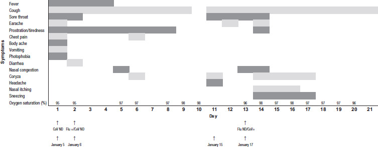
The case patient, a 57-year-old female health professional resident in Porto Alegre, State of Rio Grande do Sul, Brazil, with hypertension and obesity as risk factors, presented with symptoms of acute respiratory infection on January 5, 2022; symptoms included fever (38 °C), cough, sore throat, earache, prostration and tiredness, chest and body pain, vomiting and photophobia (Figure 1, Day 1). A nasopharyngeal swab sample was collected from the case patient on that day to investigate possible SARS-CoV-2 infection using reverse transcription–quantitative polymerase chain reaction (RT-qPCR; Kit BIOMOL OneStep/COVID-19 [IBMP, Curitiba, Brazil] in a CFX Opus Real-Time PCR System [Bio-Rad, Hercules, CA, USA]). The result was negative.
FIGURE 1. Case patient’s symptoms during two separate acute respiratory infections, by day of infection, with results of PCR tests for influenza A and SARS-CoV-2, Brazil, 2022a.

Flu: influenza A virus; CoV: severe acute respiratory syndrome coronavirus 2 (SARS-CoV-2); ND: not detected; RT-qPCR: reverse transcription––quantitative polymerase chain reaction.
Source: Figure prepared by the authors based on the results of their study.
The case patient reported that since January 3, the only person with whom she had contact was her 22-year-old daughter, who had presented on January 2 with fever (39 °C), sore throat, body aches, fatigue and headache; the daughter tested positive on January 4 for influenza A(H3N2) (cycle threshold [Ct]: 20) and SARS-CoV-2 (Ct: 29).
On January 6, the case patient reported diarrhea (Day 2), and another nasopharyngeal swab sample was collected for investigation of SARS-CoV-2, influenza A virus, influenza B virus and respiratory syncytial virus (RSV) using the Allplex SARS-CoV-2/FluA/FluB/RSV Assay kit (Seegene, Seoul, Republic of Korea) in a C1000 Touch Thermal Cycler (Bio-Rad). The sample was again negative for SARS-CoV-2, but it was positive for influenza A virus (Ct: 30), which was subtyped as A(H3N2) using the US Centers for Disease Control and Prevention protocol.
On medical advice, the patient used oseltamivir phosphate for 5 days; she was also instructed to stay home and quarantine. The case patient did not require hospitalization during the period of infection with influenza A(H3N2): her oxygen saturation was 95–98%, and the symptoms ceased after 9 days. After 10 days from the onset of symptoms, the case patient had completely recovered from all symptoms (Figure 1).
On January 15, the case patient again had symptoms, mainly cough, headache, sore throat and coryza (Figure 1, Day 11). A new nasopharyngeal swab specimen was collected on January 17 for investigation of SARS-CoV-2, influenza A and B, and RSV; only SARS-CoV-2 was confirmed (Ct: 18). During the following days, the patient experienced severe earache, sore throat, dry cough, coryza, nasal itching and intense sneezing, with oxygen saturation around 95–98%. The case patient returned to work 10 days after the onset of the second group of symptoms.
To verify whether the successive infections in the case patient originated from a single infection event during contact with her daughter or they were due to two independent events, genome sequencing was performed on SARS-CoV-2 samples from the case patient, her daughter and two of the case patient’s coworkers, who also had symptoms of acute respiratory infection during the same period. Sequencing was performed using the ARTIC nCoV-2019 V3 Amplicon Panel (Integrated DNA Technologies, Coralville, IA, USA) and the Illumina DNA Prep kit (San Diego, CA, USA) for library generation, according to the manufacturer’s instructions for the Illumina MiSeq platform, with the MiSeq Reagent Kit v.3 (600 cycle). The ViralFlow pipeline v.0.0.6 (https://github.com/dezordi/ViralFlow) was implemented to generate consensus sequences from Illumina FASTQ files; and pangolin (Phylogenetic Assignment of Named Global Outbreak Lineages) v.4.0.6 (https://github.com/cov-lineages/pangolin) was used for SARS-CoV-2 lineage assignment.
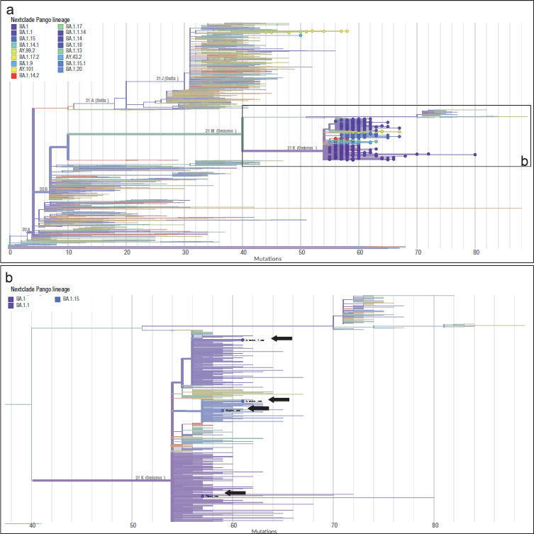
We identified distinct SARS-CoV-2 Omicron sublineages in the sequences from the case patient and her contacts. The case patient was infected by the BA.1 sublineage, whereas her daughter and one of the coworkers were infected by the BA.1.15 sublineage; the other coworker was infected by the BA.1.1 sublineage.
We used the Nextclade web tool v.2.6.21 (https://clades.nextstrain.org) to reconstruct a phylogenetic tree with the four sequenced genomes and all of the 366 SARS-CoV-2 sequences from Rio Grande do Sul sampled during January 2022 and available in the GISAID (Global Initiative on Sharing All Influenza Data) database (https://www.gisaid.org) on April 19, 2022. Figure 2 shows that the SARS-CoV-2 sequence from the patient and those from her daughter and coworkers belong to different clades, in accordance with the differences observed at the sublineage level.
FIGURE 2. Phylogeny of severe acute respiratory syndrome coronavirus 2 (SARS-CoV-2) from samples from four patients. (a) The phylogenetic tree includes 1814 SARS-CoV-2 reference sequences, 366 SARS-CoV-2 sequences from Rio Grande do Sul, Brazil, sampled in January 2022, and the four sequences from the study. (b) Detail of the four sequences generated in this study (indicated by arrows).

Source: Figure prepared by the authors based on the results of their study.
DISCUSSION
In this study, we describe an episode of influenza virus infection followed by infection with SARS-CoV-2 in a health professional within a period of 11 days. The case patient had close contact with her daughter, who had been diagnosed with both SARS-CoV-2 and influenza A(H3N2) simultaneously, with a higher viral load of influenza A than of SARS-CoV-2. Our first hypothesis was that the case patient might have been infected primarily with influenza A(H3N2) viral particles at the time of contact with her daughter, since SARS-CoV-2 was not detected in her sample. The patient had an influenza vaccination in April 2021, 9 months before becoming infected, but the main A(H3N2) strain circulating at the time she was infected – A/Darwin/9/2021(H3N2)-like (6, 7) – was different from the strain contained in the 2021 flu vaccine (8). Therefore, it is probable that the patient was not completely protected against influenza A(H3N2), as studies have reported that the effectiveness of seasonal influenza vaccines has been limited by antigenic drift (9, 10). The case patient received oseltamivir phosphate 1 day after symptom onset, which probably helped to reduce the severity of the disease and accounts for the patient’s good outcome, despite her hypertension and obesity, which are known to be risk factors both for flu and COVID-19.
During the first infection, the case patient presented with symptoms characteristic of influenza, such as fever, chest and body pain, prostration and tiredness (11, 12). The second event, which started 2 days after the influenza symptoms had ended, included symptoms characteristic of infection with the SARS-CoV-2 Omicron lineage, such as sore throat, nasal congestion, coryza, nasal itching and sneezing, and coughing (11, 12). Notably, these symptoms persisted longer than the influenza symptoms.
Sequencing revealed that the SARS-CoV-2 variant from the patient’s sample was different from the variants found in the other samples analyzed; therefore, the source of infection for the patient was not one of her contacts. It is important to note that there were no outbreaks of influenza or COVID-19 in the case patient’s work environment; the case patient had had three doses of the COVID-19 vaccine, which was the complete regimen at the time of this study; and all personal protective equipment and biosafety protocols had been adopted in the patient’s work environment. So the case patient could have been infected by another social contact who had COVID-19. As we could not identify exactly when and by whom the patient was infected, this may represent a limitation of the study. Of note, at the time of this study, Rio Grande do Sul was reporting high numbers of people with COVID-19 due to an increase in circulation of the Omicron lineage (13, 14), which spreads more easily and has higher transmissibility than other variants (15–17).
It is not uncommon to detect instances of viral coinfection or mixed infections during periods of high viral circulation in Rio Grande do Sul, as demonstrated by our group (18), because the area is characterized by a subtropical climate, with rainy autumns followed by cold winters that contribute to infections with influenza and other respiratory viruses. However, to the best of our knowledge, until the present study, there had been no reports of subsequent, separate viral respiratory infections caused by different viruses, one being SARS-CoV-2. It is known that subsequent respiratory infections can lead to chronic respiratory conditions and, consequently, to a worsening of a patient’s health.
In conclusion, we described and characterized a case who had two subsequent and clearly separated viral respiratory infections, with the first caused by influenza A(H3N2) and the second caused by SARS-CoV-2. Our findings highlight the importance of testing for more than one virus in cases with suspected respiratory disease during routine epidemiological surveillance, since common clinical manifestations of COVID-19 mimic those of other viral infections, such as influenza. Moreover, virus identification is critical to better inform patients’ treatment when specific chemoprophylactic agents or vaccines, or both, are available.
Disclaimer.
Authors hold sole responsibility for the views expressed in the manuscript, which may not necessarily reflect the opinion or policy of the Revista Panamericana de Salud Pública/Pan American Journal of Public Health or those of the Pan American Health Organization.
Acknowledgements.
The authors thank the State Health Secretariat of Rio Grande do Sul and the Brazilian Health Ministry for supporting the study.
Funding Statement
This work was supported by the Health Department of Rio Grande do Sul, by the Ministry of Health of Brazil, by Fundação de Amparo à Pesquisa do Estado do Rio Grande do Sul (grant number: FAPERGS/MS/CNPq 08/2020–PPSUS, grant process: 21/2551-0000059-7) and by Conselho Nacional de Desenvolvimento Científico e Tecnológico, Brazil (grant process: 402586/2021-2). ABGV holds a Research Fellowship from Conselho Nacional de Desenvolvimento Científico e Tecnológico (grant process: 306369/2019-2).
Footnotes
Author’ contributions.
TSG developed the original concept of the study and contributed to collecting the samples, acquiring and analyzing the data, and conducting the molecular analyses; TSG also contributed to writing the draft of the manuscript. RSS contributed to the molecular analyses and data analysis, and contributed to writing the draft of the manuscript. LFB contributed to collecting the samples, conducting the molecular analyses and analyzing the data, and writing the draft of the manuscript. CFP contributed to collecting the samples, the molecular analyses and data analysis, and to writing the draft. RBB and FMG contributed to the molecular analyses. ABGV contributed to the data analysis, and to reviewing the draft and writing the final manuscript. All authors revised and approved the final version of the manuscript.
Conflict of interest.
None declared.
Financial support.
This work was supported by the Health Department of Rio Grande do Sul, by the Ministry of Health of Brazil, by Fundação de Amparo à Pesquisa do Estado do Rio Grande do Sul (grant number: FAPERGS/MS/CNPq 08/2020–PPSUS, grant process: 21/2551-0000059-7) and by Conselho Nacional de Desenvolvimento Científico e Tecnológico, Brazil (grant process: 402586/2021-2). ABGV holds a Research Fellowship from Conselho Nacional de Desenvolvimento Científico e Tecnológico (grant process: 306369/2019-2).
Results of the PCR tests are denoted as + for positive and ND for not detected.
REFERENCES
- 1.Pan American Health Organization, World Health Organization . Organization of health services in the context of high respiratory virus circulation including COVID-19 (21 January 2022): epidemiological alert. Washington (DC): Pan American Health Organization; 2022. https://iris.paho.org/handle/10665.2/55656 [Google Scholar]; Pan American Health Organization, World Health Organization. Organization of health services in the context of high respiratory virus circulation including COVID-19 (21 January 2022): epidemiological alert. Washington (DC): Pan American Health Organization; 2022. https://iris.paho.org/handle/10665.2/55656
- 2.Ministério da Saúde, Fundação Oswaldo Cruz (FIOCRUZ) Info-Gripe aponta vírus influenza A em casos de SRAG no RJ. Rio de Janeiro: Ministério da Saúde; 2021. [cited 2021 December 9]. Internet. Portuguese. Available from: https://portal.fiocruz.br/noticia/infogripe-aponta-virus-influenza-em-casos-de-srag-no-rj. [Google Scholar]; Ministério da Saúde, Fundação Oswaldo Cruz (FIOCRUZ). Info-Gripe aponta vírus influenza A em casos de SRAG no RJ [Internet]. Rio de Janeiro: Ministério da Saúde; 2021 [cited 2021 December 9]. Portuguese. Available from: https://portal.fiocruz.br/noticia/infogripe-aponta-virus-influenza-em-casos-de-srag-no-rj
- 3.Secretaria Estadual da Saúde, Centro Estadual de Vigilância em Saúde . Alerta Epidemiológico CEVS: circulação do vírus influenza fora da sazonalidade. Porto Alegre: Rio Grande do Sul: Secretaria Estadual da Saúde; 2021. [cited 2022 March 25]. Vírus influenza. Internet. Portuguese. Available from: https://www.cevs.rs.gov.br/circulacao-do-virus-influenza-fora-da-sazonalidade. [Google Scholar]; Secretaria Estadual da Saúde, Centro Estadual de Vigilância em Saúde. Vírus influenza. Alerta Epidemiológico CEVS: circulação do vírus influenza fora da sazonalidade [Internet]. Porto Alegre: Rio Grande do Sul, Secretaria Estadual da Saúde; 2021 [cited 2022 March 25]. Portuguese. Available from: https://www.cevs.rs.gov.br/circulacao-do-virus-influenza-fora-da-sazonalidade
- 4.Dadashi M, Khaleghnejad S, Abedi Elkhichi P, Goudarzi M, Goudarzi H, Taghavi A, et al. COVID-19 and influenza co-infection: a systematic review and meta-analysis. Front Med (Lausanne) 2021;8:681469. doi: 10.3389/fmed.2021.681469. [DOI] [PMC free article] [PubMed] [Google Scholar]; Dadashi M, Khaleghnejad S, Abedi Elkhichi P, Goudarzi M, Goudarzi H, Taghavi A, et al. COVID-19 and influenza co-infection: a systematic review and meta-analysis. Front Med (Lausanne). 2021;8:681469. [DOI] [PMC free article] [PubMed]
- 5.Azekawa S, Namkoong H, Mitamura K, Kawaoka Y, Saito F. Co-infection with SARS-CoV-2 and influenza A virus. IDCases. 2020;20:e00775. doi: 10.1016/j.idcr.2020.e00775. [DOI] [PMC free article] [PubMed] [Google Scholar]; Azekawa S, Namkoong H, Mitamura K, Kawaoka Y, Saito F. Co-infection with SARS-CoV-2 and influenza A virus. IDCases. 2020;20:e00775. [DOI] [PMC free article] [PubMed]
- 6.Ministério da Saúde, Fundação Oswaldo Cruz (FIOCRUZ) H3N2 Darwin: saiba mais sobre o tipo do vírus influenza em circulação no país. Rio de Janeiro: Ministério da Saúde; 2021. [cited 2021 December 23]. Internet. Portuguese. Available from: https://portal.fiocruz.br/noticia/h3n2-darwin-saiba-mais-sobre-o-tipo-do-virus-influenza-em-circulacao-no-pais. [Google Scholar]; Ministério da Saúde, Fundação Oswaldo Cruz (FIOCRUZ). H3N2 Darwin: saiba mais sobre o tipo do vírus influenza em circulação no país [Internet]. Rio de Janeiro: Ministério da Saúde; 2021 [cited 2021 December 23]. Portuguese. Available from: https://portal.fiocruz.br/noticia/h3n2-darwin-saiba-mais-sobre-o-tipo-do-virus-influenza-em-circulacao-no-pais
- 7.Organização Pan-Americana da Saúde . Atualização epidemiológica: influenza no contexto da pandemia de COVID-19. Brasília: Organização Pan-Americana da Saúde; [28 de dezembro de 2021]. 2021. https://iris.paho.org/handle/10665.2/55599 Portuguese. [Google Scholar]; Organização Pan-Americana da Saúde. Atualização epidemiológica: influenza no contexto da pandemia de COVID-19 (28 de dezembro de 2021). Brasília: Organização Pan-Americana da Saúde; 2021. Portuguese. https://iris.paho.org/handle/10665.2/55599
- 8.World Health Organization . Recommended composition of influenza virus vaccines for use in the 2020–2021 northern hemisphere influenza season. Geneva: World Health Organization; 2020. https://www.who.int/publications/m/item/recommended-composition-of-influenza-virus-vaccines-for-use-in-the-2020-2021-northern-hemisphere-influenza-season [Google Scholar]; World Health Organization. Recommended composition of influenza virus vaccines for use in the 2020–2021 northern hemisphere influenza season. Geneva: World Health Organization; 2020. https://www.who.int/publications/m/item/recommended-composition-of-influenza-virus-vaccines-for-use-in-the-2020-2021-northern-hemisphere-influenza-season
- 9.Sant’Anna FH, Borges LGA, Fallavena PRV, Gregianini TS, Matias F, Halpin RA, et al. Genomic analysis of pandemic and post-pandemic influenza A pH1N1 viruses isolated in Rio Grande do Sul, Brazil. Arch Virol. 2014;159:621–630. doi: 10.1007/s00705-013-1855-8. [DOI] [PubMed] [Google Scholar]; Sant’Anna FH, Borges LGA, Fallavena PRV, Gregianini TS, Matias F, Halpin RA, et al. Genomic analysis of pandemic and post-pandemic influenza A pH1N1 viruses isolated in Rio Grande do Sul, Brazil. Arch Virol. 2014;159:621–30. [DOI] [PubMed]
- 10.Park BR, Kim K-H, Kotomina T, Kim M-C, Kwon Y-M, Jeeva S, et al. Broad cross protection by recombinant live attenuated influenza H3N2 seasonal virus expressing conserved M2 extracellular domain in a chimeric hemagglutinin. Sci Rep. 2021;11:4151. doi: 10.1038/s41598-021-83704-0. [DOI] [PMC free article] [PubMed] [Google Scholar]; Park BR, Kim K-H, Kotomina T, Kim M-C, Kwon Y-M, Jeeva S, et al. Broad cross protection by recombinant live attenuated influenza H3N2 seasonal virus expressing conserved M2 extracellular domain in a chimeric hemagglutinin. Sci Rep. 2021;11:4151. [DOI] [PMC free article] [PubMed]
- 11.World Health Organization . Coronavirus disease (COVID-19): similarities and differences between COVID-19 and Influenza. Geneva: World Health Organization; 2021. [cited 2022 March 7]. Internet. Available from: https://www.who.int/emergencies/diseases/novel-coronavirus-2019/question-and-answers-hub/q-a-detail/coronavirus-disease-covid-19-similarities-and-differences-with-influenza. [Google Scholar]; World Health Organization. Coronavirus disease (COVID-19): similarities and differences between COVID-19 and Influenza [Internet]. Geneva: World Health Organization; 2021 [cited 2022 March 7]. Available from: https://www.who.int/emergencies/diseases/novel-coronavirus-2019/question-and-answers-hub/q-a-detail/coronavirus-disease-covid-19-similarities-and-differences-with-influenza
- 12.Centers for Disease Control and Prevention . Similarities and differences between flu and COVID-19. Atlanta (GA): Centers for Disease Control and Prevention; 2022. [cited 2022 March 7]. Internet. Available from: https://www.cdc.gov/flu/symptoms/flu-vs-covid19.htm. [Google Scholar]; Centers for Disease Control and Prevention. Similarities and differences between flu and COVID-19 [Internet]. Atlanta (GA): Centers for Disease Control and Prevention; 2022 [cited 2022 March 7]. Available from: https://www.cdc.gov/flu/symptoms/flu-vs-covid19.htm
- 13.Secretaria Estadual da Saúde, Centro Estadual de Vigilância em Saúde . Semana Epidemiológica (SR) 07/22. Porto Alegre: Rio Grande do Sul, Secretaria Estadual da Saúde; 2022. [cited 2022 March 8]. Boletim Epidemiológico COVID-19. Internet. Portuguese. Available from: https://coronavirus.rs.gov.br/upload/arquivos/202202/25153105-boletim-epidemiologico-covid-19-se-07-2022.pdf. [Google Scholar]; Secretaria Estadual da Saúde, Centro Estadual de Vigilância em Saúde. Boletim Epidemiológico COVID-19. Semana Epidemiológica (SR) 07/22 [Internet]. Porto Alegre: Rio Grande do Sul, Secretaria Estadual da Saúde: 2022 [cited 2022 March 8]. Portuguese. Available from: https://coronavirus.rs.gov.br/upload/arquivos/202202/25153105-boletim-epidemiologico-covid-19-se-07-2022.pdf
- 14.Secretaria Estadual da Saúde, Centro Estadual de Vigilância em Saúde . Aumento nos casos confirmam a transmissão comunitária da Ômicron no Estado. Porto Alegre, Rio Grande do Sul: Secretaria Estadual da Saúde; 2022. [cited 2022 January 7]. Internet. Portuguese. Available from: https://saude.rs.gov.br/aumento-nos-casos-confirmam-a-transmissao-comunitaria-da-omicron-no-estado. [Google Scholar]; Secretaria Estadual da Saúde, Centro Estadual de Vigilância em Saúde. Aumento nos casos confirmam a transmissão comunitária da Ômicron no Estado [Internet]. Porto Alegre: Rio Grande do Sul, Secretaria Estadual da Saúde; 2022 [cited 2022 January 7]. Portuguese. Available from: https://saude.rs.gov.br/aumento-nos-casos-confirmam-a-transmissao-comunitaria-da-omicron-no-estado
- 15.He X, Hong W, Pan X, Lu G, Wei X. SARS-CoV-2 Omicron variant: characteristics and prevention. MedComm. 2021;2:838–845. doi: 10.1002/mco2.110. [DOI] [PMC free article] [PubMed] [Google Scholar]; He X, Hong W, Pan X, Lu G, Wei X. SARS-CoV-2 Omicron variant: characteristics and prevention. MedComm. 2021;2:838–45. [DOI] [PMC free article] [PubMed]
- 16.Ren S-Y, Wang W-B, Gao R-D, Zhou A-M. Omicron variant (B.1.1.529) of SARS-CoV-2: mutation, infectivity, transmission, and vaccine resistance. World J Clin Cases. 2022;10:1–11. doi: 10.12998/wjcc.v10.i1.1. [DOI] [PMC free article] [PubMed] [Google Scholar]; Ren S-Y, Wang W-B, Gao R-D, Zhou A-M. Omicron variant (B.1.1.529) of SARS-CoV-2: mutation, infectivity, transmission, and vaccine resistance. World J Clin Cases. 2022;10:1–11. [DOI] [PMC free article] [PubMed]
- 17.Centers for Disease Control and Prevention . COVID-19: variants of the virus. Atlanta (GA): Centers for Disease Control and Prevention; 2021. [cited 2023 February 6]. Internet. Available from: https://www.cdc.gov/coronavirus/2019-ncov/variants/omicron-variant.html. [Google Scholar]; Centers for Disease Control and Prevention. COVID-19: variants of the virus [Internet]. Atlanta (GA): Centers for Disease Control and Prevention; 2021 [cited 2023 February 6]. Available from: https://www.cdc.gov/coronavirus/2019-ncov/variants/omicron-variant.html
- 18.Gregianini TS, Varella IRS, Fisch P, Martins LG, Veiga ABG. Dual and triple infections with influenza A and B viruses: a case-control study in southern Brazil. J Infect Dis. 2019;220:961–968. doi: 10.1093/infdis/jiz221. [DOI] [PubMed] [Google Scholar]; Gregianini TS, Varella IRS, Fisch P, Martins LG, Veiga ABG. Dual and triple infections with influenza A and B viruses: a case-control study in southern Brazil. J Infect Dis. 2019;220:961–8. [DOI] [PubMed]


