ABSTRACT
While the airborne decay of bacterial viability has been observed for decades, an understanding of the mechanisms driving the decay has remained elusive. The airborne transport of bacteria is often a key step in their life cycle and as such, characterizing the mechanisms driving the airborne decay of bacteria is an essential step toward a more complete understanding of microbial ecology. Using the Controlled Electrodynamic Levitation and Extraction of Bioaerosols onto a Substrate (CELEBS), it was possible to systematically evaluate the impact of different physicochemical and environmental parameters on the survival of Escherichia coli in airborne droplets of Luria Bertani broth. Rather than osmotic stress driving the viability loss, as was initially considered, oxidative stress was found to play a key role. As the droplets evaporate and equilibrate with the surrounding environment, the surface-to-volume ratio increases, which in turn increased the formation of reactive oxygen species in the droplet. These reactive oxygen species appear to play a key role in driving the airborne loss of viability of E. coli.
IMPORTANCE The airborne transport of bacteria has a wide range of impacts, from disease transmission to cloud formation. By understanding the factors that influence the airborne stability of bacteria, we can better understand these processes. However, while we have known for several decades that airborne bacteria undergo a gradual loss of viability, we have not previously identified the mechanisms driving this process. In this work, we discovered that oxygen surrounding an airborne droplet facilitates the formation of reactive oxygen species within the droplet, which then gradually damage and kill bacteria within the droplet. This discovery indicates that adaptations to help bacteria deal with oxidative stress may also aid their airborne survival and be essential adaptations for bacterial airborne pathogens. Understanding the adaptations bacteria need to survive in airborne droplets could eventually lead to the development of novel antimicrobials designed to inhibit their airborne survival, helping to prevent the transmission of disease.
KEYWORDS: Escherichia coli, aerobiology, airborne microorganisms, bioaerosols, oxidative damage
INTRODUCTION
There are a variety of mechanisms by which bacteria may be transported through the environment, and understanding these mechanisms is key to understanding bacterial ecology. While many forms of motility through liquids and across solid surfaces have been characterized down to the molecular level (1–5), there remain many unknowns surrounding the transport of bacteria through the air. Airborne bacteria take on a variety of forms, ranging from respiratory pathogens and commensals contained in exhaled droplets of saliva and sputum (6–8), to soil bacteria attached to particles suspended high in the upper atmosphere (9–11). The conditions these bacteria face will be unlike anything they encounter in bulk liquid solutions and as such will be likely to induce a unique physiological response in the bacteria. This physiological response remains largely uncharacterized and poorly understood. Limitations to our understanding of the airborne physiology of bacteria has contributed to uncertainty surrounding important issues such as airborne disease transmission (12), environmental contamination (13), and the contribution of bacteria to cloud formation (14–16).
Evidence of the harmful nature of the airborne microenvironment can be seen in many studies of bacterial airborne survival. Studies using rotating drums (17–19), the Tandem Aged Respiratory Droplet Investigation System (TARDIS) (20), and Controlled Electrodynamic Levitation and Extraction of Bioaerosols onto a Substrate (CELEBS) (21, 22), have reported a time dependent loss of culturability in bacteria exposed to airborne conditions for a variety of different bacterial species under a variety of different conditions. However, the exact mechanisms driving this loss of viability remain uncertain.
It should be possible to identify likely causes of airborne loss of bacterial viability through examination of the unique physicochemical properties of aerosols and airborne droplets. For aqueous airborne droplets, the high surface area-to-volume ratio results in a rapid equilibration of the water activity of the droplet to the surrounding relative humidity (RH) (23–26). In many cases, such as in exhaled aerosols, this equilibration will result in a concurrent reduction in the size of the particle, with water quickly partitioning from the droplet until the water activity has reduced such that it is equilibrated to a drier surrounding RH. For example, a droplet of saliva with a diameter of 35 μm exposed to 50% RH (typical indoor humidity) would be expected to decrease in size to an equilibrated diameter of ~10 μm, in approximately 3 s (23), while the solute concentration and surface to volume ratio of the droplet would both increase. The lack of heterogeneous nucleation sites in an airborne droplet means that salt concentrations can become supersaturated (27, 28), exposing bacteria within the droplet to concentrations of solute that they would not encounter in any other environment. Although, it is possible that the presence of microorganisms within a droplet could provide heterogeneous nucleation sites for salt crystallization (29, 30). At a sufficiently low RH, salts can crystallize via homogenous nucleation (31), a process termed efflorescence, embedding any suspended bacteria in the airborne salt crystal (21). Bacteria in equilibrated airborne droplets could be expected to experience osmotic stress because of the low water activity, harmful bacteria-bacteria interactions, starvation, increased exposure to various forms of radiation, and increased exposure to gaseous reactants. Any of these things are potentially responsible for airborne loss of bacterial viability.
The aim of this study is to identify the primary mechanistic explanation for a loss of bacterial viability in aerosols. Escherichia coli in droplets of LB broth is the system considered, with both the bacteria and solution well characterized, allowing our study to focus on aerosol specific phenomena. The CELEBS instrument (21, 22, 32, 33) is used for the measurements of airborne stability. CELEBS allows for a high degree of control over the conditions experienced by an airborne microorganism, making it an ideal tool for systematic evaluation of the impact of a range of parameters on the airborne survival of bacteria. Through the systematic elimination of parameters that do not impact the airborne survival of E. coli in LB broth and the identification of those parameters that do have an impact, it is possible to establish a probable mechanistic cause for the loss of bacterial viability observed in airborne droplets.
RESULTS
Characterization of the response of E. coli to the airborne environment.
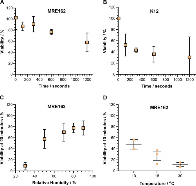
Two strains of E. coli were used in the measurements in this study: MRE162 and K12. MRE162 has been used for airborne survival studies for many years (21, 22, 34–36) and its airborne survival is particularly well characterized, making it ideal for a mechanistic study such as this. As such, MRE162 is the strain used in most of the measurements performed. K12 is also a well-characterized strain commonly used in laboratory study of E. coli and is used in this study to explore the impact of deleting various genes on the airborne survival of E. coli. The airborne viability of both strains is compared over the course of 20 min of levitation in LB broth droplets at 50% RH in Fig. 1A and B. Both strains show a loss of culturability during 20 min of levitation at 50% RH, but K12 is significantly less stable (P = 0.009 at 120 s, P = 0.0002 at 300 s, and P = 0.002 at 600 s), falling to a mean viability of 30% ± 37% in 20 min (Fig. 1B), while MRE162 decreased to a mean of 57% ± 17% (Fig. 1A). It should be noted that the 50% RH survival curve for MRE162 presented here differs from a CELEBS generated survival curve for the same conditions published in 2020. This is the result of several improvements to the CELEBS technique that have taken place since those measurements were made (Fig. S1).
FIG 1.
General characterization of the airborne survival of E. coli. CELEBS measurements of the airborne survival of E. coli MRE162 and K12 in droplets of LB broth. For A to C datapoints show the mean, error bars show the standard deviation. (A) The airborne survival of E. coli MRE162 over 20 min at 50% ± 1% RH, room temperature (18 to 21°C). For 120, and 600 s n = 3, for 300 s n = 9, for 1,200 s n = 13. (B) The airborne survival of E. coli K12 over 20 min at 50% ± 1% RH, room temperature (18 to 21°C). For 120 s n = 18, for 300, and 1,200 s n = 3, for 600 s n = 6. (C) The relationship between relative humidity and the survival of E. coli MRE162 after 20 min of levitation at room temperature (18 to 21°C). For 30% and 80% RH n = 3, for 50% RH n = 13, for 70% RH n = 6, for 90% RH n = 11. (D) The relationship between temperature and the survival of E. coli MRE162 after 10 min of levitation at 30% ± 1% RH. Each point shows the result of an individual measurement. The middle line indicates the mean with the error bars showing the standard deviation. For 10°C and 30°C n = 2, for 18°C n = 3.
A relationship with RH is observable in the airborne stability of MRE162 in LB broth droplets, with a mean viability of 9% ± 6% after 20 min at 30% RH and 78% ± 12% at 90% RH (Fig. 1C). This RH influence is in qualitative agreement with previous studies of bacterial airborne viability that show a positive correlation between RH and the viability of the bacteria (19, 37–39). Although, an opposite trend is occasionally reported in older studies (40). In addition, a clear impact of temperature on the viability of airborne MRE162 was observable. At 18°C (room temperature) in LB broth droplets at 30% RH, the mean viability after 10 min was 27% ± 13%. When the temperature was increased to 30°C, this viability decreased to 11% ± 5%, and when the temperature was decreased to 10°C the viability increased to 48% ± 12% (Fig. 1D). A similar relationship between temperature and the airborne survival of E. coli has been previously observed (41).
The observations reported in Fig. 1 provide the starting point for an investigation of the mechanism that drives the loss of viability of E. coli in airborne LB broth droplets. The relationship between temperature and the loss of viability indicates that the mechanism of viability loss is, at least in part, thermodynamically driven. The mechanism is evidently specific to the aerosol form of LB broth, as it is well established that E. coli do not lose viability in bulk solutions of LB broth at these temperatures (42). It is therefore unsurprising that the loss of viability is greater at lower relative humidity, as the factors that differentiate airborne droplets from bulk solutions are exacerbated as the RH is lowered. For example, increased loss of water from the airborne droplet will drive increased dehydration of the bacteria, increased exposure of bacteria to solutes within the droplet, and increased interaction of the droplet with gaseous reactants at the air-liquid interface, any of which could contribute the observed loss of viability. At 30% RH and below, the salts within droplets of LB broth become sufficiently concentrated to effloresce via homogenous nucleation (21, 27), which was initially suspected to drive the particularly rapid loss of viability at low RH.
Airborne droplet efflorescence does not appear to impact the viability of E. coli.
Previous CELEBS measurements with SARS-CoV-2 have demonstrated that the relationship between RH and viral airborne stability is explained, in part, by an immediate 50% to 60% loss of infectious virus upon the efflorescence of the droplet at low RH (33). However, droplet efflorescence does not appear to drive a similar loss of viability in CELEBS measurements of E. coli (21). While the loss of SARS-CoV-2 infectivity occurred almost immediately as the droplet crystallized, the loss of E. coli MRE162 viability was not readily observable until after 1 min of levitation at 30% RH, despite LB broth efflorescing within seconds at 30% RH (21, 27). To further explore the impact of phase on bacterial survival, E. coli MRE162 was levitated in PBS droplets at RHs both above and below the efflorescence threshold. PBS was used in place of LB broth as the lack of organic material in PBS means that it can be expected to effloresce in a more complete and reproducible manner than LB broth (21, 27), and, therefore, its efflorescence was expected to be more likely to drive a loss of viability.
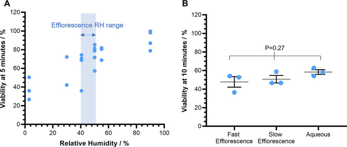
The remaining viability from 5-min levitations of E. coli in PBS droplets at gradually decreasing relative humidity are reported in Fig. 2A. The typical relationship between RH and airborne survival can be seen in PBS with survival being highest at 90% RH and lowest survival at 3% RH. There is some variation in the precise RH at which a solution will be seen to effloresce (43), and so a range in RH at which PBS may reach the threshold water activity for efflorescence to occur is indicated in Fig. 2A, with efflorescence always taking place below this range but never above it. While the relationship between RH and survival is observable, no stepwise drop in survival was observed as the RH crossed the efflorescence threshold, providing no evidence of phase change driving an increased loss of viability.
FIG 2.
The impact of efflorescence on the airborne survival of E. coli. CELEBS measurements of the airborne survival of E. coli MRE162 in droplets of PBS at room temperature (18 to 21°C). (A) The relationship between RH and the survival of E. coli MRE162 after 5 min of levitation. The RH range within which the efflorescence threshold for PBS will be crossed is indicated by the blue shaded area. Each point shows the result of an individual measurement. For 3% and 55% RH n = 3, for 30% RH n = 2, for 40%, and 90% RH n = 4, for 50% RH n = 6. (B) The impact of the physical state of the equilibrated particle on the survival of E. coli MRE162 after 10 min of levitation at room temperature. Fast efflorescence RH was set to 21.7% and all particles effloresced within 5 s of generation; slow efflorescence RH was set to 31.7% and all particles effloresced between 7 and 120 s after generation; the aqueous particles the RH was set to 46.7%. Each point shows the result of an individual measurement. The middle line indicates the mean with the error bars showing the standard deviation. n = 3 for all measurements.
To further investigate the consequences of efflorescence for viability, a similar experiment was carried out with 10-minute levitations. During this measurement, droplet phase was inferred from changes in the light scattering intensity and positional stability of the droplet observed with the camera viewing the trapped droplets from above the ring electrodes in the CELEBS (Fig. 2B). At the highest RH (47%), the droplets remained aqueous throughout the measurement, and the average viability remained at 58% ± 4%. At the intermediate RH (32%) all droplets were observed to effloresce between 7 s and 2 min after droplet generation, and the average viability after 10 min was 51% ± 7%. At the lowest RH (22%) all droplets were observed to effloresce within 5 s of generation and the average viability was 48% ± 10% after 10 min. Again, while the RH trend is observable, no significant difference was observed between the decreases in viability that take place in the effloresced droplets compared to the aqueous droplets. Rather than the efflorescence driving the viability loss, it may instead be postulated that, in the increasingly desiccated conditions as RH is lowered, an efflux of water from the bacteria could be responsible for the loss of viability.
Dehydration is unlikely to contribute to the airborne loss of viability of E. coli.
The requirement for moisture to allow bacterial growth is well understood, with E. coli needing a water activity of 0.95 or more (44), equivalent to droplets equilibrated to 95% RH. However, growth inhibition does not equate to a permanent loss of viability in bacteria. It is thought that reduced cellular hydration of bacteria results in damage to proteins, lipids, and nucleic acid through a variety of mechanisms (45, 46). However, past studies measuring desiccation induced losses of E. coli culturability tend to do so over too long a timescale to be applicable to the rapid airborne loss of viability reported here (47), making it difficult to determine whether the low water activity of airborne droplets could drive the airborne loss of viability of E. coli.
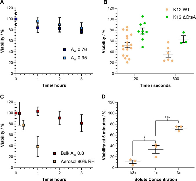
To measure the potential impact of low water activity (aw) on E. coli MRE162, the bacteria were resuspended in two bulk aqueous sodium chloride solutions, one with an aw of 0.95, which is not expected to negatively impact the survival of E. coli, and the other with salt dissolved to the bulk saturation limit, giving it an approximate aw of 0.76 (Fig. 3A). A small reduction in the mean CFU count was observable in both solutions over 3 h, both falling to approximately 80% of the initial count, but there was no significant difference in culturability of E. coli between the two suspensions. It is clear that at humidities of 76% or greater, other factors must account for a significant proportion of the loss of viability, with the airborne viability of E. coli MRE162 falling below 80% in only 20 min, even at 90% RH.
FIG 3.
Exploring the relationship between RH and E. coli airborne survival. CELEBS and bulk measurements of the survival of E. coli MRE162 and K12 at room temperature (18 to 21°C). A and C datapoints show the mean, error bars show the standard deviation. B and D datapoints show the results of individual measurements, the middle line shows the mean, the error bars show the standard deviation. (A) Survival of E. coli MRE162 over 3 h in bulk solutions of NaCl. The light blue points show the survival in a more dilute solution with a water activity of 0.95 and the dark blue points show the survival in a more concentrated solution with a water activity of 0.76. n = 5 for all measurements. (B) The airborne survival of E. coli K12 (beige points) and an OtsA deficient mutant of E. coli K12 (green points) after 2 and 10 min of levitation at 50% ± 1% RH, in LB broth droplets. (C) Comparison of the survival of E. coli MRE162 after 1 h of levitation in LB broth droplets at 80% RH (lighter beige points labeled Aerosol 80% RH) to the survival over 3 h in a bulk solution of concentrated LB broth matching the concentrations of all components in an airborne droplet equilibrated to 80% ± 1% RH (orange points labeled bulk Aw 0.8). For all measurements in the bulk solution n = 6, for the CELEBS measurement n = 3. (D) The airborne survival of E. coli MRE162 after 5 min at 30% ± 1% RH, in LB broth droplets with either a third or three times the initial solute concentration compared to the survival in LB broth with a normal initial solute concentration; *, P < 0.05; ***, P < 0.001.
The disaccharide trehalose serves as an osmoprotectant for E. coli and its production rate is increased in response to low water activity conditions (48–50). It can be hypothesized that deletion of the OtsA gene (encoding trehalose-6-phosphate synthase) in E. coli, shown to significantly lower intracellular concentrations of trehalose (51–53), would diminish airborne survival if osmotic stress is a major contributor to airborne loss of viability in E. coli. However, for the time points studied, the K12 ΔOtsA appeared to have higher viability than K12 WT (Fig. 3B) at 50% RH, remaining at 78% ± 17% viability after 2 min while K12 WT fell to 52% ± 19% under the same conditions. It is possible that the improvement to survival could be the result of increased persister cell formation in OtsA deletion mutants (54). The exact mechanism by which trehalose shields E. coli from osmotic stress is not fully characterized, although it is thought to act as a molecular chaperone, forming hydrogen bonds with biological macromolecules and stabilizing them (55). The absence of a detrimental impact of OtsA deletion on airborne survival in this experiment indicates that it is unlikely that airborne loss of viability in E. coli is caused by the same molecular damage against which trehalose should protect the cell. Taken alongside the difficulty in inducing a loss of viability through bulk exposure to lowered aw, it appears unlikely that dehydration, driven by the low water activity of airborne droplets, plays a major role in the airborne loss of viability of E. coli.
Loss of airborne viability correlated with surface area-to-volume ratio.
It is only possible in bulk LB broth solutions to replicate the concentrations of components in an airborne droplet equilibrated to high RH (equivalent to aw) of 80% and above. Solutes spontaneously form solute crystals below this water activity and the solute concentrations in bulk solution are sustained at their solubility limit. At 80% RH, the volume of a droplet of LB broth decreases by a factor of approximately 15 (21) from the typical starting volume generated at the high water activities of stock solutions used in CELEBS (aw 0.995). Similar behavior occurs in any release of aerosol from an initially dilute solution at high water activity (e.g., from an aqueous slurry or on exhalation of aerosols from the respiratory tract). The volume change concentrates all solutes within the droplet by the same factor and the concentrations of the key components of LB broth are approximately 150 g/L NaCl, 150 g/L tryptone, and 75 g/L yeast extract at aw ~0.8.
E. coli MRE162 was suspended in a concentrated broth at aw ~0.8, also with a bacterial concentration 15 times that typically prepared for levitations, with the viability monitored by CFU count. This measurement can then be compared to a CELEBS levitation of E. coli MRE162 in LB broth at 80% RH, as the concentrations of all components within the droplets were the same and should any of those components drive the airborne loss of viability, the same loss of viability would be observable in this bulk solution (Fig. 3C). However, the viability of the bacteria remained at 100% after the first hour, and only fell to 81% ± 16% after 3 h in the bulk suspension. For comparison, the viability fell to an average of 39% ± 18% after only 1 h in droplets levitated at 80% RH. This measurement provides considerable insight into the mechanism driving the airborne loss of viability of E. coli in LB broth, as the only differences between the bulk solution and levitated droplets in this experiment were the dynamic changes undergone by the airborne droplet during the initial equilibration of the droplet and the final ratio of the surface area-to-volume of the volume confining the bacteria. The 5-mL bulk solution in a test-tube with a radius of 2 cm has a surface area-to-volume ratio of approximately 2.5 × 10⁻⁴ (1:4,000). An airborne droplet with a radius of 25 μm has an initial surface area-to-volume ratio of 1.2 × 10⁻¹ (1:8), which increases to 3 × 10⁻¹ (1:3) when equilibrated to 80% RH. The increased loss of viability in airborne droplets may be related to the higher surface area-to-volume ratio. Increased loss of viability with increasing surface area-to-volume ratio could also explain the role of RH, as a lower RH will result in more water loss from the droplet, and therefore, a lower equilibrated size and higher surface area-to-volume ratio.
Disentangling the impacts of droplet size change (i.e., surface area-to-volume ratio) and solute concentration is possible by altering the initial concentration of a solution before aerosolizing it. Five-min levitations were carried out at 30% RH in LB broth droplets of either a normal concentration (25 g/L) diluted to a third of the normal concentration (8.3 g/L) or at 3 times the normal concentration (75 g/L). These three formulations levitated at the same RH all equilibrate to the same solute concentrations, but to different final droplet sizes, necessarily losing differing amounts of water to reach the same equilibrated concentration. As a minor consideration, the different starting concentrations will also lead to differences in the evaporation kinetics although these differences are small and occur over a time period during which no loss of viability is observed (see Fig. S2 for simulations of the evaporation kinetics of these different starting solution droplets using previously collected physicochemical data [21]).
The difference in equilibration size has a profound effect on the viability of the bacteria, with a clear positive correlation between equilibrated size and viability observed (Fig. 3D). In the droplets with the more dilute-starting formulation, the droplet radius at equilibration can be estimated to be 5.8 μm (surface area-to-volume ratio of 5.2 × 10⁻¹) and the viability fell to 11% ± 6% after 5 min. In the normal formulation, the radius at equilibration is 8.3 μm (surface area-to-volume ratio of 3.6 × 10⁻¹) and the viability fell to 34% ± 12% after 5 min. With the more concentrated LB broth, the radius at equilibration is 12 μm (surface area-to-volume ratio of 2.5 × 10⁻¹) and the average viability remained at 73% ± 5% after 5 min. This same trend has also previously been observed using the CELEBS technique at 50% RH (21). All the evidence collected at this point indicates that the surface area-to-volume ratio of the airborne droplet is a key parameter that correlates with the rate of loss of viability for E. coli.
E. coli airborne loss of viability is reduced in a hypoxic environment.
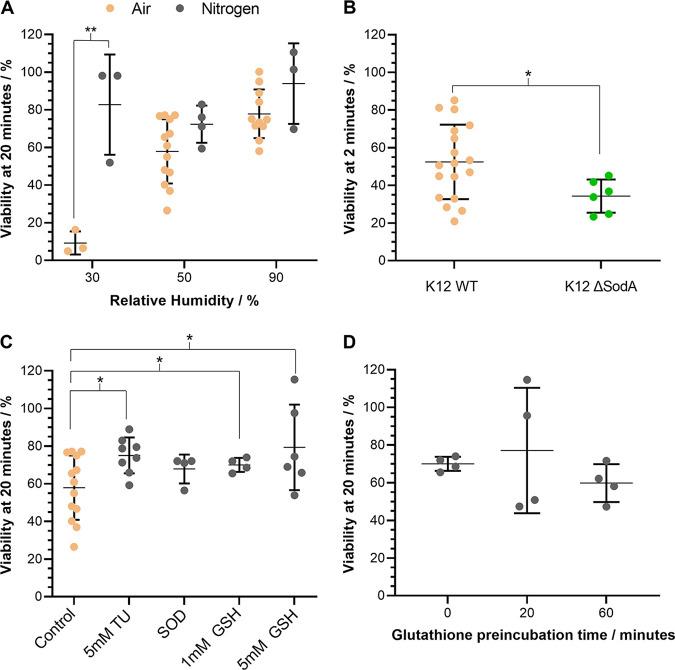
The apparent link between the surface area-to-volume ratio of the equilibrated droplet and the rate of viability loss suggests that exposure to a gas phase component may drive the loss of viability. To determine if the presence of oxygen has an impact on the loss of viability, CELEBS measurements were repeated with the airflow replaced by a flow of pure nitrogen (Fig. 4A). In all cases, replacing the gas flow with nitrogen increased the average airborne viability of E. coli MRE162, but only at 30% RH was a significant difference observable (P < 0.01). While a clear impact of RH is observable in the presence of air, this trend is not observed in the nitrogen gas flow. Thus, the clearest impact of changing the gas phase from air to nitrogen is observed at the lowest RH, with the average viability after 20 min in 30% RH air being 9% ± 6% and the average viability in 30% RH nitrogen being 83% ± 27%. These results are in agreement with the findings of early rotating drum studies, in which a decreased airborne loss of viability was observed in the absence of oxygen for both E. coli and Serratia marcescens (17–19, 56), as well as a more recent study of the effects of spray drying on Lactococcus lactis (57).
FIG 4.
The impact of oxidative stress on the airborne survival of E. coli. CELEBS measurements of the airborne survival of E. coli MRE162 and K12 in droplets of LB broth at room temperature (18 to 21°C). Datapoints show the results of individual measurements, the middle line shows the mean, error bars show the standard deviation. (A) Comparison of the airborne survival of E. coli MRE162 after 20 min of levitation in air (beige points), to levitations for 20 min in nitrogen (gray points), at 30% ± 1%, 50% ± 1%, and 90% ± 1% RH. **, P < 0.01. (B) The airborne survival of E. coli K12 (beige points) and a SodA deficient mutant of E. coli K12 (green points) after 2 min of levitation at 50% ± 1% RH. *, P < 0.05. (C) The effect of the presence of 5 mM thiourea (5 mM TU), 1 unit/mL superoxide dismutase (SOD), 1 mM glutathione (1 mM GSH), and 5 mM glutathione (5 mM GSH) on the airborne survival of E. coli MRE162 after 20 min of levitation at 50% ± 1% RH. Measurements are compared to 20 min of levitation at 50% ± 1% RH in LB broth without antioxidants (Control). *, P < 0.05. (D) The effect of preincubation at 37°C of a suspension of E. coli MRE162 in LB broth with 1 mM glutathione, on subsequent airborne survival measurements of 20 min at 50% ± 1% RH using the suspension.
The apparently intertwined impacts of droplet size and oxygen presence on the loss of viability further narrows down the potential causes of the loss of viability. It has been demonstrated in a bulk phase study that aerobic conditions increase E. coli loss of viability driven by high salt concentrations and low pH (58). It could be suggested that the high surface area to volume ratio results in a higher concentration of oxygen in smaller droplets. However, the diffusion rate of oxygen at room temperature within a droplet of the sizes studied here is such that the concentration of oxygen will be maintained at saturation concentration (determined by Henry’s law solubility) throughout the levitation and homogeneously throughout the droplet, regardless of droplet size and bacterial metabolism within the droplet (59). Thus, the apparent impact of droplet size on survival cannot be explained by differences in the concentration of dissolved oxygen (Fig. 3D). Furthermore, the loss of viability observed in the bulk solutions (Fig. 3A and C) was very slow. While the oxygen concentration in those bulk solutions will potentially have been below saturation, they will not have been anaerobic. If the loss of viability was a combination of the high concentration of solutes, bacteria, and the presence of oxygen, a more significant loss of viability would likely have been observed in the bulk solution of Fig. 3C. It seems instead that the mechanism of airborne viability loss requires a combination of the presence of oxygen and the high surface area-to-volume ratio of airborne droplets. Previous studies have demonstrated unique chemistry at air liquid interfaces (60–62), and it is possible that oxygen is able to form products harmful to the bacteria through such surface chemistry.
Reactive oxygen species formation drives airborne loss of viability in E. coli.
A K12 ΔSodA mutant was found to have significantly reduced survival after a 2-min levitation at 50% RH compared to K12 WT, with a reduction in viability from 52% ± 20% to 34% ± 9% (P < 0.05) (Fig. 4B). SodA encodes one of three superoxide dismutases expressed by E. coli and its deletion renders the bacteria less able to process superoxide radicals, as demonstrated by an increased susceptibility to paraquat (63, 64). However, while an increased loss of airborne viability when SodA is deleted could indicate a role of superoxide radicals in the airborne loss of viability, it is also possible that reducing the capacity of the bacteria to process reactive oxygen species (ROS) merely introduced oxidative stress as an additional mechanism of viability loss. It was, however, noteworthy that of all the isogenic mutants of K12 studied, it was only the ΔSodA mutant that demonstrated reduced airborne viability (Fig. S3).
To further explore the role of ROS in the airborne loss of viability of E. coli, several antioxidants were added to the droplets prior to levitation. The antioxidants tested were: thiourea, a scavenger of superoxide and hydroxyl radicals (65); purified superoxide dismutase from bovine liver; and glutathione, a naturally occurring thiol capable of quenching reactive oxygen species both directly and through enzyme catalyzed mechanisms (66). The addition of 5 mM thiourea and 1 mM glutathione significantly (P-values compared to the control of 0.018 and 0.032) increased the mean survival of E. coli MRE162 when levitated for 20 min in LB broth droplets in 50% RH air (Fig. 4C). The addition of water alone did not influence airborne viability (Fig. S4). The consistent improvement in survival upon antioxidant addition, coupled with the diminished airborne survival of K12 ΔSodA, indicate a role of ROS in the airborne loss of viability of E. coli within the 20-minute measurement time.
Many forms of stress have been demonstrated to result in the formation of ROS within bacterial cells (46, 67–69), but the measurements with antioxidants indicate that the ROS causing damage to the bacteria were being formed extracellularly. While uptake of small molecules such as thiourea and glutathione by E. coli is possible, and is a well-characterized process for glutathione (70), it seemed doubtful that the 20 min of levitation would allow sufficient time for this uptake to impact the viability. To further verify the extracellular nature of the ROS formation, the measurements with 1 mM glutathione were repeated. However, the suspension of bacteria with the glutathione was placed in a 37°C incubator for varying lengths of time prior to levitation to allow the antioxidant to enter the bacteria prior to levitation (Fig. 4D). This preincubation did not result in an improvement to the airborne viability, perhaps indicating that the impact of glutathione was not contingent of the glutathione needing to enter the bacterial cell prior to levitation. Taken together, the measurements with antioxidants suggest a role of ROS in the airborne loss of viability of E. coli and that the ROS causing the loss of viability could be formed in the droplet rather than within the bacteria.
DISCUSSION
The measurements presented provide insights into the mechanisms driving airborne loss of viability in E. coli. Osmotic stress, initially an obvious candidate for driving the loss of viability, appears unlikely to drive a loss of viability at the rates observed in aerosols, especially at higher RHs. While in previous measurements with SARS-CoV-2, efflorescence of the droplet appeared to correspond to a large loss of virus in the droplet (33), this was not the case for E. coli, with no significant differences in survival in aqueous versus effloresced droplets being observable. Instead, it appears that the presence of oxygen around the droplet is one of the contributing factors, allowing the formation of ROS at the air-liquid interface, which gradually damage and kill the bacteria. The rate at which this happens is determined both by the temperature and the surface area-to-volume ratio of the droplet, with smaller droplets facilitating greater exposure of the bacteria to potential oxidative damage. It can be speculated that while the low water activity of the droplet alone does not have an effect, it could play a role in increasing the rate of oxidative damage by preventing the bacteria from producing the enzymes needed to protect itself against oxidants and by reducing the activity of those enzymes already present within the bacteria. Previous studies have demonstrated increased ROS concentrations in plants experiencing dehydration (71–74), and it is thought that this also occurs in bacterial cells (45).
There are still several unanswered questions surrounding the loss of viability in airborne E. coli. While it can be speculated that unique air-liquid interface chemistry will facilitate the formation of ROS from gaseous oxygen at the surface of the droplet, the precise nature of this mechanism remains a mystery. There are also potential mechanisms of bacterial airborne viability loss that were not explored in this study. As airborne droplets evaporate and shrink, it is likely that their pH will change. While relatively stable from pH 4 to 10, E. coli will likely experience a loss of viability in airborne droplets that reach extremes of acidity or alkalinity (75, 76). However, the pH change an evaporating droplet of LB broth will undergo upon aerosolization is not yet known, and difficult to measure making the influence of pH on viability loss in the system studies here difficult to investigate. Exhaled aerosol becomes alkaline upon exposure to ambient CO2 concentrations due to the bicarbonate content of respiratory secretions (33, 77, 78), which is a process not replicated in LB broth, but could be explored in future studies of bacteria more likely to be present in exhaled aerosol, such as Streptococcus spp. It is also unknown if an enrichment of bacterial cells at the surface of the droplet could play a role in their loss of viability. ROS have very short half-lives (79), meaning that if they are formed at the air-liquid interface, the risk to the bacteria is likely greater at the droplet surface. The role of the surface area-to-volume ratio of the droplet could therefore be 2-fold, both presenting a greater proportion of the droplet to the ROS forming conditions and increasing the likelihood of the bacteria being exposed to the ROS. The degree of contact between bacteria and the droplet surface will also be affected both by the viscosity of the droplet and the motility of the bacteria.
This systematic approach provides clear insights into the possible mechanisms of airborne damage to bacteria, a phenomenon that has been observed for over 70 years (40) but does not yet have an agreed upon mechanistic explanation. While E. coli in droplets of LB broth is limited in terms of its relevance to real world biological phenomena, the focus on a well understood and highly controlled system has made possible deeper insights into relationships between the physicochemical properties of the droplet and the biological response of the bacteria. The results of this study provide a starting point for the exploration of more complex biologically relevant systems and will allow for future measurements with different bacterial species in different droplet compositions, to be interpreted in the context of specific processes, such as oxidative and osmotic stress. Such measurements will improve our understanding of what remains among the most mysterious processes in microbiology, and will not only give us useful insights into airborne disease transmission, but will also deepen our understanding of bacterial ecology as a whole.
MATERIALS AND METHODS
Strains and growth conditions.
E. coli MRE162 and K12 were used for these experiments. K12 isogenic mutants were obtained from the Yale University Keio collection. Bacteria were typically grown at 37°C for 18 h to 24 h in LB broth (NaCl 10 g L−1, tryptone 10 g L−1, yeast extract 5 g L−1, dissolved in distilled water). Broth cultures were incubated for 20 h to 24 h, such that bacteria would reach early to midstationary phase of growth prior to measurement.
Measuring viability in aerosols.
Bacterial culture was diluted to OD600 0.5 in LB broth (or PBS) prior to the experiment. The diluted culture was then loaded into the droplet dispenser, which was aligned with the electrodynamic balance (EDB) in the center of the CELEBS chamber. Several droplets (typically four per measurement) with a radius of 24 to 28 μm each, were injected through an induction electrode and into the EDB. The charge applied to the droplets by the induction electrode allowed for the droplets to be trapped in an oscillating electric field created by the ring electrodes of the EDB. The LabVIEW software used to control CELEBS changes the frequency of the field over time, which allowed for droplets to be kept in the field while they evaporated, and their mass to charge ratio changed. This principle has been previously described (3, 4).
The atmosphere around the EDB was maintained at the desired relative humidity through an air inlet angled toward the EDB, the humidity of which can be adjusted by altering the ratio of dry to wet air. The RH of the airflow was measured by a Vaisala HMT330 RH probe prior to entering the levitation chamber, which was used to ensure the airflow entering the chamber was within 1% of the desired RH. The air supply from the compressor could be replaced with a supply of pure nitrogen from a pressurized cylinder to create a hypoxic environment around the droplets. A plate containing LB agar was kept in a compartment beneath the EDB, and a small amount of liquid LB broth was applied to the surface of the plate. After the droplets had been suspended for the desired length of time, the compartment was opened, causing the droplets to deposit into the broth on the surface of the plate. The plate was then incubated at room temperature for approximately 5 min before the broth was spread evenly across the surface and placed into an incubator overnight at 37°C.
Calculating bacteria per droplet and percentage survival.
The number of bacterial colonies on the agar plate for each levitation was counted the day after the experiment to give the CFU for each levitation. A camera was positioned above the EDB, allowing for the number of droplets trapped for each measurement to be counted and recorded. The CFU per droplet for each levitation was calculated by dividing the number of CFU on the plate by the number of droplets deposited onto the plate. For each experiment, several 5-s levitations were performed to measure the initial CFU per droplet allowing for the results of each experiment to be expressed in terms of percentage survival, making comparison of results between experiments easier.
Measuring bulk phase changes in viability.
Bacterial cultures were grown overnight as standard. The cultures were then centrifuged for 5 min with a relative centrifugal force of 8,200 × g. The resulting pellets were then resuspended in the test solutions. The viability of bacteria in the solution over time was assessed by removing 100 μL from the solution at each time point and diluting it into LB broth at a factor of 106. The number of bacteria within this solution was then assessed by CFU counting. The CFU counts were normalized to a measurement taken at the start of the experiment to allow for values to be expressed in terms of percentage survival.
Statistical analysis.
For each comparison, an F-test was used to determine if the variance of the two data sets was equal. Depending on the results of the F-test, P values were calculated using a Student's t test either accounting for equal or unequal variance. In the case of multiple comparisons to a control or comparisons between time course data sets, an ANOVA is first carried out to test for significance, followed by multiple t-tests with alpha values adjusted using the Bonferroni-Holm correction for multiple comparisons.
Data availability.
The txt file data have been deposited in data.bris, the University of Bristol Research Data repository: https://doi.org/10.5523/bris.1u96yv19o3ojh2ow3zpttwp3e4.
Supplementary Material
ACKNOWLEDGMENTS
This work was supported by funding from EPSRC and DSTL. We thank Thomas Hilditch for his suggestion to alter initial solute concentration, and Alexander Hughes-Games for his advice regarding potential airborne death mechanisms.
We declare no conflicts of interest.
Footnotes
Supplemental material is available online only.
Contributor Information
Henry P. Oswin, Email: ho1451@bristol.ac.uk.
Jonathan P. Reid, Email: J.P.Reid@bristol.ac.uk.
Jannell V. Bazurto, University of Minnesota Twin Cities
REFERENCES
- 1.Darzins A, Russell MA. 1997. Molecular genetic analysis of type-4 pilus biogenesis and twitching motility using Pseudomonas aeruginosa as a model system - a review. Gene 192:109–115. doi: 10.1016/S0378-1119(97)00037-1. [DOI] [PubMed] [Google Scholar]
- 2.Kitao A, Hata H. 2018. Molecular dynamics simulation of bacterial flagella. Biophys Rev 10:617–629. doi: 10.1007/s12551-017-0338-7. [DOI] [PMC free article] [PubMed] [Google Scholar]
- 3.Patrick JE, Kearns DB. 2012. Swarming motility and the control of master regulators of flagellar biosynthesis. Mol Microbiol 83:14–23. doi: 10.1111/j.1365-2958.2011.07917.x. [DOI] [PMC free article] [PubMed] [Google Scholar]
- 4.Nan B, Zusman DR. 2016. Novel mechanisms power bacterial gliding motility. Mol Microbiol 101:186–193. doi: 10.1111/mmi.13389. [DOI] [PMC free article] [PubMed] [Google Scholar]
- 5.Wadhwa N, Berg HC. 2021. Bacterial motility: machinery and mechanisms. Nat Rev Microbiol 20:161–173. [DOI] [PubMed] [Google Scholar]
- 6.Wood ME, Stockwell RE, Johnson GR, Ramsay KA, Sherrard LJ, Kidd TJ, Cheney J, Ballard EL, O'Rourke P, Jabbour N, Wainwright CE, Knibbs LD, Sly PD, Morawska L, Bell SC. 2019. Cystic fibrosis pathogens survive for extended periods within cough-generated droplet nuclei. Thorax 74:87–90. doi: 10.1136/thoraxjnl-2018-211567. [DOI] [PubMed] [Google Scholar]
- 7.Jones-López EC, Namugga O, Mumbowa F, Ssebidandi M, Mbabazi O, Moine S, Mboowa G, Fox MP, Reilly N, Ayakaka I, Kim S, Okwera A, Joloba M, Fennelly KP. 2013. Cough aerosols of Mycobacterium tuberculosis predict new infection: a household contact study. Am J Respir Crit Care Med 187:1007–1015. doi: 10.1164/rccm.201208-1422OC. [DOI] [PMC free article] [PubMed] [Google Scholar]
- 8.Eames I, Tang JW, Li Y, Wilson P. 2009. Airborne transmission of disease in hospitals. J R Soc Interface 6:S697–S702. [DOI] [PMC free article] [PubMed] [Google Scholar]
- 9.Bryan NC, Christner BC, Guzik TG, Granger DJ, Stewart MF. 2019. Abundance and survival of microbial aerosols in the troposphere and stratosphere. ISME J 13:2789–2799. doi: 10.1038/s41396-019-0474-0. [DOI] [PMC free article] [PubMed] [Google Scholar]
- 10.Harris MJ, Wickramasinghe NC, Lloyd D, Narlikar JV, Rajaratnam P, Turner MP, Al-Mufti S, Wallis MK, Ramadurai S, Hoyle F. 2002. The detection of living cells in stratospheric samples, p 192–198. Instruments, Methods, and Missions for Astrobiology IV. [Google Scholar]
- 11.Smith DJ, Ravichandar JD, Jain S, Griffin DW, Yu H, Tan Q, Thissen J, Lusby T, Nicoll P, Shedler S, Martinez P, Osorio A, Lechniak J, Choi S, Sabino K, Iverson K, Chan L, Jaing C, McGrath J. 2018. Airborne bacteria in earth’s lower stratosphere resemble taxa detected in the troposphere: results from a new NASA Aircraft Bioaerosol Collector (ABC). Front Microbiol 9:1–20. doi: 10.3389/fmicb.2018.01752. [DOI] [PMC free article] [PubMed] [Google Scholar]
- 12.Walther BA, Ewald PW. 2004. Pathogen survival in the external environment and the evolution of virulence. Biol Rev Camb Philos Soc 79:849–869. doi: 10.1017/s1464793104006475. [DOI] [PMC free article] [PubMed] [Google Scholar]
- 13.Lindow SE, Knudsen GR, Seidler RJ, Walter MV, Lambou VW, Amy PS, Schmedding D, Prince V, Hern S. 1988. Aerial dispersal and epiphytic survival of Pseudomonas syringae during a pretest for the release of genetically engineered strains into the environment. Appl Environ Microbiol 54:1557–1563. doi: 10.1128/aem.54.6.1557-1563.1988. [DOI] [PMC free article] [PubMed] [Google Scholar]
- 14.Möhler O, DeMott PJ, Vali G, Levin Z. 2007. Microbiology and atmospheric processes: the role of biological particles in cloud physics. Biogeosciences 4:1059–1071. doi: 10.5194/bg-4-1059-2007. [DOI] [Google Scholar]
- 15.Després VR, Alex Huffman J, Burrows SM, Hoose C, Safatov AS, Buryak G, Fröhlich-Nowoisky J, Elbert W, Andreae MO, Pöschl U, Jaenicke R. 2012. Primary biological aerosol particles in the atmosphere: a review. Tellus Chem Phys Meteorol 64. [Google Scholar]
- 16.Franc GD, Demott PJ. 1998. Cloud activation characteristics of airborne Erwinia carotovora cells. J Appl Meteorol 37:1293–1300. doi:. [DOI] [Google Scholar]
- 17.Cox CS, Baldwin F. 1967. The toxic effect of oxygen upon the aerosol survival of Escherichia coli B. J Gen Microbiol 49:115–117. doi: 10.1099/00221287-49-1-115. [DOI] [PubMed] [Google Scholar]
- 18.Cox CS. 1968. The aerosol survival of Escherichia coli B in nitrogen, argon and helium atmospheres and the influence of relative humidity. J Gen Microbiol 50:139–147. doi: 10.1099/00221287-50-1-139. [DOI] [PubMed] [Google Scholar]
- 19.Hess GE. 1965. Effects of oxygen on aerosolized Serratia marcescens. Appl Microbiol 13:781–787. doi: 10.1128/am.13.5.781-787.1965. [DOI] [PMC free article] [PubMed] [Google Scholar]
- 20.Johnson GR, Knibbs LD, Kidd TJ, Wainwright CE, Wood ME, Ramsay KA, Bell SC, Morawska L. 2016. A novel method and its application to measuring pathogen decay in bioaerosols from patients with respiratory disease. PLoS One 11:1–20. doi: 10.1371/journal.pone.0158763. [DOI] [PMC free article] [PubMed] [Google Scholar]
- 21.Fernandez MO, Thomas RJ, Oswin H, Haddrell AE, Reid JP. 2020. Transformative approach to investigate the microphysical factors influencing airborne transmission of pathogens. Appl Environ Microbiol 86:e01543-20. [DOI] [PMC free article] [PubMed] [Google Scholar]
- 22.Fernandez MO, Thomas RJ, Garton NJ, Hudson A, Haddrell A, Reid JP. 2019. Assessing the airborne survival of bacteria in populations of aerosol droplets with a novel technology. J R Soc Interface 16:20180779. doi: 10.1098/rsif.2018.0779. [DOI] [PMC free article] [PubMed] [Google Scholar]
- 23.Walker JS, Archer J, Gregson FKA, Michel SES, Bzdek BR, Reid JP. 2021. Accurate representations of the microphysical processes occurring during the transport of exhaled aerosols and droplets. ACS Cent Sci 7:200–209. doi: 10.1021/acscentsci.0c01522. [DOI] [PMC free article] [PubMed] [Google Scholar]
- 24.Hargreaves G, Kwamena N-OA, Zhang YH, Butler JR, Rushworth S, Clegg SL, Reid JP. 2010. Measurements of the equilibrium size of supersaturated aqueous sodium chloride droplets at low relative humidity using aerosol optical tweezers and an electrodynamic balance. J Phys Chem A 114:1806–1815. doi: 10.1021/jp9095985. [DOI] [PubMed] [Google Scholar]
- 25.Tong HJ, Reid JP, Bones DL, Luo BP, Krieger UK. 2011. Measurements of the timescales for the mass transfer of water in glassy aerosol at low relative humidity and ambient temperature. Atmos Chem Phys 11:4739–4754. doi: 10.5194/acp-11-4739-2011. [DOI] [Google Scholar]
- 26.Hanford KL, Mitchem L, Reid JP, Clegg SL, Topping DO, McFiggans GB. 2008. Comparative thermodynamic studies of aqueous glutaric acid, ammonium sulfate and sodium chloride aerosol at high humidity. J Phys Chem A 112:9413–9422. doi: 10.1021/jp802520d. [DOI] [PubMed] [Google Scholar]
- 27.Huynh E, Olinger A, Woolley D, Kohli RK, Choczynski JM, Davies JF, Lin K, Marr LC, Davis RD. 2022. Evidence for a semisolid phase state of aerosols and droplets relevant to the airborne and surface survival of pathogens. Proc Natl Acad Sci 119:e2109750119. doi: 10.1073/pnas.2109750119. [DOI] [PMC free article] [PubMed] [Google Scholar]
- 28.Gregson FKA, Robinson JF, Miles REH, Royall CP, Reid JP. 2019. Drying kinetics of salt solution droplets: water evaporation rates and crystallization. J Phys Chem B 123:266–276. doi: 10.1021/acs.jpcb.8b09584. [DOI] [PubMed] [Google Scholar]
- 29.Qian C, Rui Y, Wang C, Wang X, Xue B, Yi H. 2021. Bio-mineralization induced by Bacillus mucilaginosus in crack mouth and pore solution of cement-based materials. Mater Sci Eng C Mater Biol Appl 126:112120. doi: 10.1016/j.msec.2021.112120. [DOI] [PubMed] [Google Scholar]
- 30.Lopez-Cortes A, Ochoa JL. 1999. The biological significance of Halobacteria on nucleation and sodium chloride crystal growth. Stud Surf Sci Catal 7:903–923. [Google Scholar]
- 31.Khvorostyanov VI, Curry JA. 2014. Deliquescence and efflorescence in atmospheric aerosols, pp 547–576. Thermodyn Kinet Microphys Clouds. Cambridge University Press, Cambridge, United Kingdom. doi: 10.1017/cbo9781139060004.012. [DOI] [Google Scholar]
- 32.Oswin HP, Haddrell AE, Otero-Fernandez M, Cogan TA, Mann JFS, Morley CH, Hill DJ, Davidson AD, Finn A, Thomas RJ, Reid JP. 2021. Measuring stability of virus in aerosols under varying environmental conditions. Aerosol Sci Technol 55:1315–1320. doi: 10.1080/02786826.2021.1976718. [DOI] [Google Scholar]
- 33.Oswin HP, Haddrell AE, Otero-Fernandez M, Mann JFS, Cogan TA, Hilditch TG, Tian J, Hardy DA, Hill DJ, Finn A, Davidson AD, Reid JP. 2022. The dynamics of SARS-CoV-2 infectivity with changes in aerosol microenvironment. Proc Natl Acad Sci USA 119:1–42. [DOI] [PMC free article] [PubMed] [Google Scholar]
- 34.Strange RE, Benbough JE, Hambleton P, Martin KL. 1972. Methods for the assessment of microbial populations recovered from enclosed aerosols. J Gen Microbiol 72:117–125. doi: 10.1099/00221287-72-1-117. [DOI] [PubMed] [Google Scholar]
- 35.Benbough JE, Hood AM. 1971. Viricidal activity of open air. J Hyg (Lond) 69:619–626. doi: 10.1017/s0022172400021896. [DOI] [PMC free article] [PubMed] [Google Scholar]
- 36.Handley BA, Webster AJF. 1993. Some factors affecting airborne survival of Pseudomonas fluorescens indoors. J Appl Bacteriol 75:35–42. doi: 10.1111/j.1365-2672.1993.tb03404.x. [DOI] [PubMed] [Google Scholar]
- 37.Stersky AK, Heldman DR, Hedrick TI. 1972. Viability of airborne Salmonella Newbrunswick under various conditions. J Dairy Sci 55:14–18. doi: 10.3168/jds.S0022-0302(72)85424-9. [DOI] [PubMed] [Google Scholar]
- 38.Wathes CM, Howard K, Webster AJF. 1986. The survival of Escherichia coli in an aerosol at air temperatures of 15 and 30°C and a range of humidities. J Hyg (Lond) 97:489–496. doi: 10.1017/s0022172400063671. [DOI] [PMC free article] [PubMed] [Google Scholar]
- 39.Cox CS. 1966. The survival of Escherichia coli sprayed into air and into nitrogen from distilled water and from solutions of protecting agents, as a function of relative humidity. J Gen Microbiol 43:383–399. doi: 10.1099/00221287-43-3-383. [DOI] [PubMed] [Google Scholar]
- 40.Dunklin EW, Puck TT. 1948. The lethal effect of relative humidity on airborne bacteria. J Exp Med 87:87–101. doi: 10.1084/jem.87.2.87. [DOI] [PMC free article] [PubMed] [Google Scholar]
- 41.Ehrlich R, Miller S, Walker RL. 1970. Relationship between atmospheric temperature and survival of airborne bacteria. Appl Microbiol 19:245–249. doi: 10.1128/am.19.2.245-249.1970. [DOI] [PMC free article] [PubMed] [Google Scholar]
- 42.Sezonov G, Joseleau-Petit D, D'Ari R. 2007. Escherichia coli physiology in Luria-Bertani broth. J Bacteriol 189:8746–8749. doi: 10.1128/JB.01368-07. [DOI] [PMC free article] [PubMed] [Google Scholar]
- 43.Sear RP. 2014. Quantitative studies of crystal nucleation at constant supersaturation: experimental data and models. CrystEngComm 16:6506–6522. doi: 10.1039/C4CE00344F. [DOI] [Google Scholar]
- 44.Marshall BJ, Ohye DF, Christian JH. 1971. Tolerance of bacteria to high concentrations of NaCl and glycerol in the growth medium. Appl Microbiol 21:363–364. doi: 10.1128/am.21.2.363-364.1971. [DOI] [PMC free article] [PubMed] [Google Scholar]
- 45.Lebre PH, De Maayer P, Cowan DA. 2017. Xerotolerant bacteria: surviving through a dry spell. Nat Rev Microbiol 15:285–296. doi: 10.1038/nrmicro.2017.16. [DOI] [PubMed] [Google Scholar]
- 46.Laskowska E, Kuczyńska-Wiśnik D. 2020. New insight into the mechanisms protecting bacteria during desiccation. Curr Genet 66:313–318. doi: 10.1007/s00294-019-01036-z. [DOI] [PMC free article] [PubMed] [Google Scholar]
- 47.Potts M. 1994. Desiccation tolerance of prokaryotes. Microbiol Rev 58:755–805. doi: 10.1128/mr.58.4.755-805.1994. [DOI] [PMC free article] [PubMed] [Google Scholar]
- 48.Iturriaga G, Suárez R, Nova-Franco B. 2009. Trehalose metabolism: from osmoprotection to signaling. Int J Mol Sci 10:3793–3810. doi: 10.3390/ijms10093793. [DOI] [PMC free article] [PubMed] [Google Scholar]
- 49.Welsh DT, Reed RH, Herbert RA. 1991. The role of trehalose in the osmoadaptation of Escherichia coli NCIB 9484: interaction of trehalose, K+ and glutamate during osmoadaptation in continuous culture. J Gen Microbiol 137:745–750. doi: 10.1099/00221287-137-4-745. [DOI] [PubMed] [Google Scholar]
- 50.Purvis JE, Yomano LP, Ingram LO. 2005. Enhanced trehalose production improves growth of Escherichia coli under osmotic stress. Appl Environ Microbiol 71:3761–3769. doi: 10.1128/AEM.71.7.3761-3769.2005. [DOI] [PMC free article] [PubMed] [Google Scholar]
- 51.Giaever HM, Styrvold OB, Kaasen I, Strøm AR. 1988. Biochemical and genetic characterization of osmoregulatory trehalose synthesis in Escherichia coli. J Bacteriol 170:2841–2849. doi: 10.1128/jb.170.6.2841-2849.1988. [DOI] [PMC free article] [PubMed] [Google Scholar]
- 52.Kaasen I, Falkenberg P, Styrvold OB, Strom AR. 1992. Molecular cloning and physical mapping of the otsBA genes, which encode the osmoregulatory trehalose pathway of Escherichia coli: evidence that transcription is activated by KatF (AppR). J Bacteriol 174:3422–3422. [DOI] [PMC free article] [PubMed] [Google Scholar]
- 53.Kandror O, DeLeon A, Goldberg AL. 2002. Trehalose synthesis is induced upon exposure of Escherichia coli to cold and is essential for viability at low temperatures. Proc Natl Acad Sci USA 99:9727–9732. doi: 10.1073/pnas.142314099. [DOI] [PMC free article] [PubMed] [Google Scholar]
- 54.Kuczyńska-Wiśnik D, Stojowska K, Matuszewska E, Leszczyńska D, Algara MM, Augustynowicz M, Laskowska E. 2015. Lack of intracellular trehalose affects formation of Escherichia coli persister cells. Microbiology (Reading) 161:786–796. doi: 10.1099/mic.0.000012. [DOI] [PubMed] [Google Scholar]
- 55.Jain NK, Roy I. 2009. Effect of trehalose on protein structure. Protein Sci 18:24–36. doi: 10.1002/pro.3. [DOI] [PMC free article] [PubMed] [Google Scholar]
- 56.Benbough JE. 1969. Factors affecting the toxicity of oxygen towards airborne coliform bacteria. J Gen Microbiol 56:241–250. doi: 10.1099/00221287-56-2-241. [DOI] [PubMed] [Google Scholar]
- 57.Ghandi A, Powell IB, Howes T, Chen XD, Adhikari B. 2012. Effect of shear rate and oxygen stresses on the survival of Lactococcus lactis during the atomization and drying stages of spray drying: a laboratory and pilot scale study. J Food Eng 113:194–200. doi: 10.1016/j.jfoodeng.2012.06.005. [DOI] [Google Scholar]
- 58.Kreske AC, Bjornsdottir K, Breidt F, Hassan H. 2008. Effects of pH, dissolved oxygen, and ionic strength on the survival of Escherichia coli O157:H7 in organic acid solutions. J Food Prot 71:2404–2409. doi: 10.4315/0362-028x-71.12.2404. [DOI] [PubMed] [Google Scholar]
- 59.Han P, Bartels DM. 1996. Temperature dependence of oxygen diffusion in H 2 O and D 2 O. J Phys Chem 100:5597–5602. doi: 10.1021/jp952903y. [DOI] [Google Scholar]
- 60.Valsaraj KT. 2012. A review of the aqueous aerosol surface chemistry in the atmospheric context. Open J Phys Chem 2:58–66. doi: 10.4236/ojpc.2012.21008. [DOI] [Google Scholar]
- 61.Sui X, Xu B, Yao J, Kostko O, Ahmed M, Yu X-Y. 2021. New insights into secondary organic aerosol formation at the air-liquid interface. J Phys Chem Lett 12:324–329. doi: 10.1021/acs.jpclett.0c03319. [DOI] [PubMed] [Google Scholar]
- 62.Zhang F, Yu X, Chen J, Zhu Z, Yu XY. 2019. Dark air–liquid interfacial chemistry of glyoxal and hydrogen peroxide. Npj Clim Atmos Sci 2:3–10. [Google Scholar]
- 63.Steinman HM. 1992. Construction of an Escherichia coli K-12 strain deleted for manganese and iron superoxide dismutase genes and its use in cloning the iron superoxide dismutase gene of Legionella pneumophila. Mol Gen Genet 232:427–430. doi: 10.1007/BF00266247. [DOI] [PubMed] [Google Scholar]
- 64.Carlioz A, Touati D. 1986. Isolation of superoxide dismutase mutants in Escherichia coli: is superoxide dismutase necessary for aerobic life? EMBO J 5:623–630. doi: 10.1002/j.1460-2075.1986.tb04256.x. [DOI] [PMC free article] [PubMed] [Google Scholar]
- 65.Huong DQ, Van Bay M, Nam PC. 2021. Antioxidant activity of thiourea derivatives: an experimental and theoretical study. J Mol Liq 340:117149. doi: 10.1016/j.molliq.2021.117149. [DOI] [Google Scholar]
- 66.Gaucher C. 2018. Glutathione: antioxidant properties dedicated to nanotechnologies. Antioxidants 7. doi: 10.3390/antiox7050062. [DOI] [PMC free article] [PubMed] [Google Scholar]
- 67.Van Acker H, Coenye T. 2017. The role of reactive oxygen species in antibiotic-mediated killing of bacteria. Trends Microbiol 25:456–466. doi: 10.1016/j.tim.2016.12.008. [DOI] [PubMed] [Google Scholar]
- 68.Hong Y, Li L, Luan G, Drlica K, Zhao X. 2017. Contribution of reactive oxygen species to thymineless death in Escherichia coli. Nat Microbiol 2:1667–1675. doi: 10.1038/s41564-017-0037-y. [DOI] [PMC free article] [PubMed] [Google Scholar]
- 69.Zhao X, Drlica K. 2014. Reactive oxygen species and the bacterial response to lethal stress. Curr Opin Microbiol 21:1–6. doi: 10.1016/j.mib.2014.06.008. [DOI] [PMC free article] [PubMed] [Google Scholar]
- 70.Suzuki H, Koyanagi T, Izuka S, Onishi A, Kumagai H. 2005. The yliA, -B, -C, and -D genes of Escherichia coli K-12 encode a novel glutathione importer with an ATP-binding cassette. J Bacteriol 187:5861–5867. doi: 10.1128/JB.187.17.5861-5867.2005. [DOI] [PMC free article] [PubMed] [Google Scholar]
- 71.Sgherri CLM, Navari-Izzo F. 1995. Sunflower seedlings subjected to increasing water deficit stress: oxidative stress and defence mechanisms. Physiologia Plantarum 93:25–30. doi: 10.1034/j.1399-3054.1995.930105.x. [DOI] [Google Scholar]
- 72.Sgherri CLM, Pinzino C, Navari-Izzo F. 1993. Chemical changes and O2.-production in thylakoid membranes under water stress. Physiol Plant 87:211–216. doi: 10.1034/j.1399-3054.1993.870213.x. [DOI] [Google Scholar]
- 73.Cruz De Carvalho MH. 2008. Drought stress and reactive oxygen species: production, scavenging and signaling. Plant Signal Behav 3:156–165. doi: 10.4161/psb.3.3.5536. [DOI] [PMC free article] [PubMed] [Google Scholar]
- 74.Moran JF, et al. 1994. Drought induces oxidative stress in pea plants. Planta 194:346–352. doi: 10.1007/BF00197534. [DOI] [Google Scholar]
- 75.Parhad NM, Rao NU. 1974. Effect of pH on survival of Escherichia coli. J Water Pollut Control Fed 46:980–986. [Google Scholar]
- 76.Starliper CE, Watten BJ. 2013. Bactericidal efficacy of elevated pH on fish pathogenic and environmental bacteria. J Adv Res 4:345–353. doi: 10.1016/j.jare.2012.06.003. [DOI] [PMC free article] [PubMed] [Google Scholar]
- 77.Horváth I, Hunt J, Barnes PJ, Alving K, Antczak A, Baraldi E, Becher G, van Beurden WJC, Corradi M, Dekhuijzen R, Dweik RA, Dwyer T, Effros R, Erzurum S, Gaston B, Gessner C, Greening A, Ho LP, Hohlfeld J, Jöbsis Q, Laskowski D, Loukides S, Marlin D, Montuschi P, Olin AC, Redington AE, Reinhold P, van Rensen ELJ, Rubinstein I, Silkoff P, Toren K, Vass G, Vogelberg C, Wirtz H, ATS/ERS Task Force on Exhaled Breath Condensate . 2005. Exhaled breath condensate: methodological recommendations and unresolved questions. Eur Respir J 26:523–548. doi: 10.1183/09031936.05.00029705. [DOI] [PubMed] [Google Scholar]
- 78.Vaughan J, Ngamtrakulpanit L, Pajewski TN, Turner R, Nguyen TA, Smith A, Urban P, Hom S, Gaston B, Hunt J. 2003. Exhaled breath condensate pH is a robust and reproducible assay of airway acidity. Eur Respir J 22:889–894. doi: 10.1183/09031936.03.00038803. [DOI] [PubMed] [Google Scholar]
- 79.Mittler R. 2017. ROS are good. Trends Plant Sci 22:11–19. doi: 10.1016/j.tplants.2016.08.002. [DOI] [PubMed] [Google Scholar]
Associated Data
This section collects any data citations, data availability statements, or supplementary materials included in this article.
Supplementary Materials
Supplemental material. Download spectrum.03347-22-s0001.pdf, PDF file, 0.2 MB (194KB, pdf)
Data Availability Statement
The txt file data have been deposited in data.bris, the University of Bristol Research Data repository: https://doi.org/10.5523/bris.1u96yv19o3ojh2ow3zpttwp3e4.


