

Abstract
OBJECTIVE: To describe and quantify the morphological characteristics of nailfold capillaries that distinguish different forms of connective tissue disease from healthy controls. METHODS: A CCD video microscope with fibreoptic illumination and PC based image processing was used to visualise nailfold capillaries and to quantify findings in 23 patients with systemic sclerosis (SSc), 22 patients with systemic lupus erythematosus (SLE), 21 patients with undifferentiated connective tissue disease (UCTD), and 38 healthy controls. RESULTS: Capillary density was reduced in SSc (5.2 (SD 1.3) capillaries/mm) compared with other patient groups and controls. The average number of enlarged capillaries/finger was high in all disease groups (5.5-6.6) compared with controls (2). However, giant capillaries were most frequent in SSc (43%) and were not present in controls. Mild and moderate avascular areas were present in all groups (35%-68%), but severe avascularity was most frequent in SSc (44%) compared with other patients (18%-19%) and controls (0%). The greatest frequency of extensive haemorrhage was in SSc (35%). CONCLUSIONS: There is a range of abnormal capillary findings in patients with connective tissue disease and healthy controls. However, certain abnormalities such as a reduced number of capillaries, severe avascularity, giant capillaries, and haemorrhage are most commonly associated with SSc. Videomicroscopy with image processing offers many technical advantages that can be exploited in further studies of nailfold capillaries.
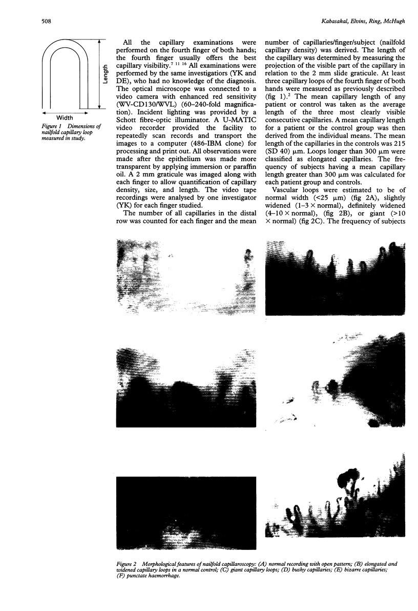
Full text
PDF

Images in this article
Selected References
These references are in PubMed. This may not be the complete list of references from this article.
- Andrade L. E., Gabriel Júnior A., Assad R. L., Ferrari A. J., Atra E. Panoramic nailfold capillaroscopy: a new reading method and normal range. Semin Arthritis Rheum. 1990 Aug;20(1):21–31. doi: 10.1016/0049-0172(90)90091-s. [DOI] [PubMed] [Google Scholar]
- Granier F., Vayssairat M., Priollet P., Housset E. Nailfold capillary microscopy in mixed connective tissue disease. Comparison with systemic sclerosis and systemic lupus erythematosus. Arthritis Rheum. 1986 Feb;29(2):189–195. doi: 10.1002/art.1780290206. [DOI] [PubMed] [Google Scholar]
- Grassi W., Core P., Carlino G., Cervini C. Nailfold capillary permeability in psoriatic arthritis. Scand J Rheumatol. 1992;21(5):226–230. doi: 10.3109/03009749209099229. [DOI] [PubMed] [Google Scholar]
- Harper F. E., Maricq H. R., Turner R. E., Lidman R. W., Leroy E. C. A prospective study of Raynaud phenomenon and early connective tissue disease. A five-year report. Am J Med. 1982 Jun;72(6):883–888. doi: 10.1016/0002-9343(82)90846-4. [DOI] [PubMed] [Google Scholar]
- LeRoy E. C., Maricq H. R., Kahaleh M. B. Undifferentiated connective tissue syndromes. Arthritis Rheum. 1980 Mar;23(3):341–343. doi: 10.1002/art.1780230312. [DOI] [PubMed] [Google Scholar]
- Lee P., Leung F. Y., Alderdice C., Armstrong S. K. Nailfold capillary microscopy in the connective tissue diseases: a semiquantitative assessment. J Rheumatol. 1983 Dec;10(6):930–938. [PubMed] [Google Scholar]
- Lovy M., MacCarter D., Steigerwald J. C. Relationship between nailfold capillary abnormalities and organ involvement in systemic sclerosis. Arthritis Rheum. 1985 May;28(5):496–501. doi: 10.1002/art.1780280505. [DOI] [PubMed] [Google Scholar]
- Maricq H. R., LeRoy E. C., D'Angelo W. A., Medsger T. A., Jr, Rodnan G. P., Sharp G. C., Wolfe J. F. Diagnostic potential of in vivo capillary microscopy in scleroderma and related disorders. Arthritis Rheum. 1980 Feb;23(2):183–189. doi: 10.1002/art.1780230208. [DOI] [PubMed] [Google Scholar]
- Maricq H. R., LeRoy E. C. Patterns of finger capillary abnormalities in connective tissue disease by "wide-field" microscopy. Arthritis Rheum. 1973 Sep-Oct;16(5):619–628. doi: 10.1002/art.1780160506. [DOI] [PubMed] [Google Scholar]
- Maricq H. R. Wide-field capillary microscopy. Arthritis Rheum. 1981 Sep;24(9):1159–1165. doi: 10.1002/art.1780240907. [DOI] [PubMed] [Google Scholar]
- Preliminary criteria for the classification of systemic sclerosis (scleroderma). Subcommittee for scleroderma criteria of the American Rheumatism Association Diagnostic and Therapeutic Criteria Committee. Arthritis Rheum. 1980 May;23(5):581–590. doi: 10.1002/art.1780230510. [DOI] [PubMed] [Google Scholar]
- Redisch W., Messina E. J., Hughes G., McEwen C. Capillaroscopic observations in rheumatic diseases. Ann Rheum Dis. 1970 May;29(3):244–253. doi: 10.1136/ard.29.3.244. [DOI] [PMC free article] [PubMed] [Google Scholar]
- Rouen L. R., Terry E. N., Doft B. H., Clauss R. H., Redisch W. Classification and measurement of surface microvessels in man. Microvasc Res. 1972 Jul;4(3):285–292. doi: 10.1016/0026-2862(72)90040-4. [DOI] [PubMed] [Google Scholar]
- Sato S., Takehara K., Soma Y., Tsuchida T., Ishibashi Y. Diagnostic significance of nailfold bleeding in scleroderma spectrum disorders. J Am Acad Dermatol. 1993 Feb;28(2 Pt 1):198–203. doi: 10.1016/0190-9622(93)70027-q. [DOI] [PubMed] [Google Scholar]
- Studer A., Hunziker T., Lütolf O., Schmidli J., Chen D., Mahler F. Quantitative nailfold capillary microscopy in cutaneous and systemic lupus erythematosus and localized and systemic scleroderma. J Am Acad Dermatol. 1991 Jun;24(6 Pt 1):941–945. doi: 10.1016/0190-9622(91)70150-z. [DOI] [PubMed] [Google Scholar]
- Tan E. M., Cohen A. S., Fries J. F., Masi A. T., McShane D. J., Rothfield N. F., Schaller J. G., Talal N., Winchester R. J. The 1982 revised criteria for the classification of systemic lupus erythematosus. Arthritis Rheum. 1982 Nov;25(11):1271–1277. doi: 10.1002/art.1780251101. [DOI] [PubMed] [Google Scholar]
- Wong M. L., Highton J., Palmer D. G. Sequential nailfold capillary microscopy in scleroderma and related disorders. Ann Rheum Dis. 1988 Jan;47(1):53–61. doi: 10.1136/ard.47.1.53. [DOI] [PMC free article] [PubMed] [Google Scholar]