Abstract
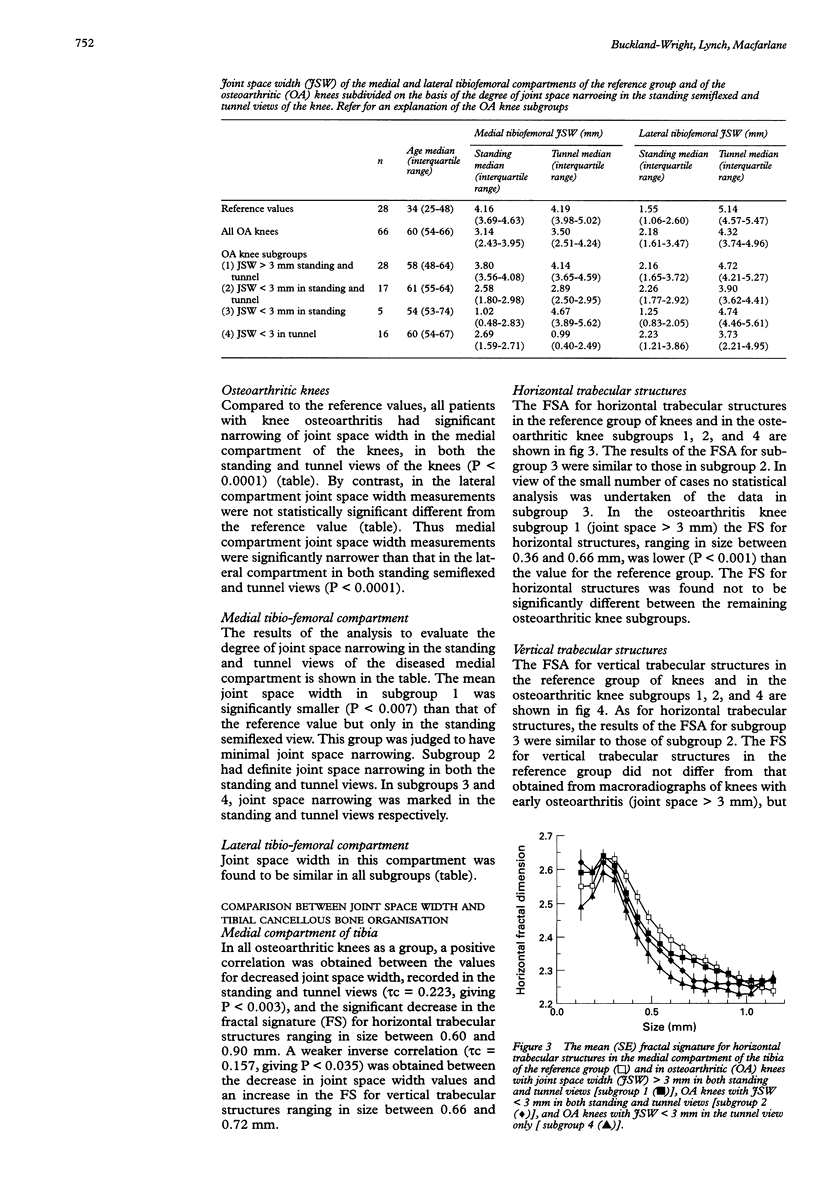
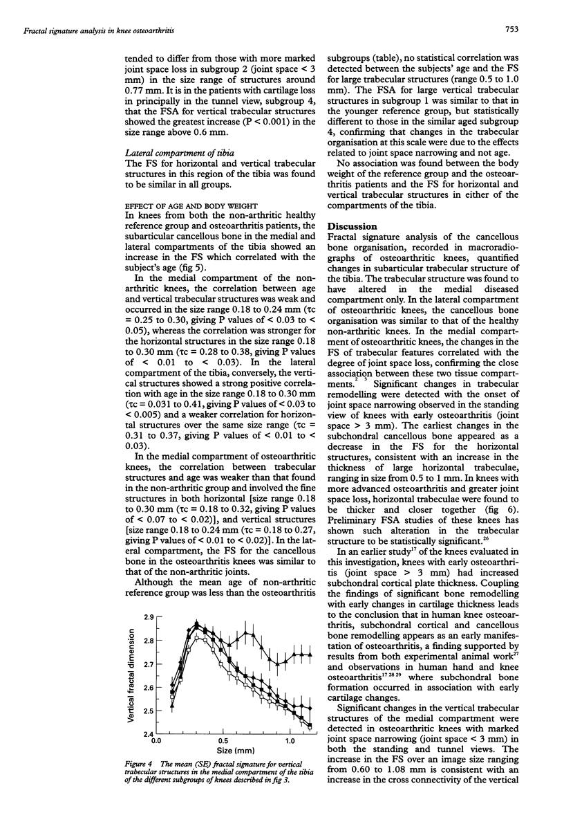
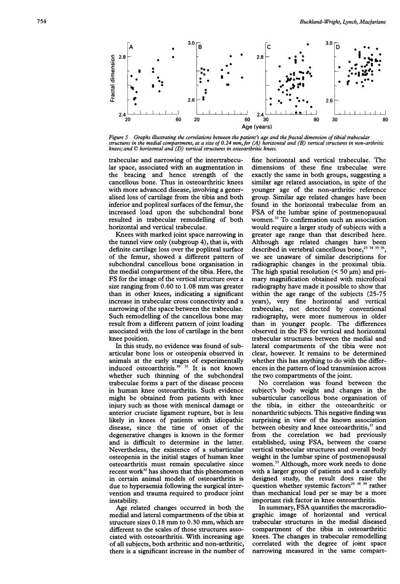
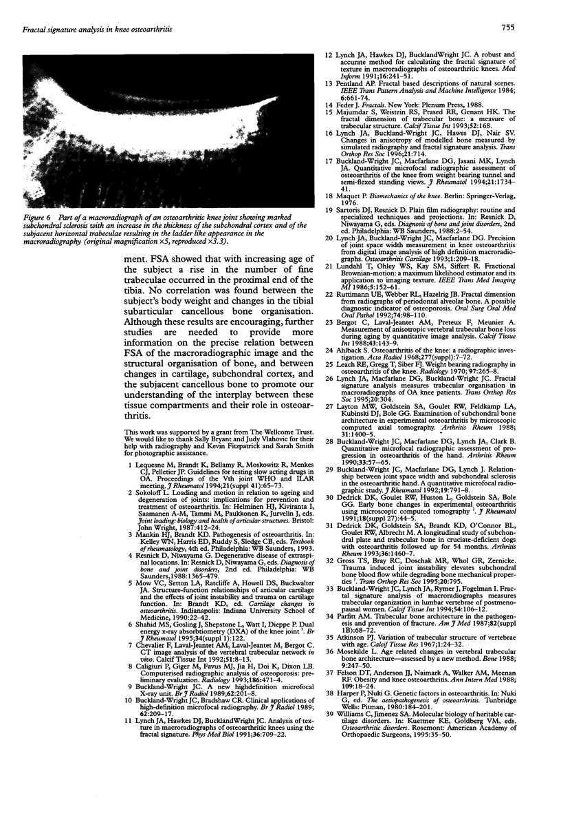
OBJECTIVE: To determine whether fractal signature analysis (FSA) of digitised macroradiographs of knees quantifies alterations in trabecular structure in the tibial cancellous bone of osteoarthritic patients with either early or definite joint space narrowing compared with non-arthritic subjects. METHODS: 90 osteoarthritic knees had macroradiographs at x5 magnification. Joint space width and FSA of horizontal and vertical trabecular organisation in the tibial subarticular cancellous bone were measured in the medial and lateral tibio-femoral compartments and compared to reference values obtained from the knees of 14 healthy non-arthritic volunteers, and to the subject's age and weight. RESULTS: Compared to the non-arthritic joints, FSA of the trabecular structure of the medial diseased compartment of the tibia was significantly different and correlated with the degree of joint space narrowing (P < 0.003); FSA of horizontal trabecular structures decreased (P < 0.001) in knees with early osteoarthritis (joint space > 3 mm) and vertical trabecular FSA increased in knees with marked joint space narrowing (joint space < 3 mm). In the lateral compartment of the tibia, FSA did not show a difference between any of the categories. With increasing age of all subjects, the changes in FSA indicated a significant increase in the number of fine horizontal and vertical trabeculae. No correlation was found between the subjects' body weight and changes in the subarticular cancellous bone organisation. CONCLUSIONS: FSA quantifies changes in cancellous bone organisation in knee osteoarthritis. In the diseased compartment, increased horizontal trabecular thickness occurred early and preceded the later changes in the vertical structures.
Full text
PDF
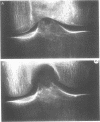
Images in this article
Selected References
These references are in PubMed. This may not be the complete list of references from this article.
- Bergot C., Laval-Jeantet A. M., Prêteux F., Meunier A. Measurement of anisotropic vertebral trabecular bone loss during aging by quantitative image analysis. Calcif Tissue Int. 1988 Sep;43(3):143–149. doi: 10.1007/BF02571311. [DOI] [PubMed] [Google Scholar]
- Buckland-Wright J. C. A new high-definition microfocal X-ray unit. Br J Radiol. 1989 Mar;62(735):201–208. doi: 10.1259/0007-1285-62-735-201. [DOI] [PubMed] [Google Scholar]
- Buckland-Wright J. C., Bradshaw C. R. Clinical applications of high-definition microfocal radiography. Br J Radiol. 1989 Mar;62(735):209–217. doi: 10.1259/0007-1285-62-735-209. [DOI] [PubMed] [Google Scholar]
- Buckland-Wright J. C., Lynch J. A., Rymer J., Fogelman I. Fractal signature analysis of macroradiographs measures trabecular organization in lumbar vertebrae of postmenopausal women. Calcif Tissue Int. 1994 Feb;54(2):106–112. doi: 10.1007/BF00296060. [DOI] [PubMed] [Google Scholar]
- Buckland-Wright J. C., Macfarlane D. G., Jasani M. K., Lynch J. A. Quantitative microfocal radiographic assessment of osteoarthritis of the knee from weight bearing tunnel and semiflexed standing views. J Rheumatol. 1994 Sep;21(9):1734–1741. [PubMed] [Google Scholar]
- Buckland-Wright J. C., Macfarlane D. G., Lynch J. A., Clark B. Quantitative microfocal radiographic assessment of progression in osteoarthritis of the hand. Arthritis Rheum. 1990 Jan;33(1):57–65. doi: 10.1002/art.1780330107. [DOI] [PubMed] [Google Scholar]
- Caligiuri P., Giger M. L., Favus M. J., Jia H., Doi K., Dixon L. B. Computerized radiographic analysis of osteoporosis: preliminary evaluation. Radiology. 1993 Feb;186(2):471–474. doi: 10.1148/radiology.186.2.8421753. [DOI] [PubMed] [Google Scholar]
- Chevalier F., Laval-Jeantet A. M., Laval-Jeantet M., Bergot C. CT image analysis of the vertebral trabecular network in vivo. Calcif Tissue Int. 1992 Jul;51(1):8–13. doi: 10.1007/BF00296208. [DOI] [PubMed] [Google Scholar]
- Dedrick D. K., Goldstein S. A., Brandt K. D., O'Connor B. L., Goulet R. W., Albrecht M. A longitudinal study of subchondral plate and trabecular bone in cruciate-deficient dogs with osteoarthritis followed up for 54 months. Arthritis Rheum. 1993 Oct;36(10):1460–1467. doi: 10.1002/art.1780361019. [DOI] [PubMed] [Google Scholar]
- Dedrick D. K., Goulet R., Huston L., Goldstein S. A., Bole G. G. Early bone changes in experimental osteoarthritis using microscopic computed tomography. J Rheumatol Suppl. 1991 Feb;27:44–45. [PubMed] [Google Scholar]
- Felson D. T., Anderson J. J., Naimark A., Walker A. M., Meenan R. F. Obesity and knee osteoarthritis. The Framingham Study. Ann Intern Med. 1988 Jul 1;109(1):18–24. doi: 10.7326/0003-4819-109-1-18. [DOI] [PubMed] [Google Scholar]
- Layton M. W., Goldstein S. A., Goulet R. W., Feldkamp L. A., Kubinski D. J., Bole G. G. Examination of subchondral bone architecture in experimental osteoarthritis by microscopic computed axial tomography. Arthritis Rheum. 1988 Nov;31(11):1400–1405. doi: 10.1002/art.1780311109. [DOI] [PubMed] [Google Scholar]
- Leach R. E., Gregg T., Siber F. J. Weight-bearing radiography in osteoarthritis of the knee. Radiology. 1970 Nov;97(2):265–268. doi: 10.1148/97.2.265. [DOI] [PubMed] [Google Scholar]
- Lequesne M., Brandt K., Bellamy N., Moskowitz R., Menkes C. J., Pelletier J. P., Altman R. Guidelines for testing slow acting drugs in osteoarthritis. J Rheumatol Suppl. 1994 Sep;41:65–73. [PubMed] [Google Scholar]
- Lynch J. A., Buckland-Wright J. C., Macfarlane D. G. Precision of joint space width measurement in knee osteoarthritis from digital image analysis of high definition macroradiographs. Osteoarthritis Cartilage. 1993 Oct;1(4):209–218. doi: 10.1016/s1063-4584(05)80327-9. [DOI] [PubMed] [Google Scholar]
- Lynch J. A., Hawkes D. J., Buckland-Wright J. C. A robust and accurate method for calculating the fractal signature of texture in macroradiographs of osteoarthritic knees. Med Inform (Lond) 1991 Apr-Jun;16(2):241–251. doi: 10.3109/14639239109012130. [DOI] [PubMed] [Google Scholar]
- Lynch J. A., Hawkes D. J., Buckland-Wright J. C. Analysis of texture in macroradiographs of osteoarthritic knees using the fractal signature. Phys Med Biol. 1991 Jun;36(6):709–722. doi: 10.1088/0031-9155/36/6/001. [DOI] [PubMed] [Google Scholar]
- Mosekilde L. Age-related changes in vertebral trabecular bone architecture--assessed by a new method. Bone. 1988;9(4):247–250. doi: 10.1016/8756-3282(88)90038-5. [DOI] [PubMed] [Google Scholar]
- Parfitt A. M. Trabecular bone architecture in the pathogenesis and prevention of fracture. Am J Med. 1987 Jan 26;82(1B):68–72. doi: 10.1016/0002-9343(87)90274-9. [DOI] [PubMed] [Google Scholar]
- Ruttimann U. E., Webber R. L., Hazelrig J. B. Fractal dimension from radiographs of peridental alveolar bone. A possible diagnostic indicator of osteoporosis. Oral Surg Oral Med Oral Pathol. 1992 Jul;74(1):98–110. doi: 10.1016/0030-4220(92)90222-c. [DOI] [PubMed] [Google Scholar]


