Full text
PDF
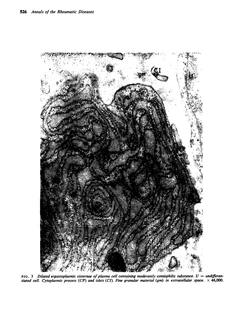

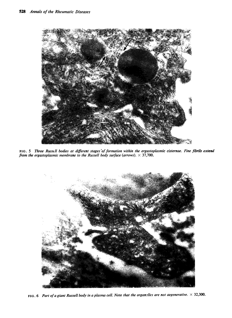
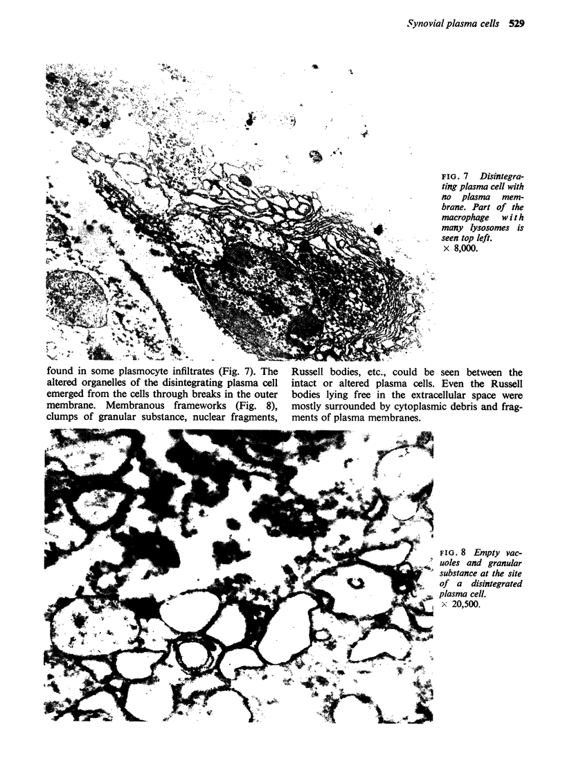
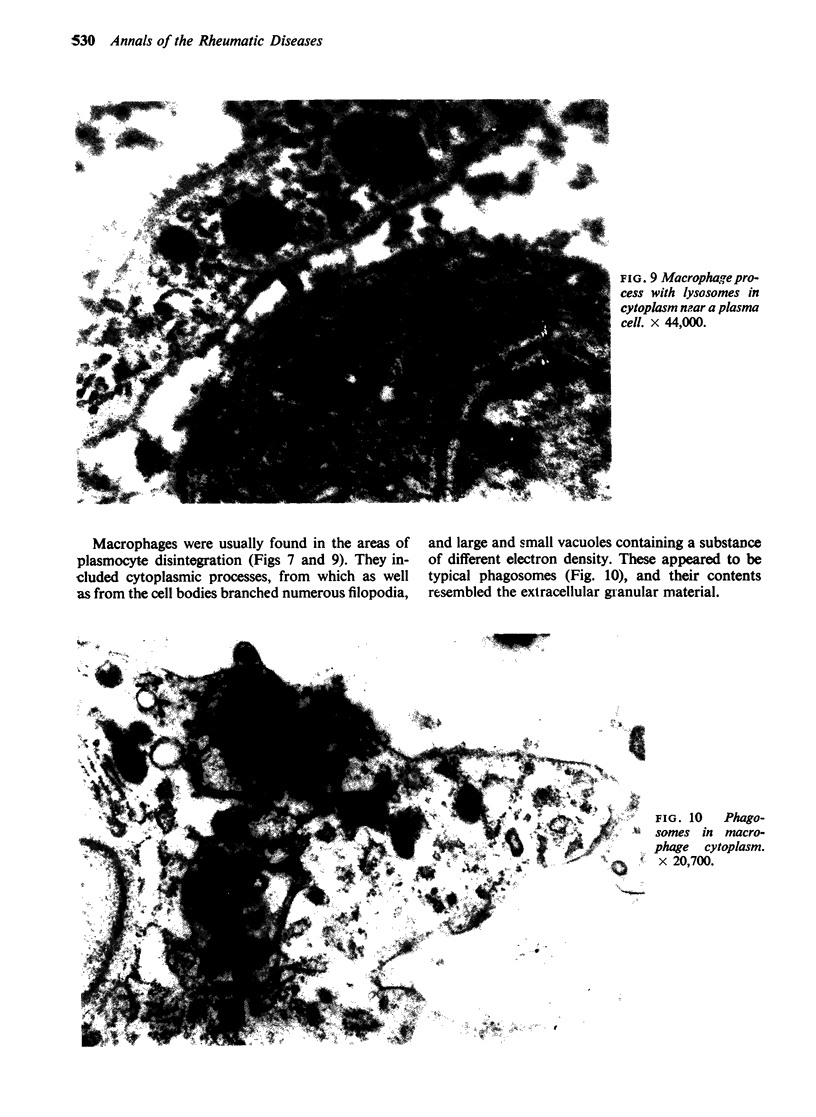
Images in this article
Selected References
These references are in PubMed. This may not be the complete list of references from this article.
- BARLAND P., NOVIKOFF A. B., HAMERMAN D. FINE STRUCTURE AND CYTOCHEMISTRY OF THE RHEUMATOID SYNOVIAL MEMBRANE, WITH SPECIAL REFERENCE TO LYSOSOMES. Am J Pathol. 1964 May;44:853–866. [PMC free article] [PubMed] [Google Scholar]
- Ghadially F. N., Roy S. Ultrastructure of synovial membrane in rheumatoid arthritis. Ann Rheum Dis. 1967 Sep;26(5):426–443. doi: 10.1136/ard.26.5.426. [DOI] [PMC free article] [PubMed] [Google Scholar]
- HIROHATA K., KOBAYASHI I. FINE STRUCTURES OF THE SYNOVIAL TISSUES IN RHEUMATOID ARTHRITIS. Kobe J Med Sci. 1964 Dec;10:195–225. [PubMed] [Google Scholar]
- Highton T. C., Caughey D. E., Rayns D. G. A new inclusion body in rheumatoid synovia. Ann Rheum Dis. 1966 Mar;25(2):149–155. doi: 10.1136/ard.25.2.149. [DOI] [PMC free article] [PubMed] [Google Scholar]
- MELLORS R. C., NOWOSLAWSKI A., KORNGOLD L., SENGSON B. L. Rheumatoid factor and the pathogenesis of rheumatoid arthritis. J Exp Med. 1961 Feb 1;113:475–484. doi: 10.1084/jem.113.2.475. [DOI] [PMC free article] [PubMed] [Google Scholar]
- ORTEGA L. G., MELLORS R. C. Cellular sites of formation of gamma globulin. J Exp Med. 1957 Nov 1;106(5):627–640. doi: 10.1084/jem.106.5.627. [DOI] [PMC free article] [PubMed] [Google Scholar]
- REYNOLDS E. S. The use of lead citrate at high pH as an electron-opaque stain in electron microscopy. J Cell Biol. 1963 Apr;17:208–212. doi: 10.1083/jcb.17.1.208. [DOI] [PMC free article] [PubMed] [Google Scholar]
- SABATINI D. D., BENSCH K., BARRNETT R. J. Cytochemistry and electron microscopy. The preservation of cellular ultrastructure and enzymatic activity by aldehyde fixation. J Cell Biol. 1963 Apr;17:19–58. doi: 10.1083/jcb.17.1.19. [DOI] [PMC free article] [PubMed] [Google Scholar]
- THIERY J. P. Etude sur le plasmocyte en contraste de phase et en microscopie électronique. III. Plasmocytes à corps de Russell et à cristaux. Rev Hematol. 1958 Jan-Mar;13(1):61–78. [PubMed] [Google Scholar]
- WELLENSIEK H. J. Zur submikroskopischen Morphologie von Plasmazellen mit Russelschen Korperchen und Eiweisskristallen. Beitr Pathol Anat. 1957;118(2):173–202. [PubMed] [Google Scholar]
- WELSH R. A. Electron microscopic localization of Russell bodies in the human plasma cell. Blood. 1960 Sep;16:1307–1312. [PubMed] [Google Scholar]
- Wyllie J. C., Haust M. D., More R. H. The fine structure of synovial lining cells in rheumatoid arthritis. Lab Invest. 1966 Mar;15(3):519–529. [PubMed] [Google Scholar]


