Abstract
Cilia are ubiquitous eukaryotic organelles responsible for cellular motility and sensory functions. The ciliary axoneme is a microtubule-based cytoskeleton consisting of two central singlets and nine outer doublet microtubules. Cryo-electron microscopy-based studies have revealed a complex network inside the lumen of both tubules composed of microtubule-inner proteins (MIPs). However, the functions of most MIPs remain unknown. Here, we present single-particle cryo-EM-based analyses of the Tetrahymena thermophila native doublet microtubule and identify 42 MIPs. These data shed light on the evolutionarily conserved and diversified roles of MIPs. In addition, we identified MIPs potentially responsible for the assembly and stability of the doublet outer junction. Knockout of the evolutionarily conserved outer junction component CFAP77 moderately diminishes Tetrahymena swimming speed and beat frequency, indicating the important role of CFAP77 and outer junction stability in cilia beating generation and/or regulation.
Subject terms: Cryoelectron microscopy, Cilia, Bacterial structural biology
Using cryo-EM, the authors identified 42 MIPs, including outer junction protein CFAP77 and outer dense fibers, in native doublet microtubules of Tetrahymena thermophila. Knockout of CFAP77 reduced ciliary beat frequency and led to outer junction damage.
Introduction
Cilia are hair-like structures protruding from the cell surface, conserved from protists to humans. Immotile sensory cilia play an essential role in sensing and transducing external signals such as sonic hedgehog from the surrounding environment to the cell. Coordinated beating of motile cilia enables transport of cells or fluids along the surface of ciliated cells, and thus cilia are key factors in clearing mucus out of the respiratory tract or circulating cerebrospinal fluid1. The core structure of motile cilia, the axoneme, consists of two central singlet microtubules and nine peripherally positioned doublet microtubules (DMT)2,3. The assembly and stability of the DMTs are indispensable for the ciliary function as they serve as a scaffold for numerous ciliary complexes, the force transducers for ciliary bending4, and tracks for intraflagellar transport5.
The inner (luminal) wall of the DMT is supported by an intricate network of microtubule inner proteins (MIPs)6–8. Using single-particle cryo-electron microscopy (cryo-EM), the identities of over 30 MIPs have been revealed in the ciliate Tetrahymena thermophila4,9,10, the green algae Chlamydomonas reinhardtii11, and bovine respiratory cilia12.
At least half of the MIPs identified in those studies are conserved, including the inner junction proteins FAP20, PACRG9,13,14, FAP45, FAP5215, and the protofilament (PF) ribbon proteins, RIB43a and RIB7216,17. Interestingly, some MIPs show poor conservation. For example, the tektin bundles of the PF ribbon were identified in bovine respiratory cilia, but not in Tetrahymena or Chlamydomonas12.
There are few studies exploring the functions of MIPs. RIB72/EFHC1, an EF-hand protein, is implicated in juvenile epilepsy disorder18,19 and is important for Tetrahymena cell motility20,21. FAP45 and FAP52 play a role in anchoring the B-tubule to the A-tubule wall; a lack of FAP45/FAP52 leads to B-tubule instability15. Pierce1 and 2, the orthologs of Chlamydomonas FAP18211, are important for outer dynein arm assembly in mammals and zebrafish12.
Until now, the identity of MIPs at the outer junction of the DMT remains unknown. The outer junction is thought to be the site of B-tubule assembly22. Disruption of the outer junction leads to DMT damage and consequently dysfunctional cilia. At the outer junction, α- and β-tubulins from PF B1 form a non-canonical interaction with tubulins of A-tubule8. Cleavage of the C-terminal tails of tubulins by subtilisin enables B-tubule formation in vitro22. On the other hand, C-terminal tails of tubulins are essential for ciliary function in vivo23,24. Therefore, there must be a mechanism that suppresses the C-terminal tails of tubulins of the A-tubule enabling the formation of the B-tubule.
We hypothesized that MIPs belong to one of two groups: evolutionarily conserved “core” MIPs, which are ubiquitous in cilia function; or species or lineage-specific MIPs that contribute to the stability required for some specific movement of cilia and flagella in some species or tissues. Here, we used an integrated approach combining single-particle cryo-EM, mass spectrometry, and artificial intelligence to model MIPs in the DMT lumen of the ciliate Tetrahymena thermophila. We showed that nearly half of the MIPs identified in Tetrahymena are evolutionarily conserved. In contrast to DMTs in Chlamydomonas reinhardtii flagella11 and bovine respiratory cilia12 where the MIP network adopts a 48-nm periodicity, the Tetrahymena MIP network repeats every 96-nm. In addition, we observed filaments on the outer surface of the DMT, which have implications for intraflagellar transport. Finally, we used a combination of genetics and microscopy techniques to show that CFAP77, the conserved MIP found at the outer junction of the DMT, is important for DMT stability and ciliary motility.
Results
Tetrahymena DMT consists of conserved and non-conserved MIPs
We optimized the purification of DMTs from isolated Tetrahymena cilia to preserve all the associated structures (“Methods”). This approach was successfully used previously to obtain the outer dynein arm-bound DMT structure25. The single-particle cryo-EM analyses of the DMT from wild-type (WT) and K40R cells allowed us to obtain a 48-nm repeat structure with a global resolution of 4.1 and 3.7 Å (Fig. 1A and Supplementary Fig. 1A). Focused refinement of overlapping regions that collectively encompass the WT DMT resulted in maps with a resolution range of 3.6 to 3.9 Å (Supplementary Fig. 1B, Table 1, and “Methods”). In addition, we obtained the focused refined DMT structures at 3.2–3.6 Å resolution of the K40R strain, in which lysine residue 40 of α-tubulin is mutated to arginine to prevent acetylation25,26. Since the mutation does not seem to affect the overall MIP decoration, we performed many of our analyses in the K40R maps due to the higher resolution.
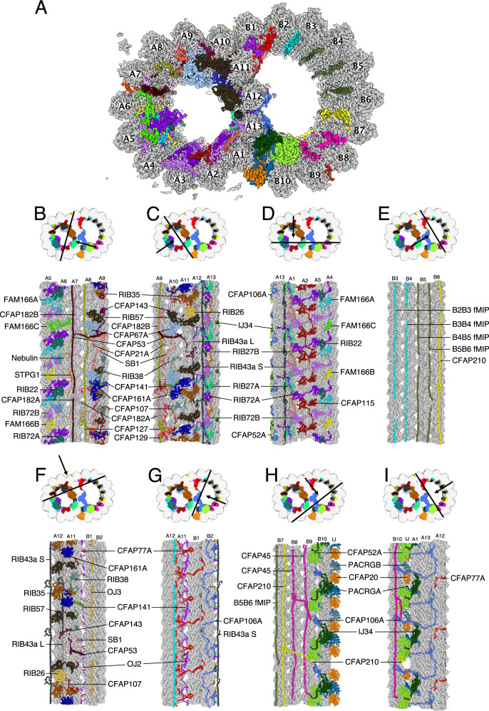
Fig. 1. The structure of the native DMT from Tetrahymena thermophila.
A A cross-section of the DMT map. Each color denotes an individual MIP. Tubulins are in gray. B–I Views of the lumen of the DMT from different angles as indicated by the black arrow. The cutting plane indicated by black lines.
Table 1.
Cryo-EM data collection and refinement parameters for all datasets used in this study
| Method | Single particle analysis | Subtomogram averaging | |||
|---|---|---|---|---|---|
| Dataset | WT (CU428) | K40R | 96 nm combined | WT (CU428) | CFAP77AB-KO |
| Microscope | Titan Krios | Titan Krios | Titan Krios | Titan Krios | Titan Krios |
| Electron Detector | Gatan K3 | Gatan K3 | Gatan G3 | Gatan K3 | Gatan K3 |
| Zero-loss filter (eV) | 30 | 30 | 30 | 20 | 20 |
| Magnification | 64,000 | 64,000 | 64,000 | 42,000 | 42,000 |
| Voltage (keV) | 300 | 300 | 300 | 300 | 300 |
| Electron exposure (e/A2) | 45 | 45 & 73 | 45 & 73 | 160 | 160 |
| Defocus range (μm) | 1.0–3.0 | 1.0–3.0 | 1.0–3.0 | 2.5–3.5 | 2.5-3.5 |
| Pixel size (Å) | 1.37 | 1.37 | 1.37 | 2.12 | 2.12 |
| Tilt range (increment) | – | – | – | −60° to 60° (3°) | −60° to 60° (3°) |
| Tilt scheme | – | – | – | Dose symmetric | Dose symmetric |
| Movies acquired | 18,384 | 25,610 | 43,994 | – | – |
| Particles number | 148,365 | 182,355 | 172,223 | – | – |
| Tilt series acquired | – | – | – | 58 | 20 |
| Subtomograms averaged | – | – | – | 2608 | 1702 |
| Symmetry imposed | C1 | C1 | C1 | C1 | C1 |
| Repeat unit (nm) | 48 | 48 | 96 | 96 | 96 |
| Map resolution | 3.6 to 4.0 | 3.3 to 3.5 | 3.75 | 19 | 22 |
To identify and model the MIP network, we devised a strategy that employed: (i) homology modeling of conserved MIPs; (ii) de novo identification and modeling of unknown MIPs using artificial intelligent backbone tracing DeepTracer27 of densities in our map, artificial intelligent structure prediction ColabFold28,29 of all proteins present in the proteome of the DMT, and structure similarity search of protein backbones within the predicted structures of the proteome of the DMT; and (iii) automatic determination of sequences of a backbone trace using the FindMySequence program30 (“Methods” and Supplementary Fig. 1C). To validate the protein identities, we used multiple techniques and independently arrived at the same identity (Supplementary Tables 1–3). We used FindMySequence to compare the E-value of our candidate and the next best hits (Supplementary Table 4). We also examined side-chain fitting of all protein models and cross-links identified from cross-linking mass spectrometry of the cilia in situ (Supplementary Fig. 2A, B and Supplementary Table 5)31. With such an approach, we could identify and validate almost all Tetrahymena MIPs and show that they are forming a weaving network inside the DMT (Table 2 and Supplementary Tables 1–4). Many of those MIP densities were missing from our previously reported structure of salt-treated DMT4 such as TtPACRGA/B, TtCFAP20, TtOJ3, TtRIB22, and TtRIB27A/B.
Table 2.
Refinement statistics of WT and K40R 48 nm models
| WT (CU428) | K40R | |
|---|---|---|
| Model-to-Map fit, CCmask | 0.8009 | 0.7961 |
| All-atom clashscore | 16.61 | 13.21 |
| Ramachandran plot | ||
| Outliers [%] | 0.14 | 0.14 |
| Allowed [%] | 3.70 | 3.60 |
| Favored [%] | 96.16 | 96.26 |
| Rotamer outliers [%] | 0.03 | 0.04 |
| Cbeta deviations [%] | 0.00 | 0.00 |
| Cis-proline [%] | 4.56 | 4.56 |
| Cis-general [%] | 0.01 | 0.01 |
| Twisted proline [%] | 0.17 | 0.18 |
| Twisted general [%] | 0.02 | 0.01 |
Among the 42 identified proteins (Fig. 1B–I, Supplementary Fig. 2, Supplementary Tables 1–3, and Supplementary Movie 1), approximately half are conserved in different species (the “core” MIPs) while the rest are species-specific MIPs. Interestingly, many MIPs represented by a single ortholog in Chlamydomonas, and bovine had two or more paralogs (denoted by A, B and C) in Tetrahymena (see Supplementary Table 1–3 and Supplementary Fig. 3A–H). Many paralogs have alternating patterns along the DMT; for example, we were able to discern between the paralog densities in our map when we modeled TtRIB72, TtPACRG, TtCFAP182, TtRIB27, and TtFAM166. In some instances, such as TtCFAP106 and TtCFAP77, both paralogs were detected in our ciliary mass spectrometry data, but we could not distinguish their densities using cryo-EM due to sequence similarity. In other cases, certain paralogs were present in significantly lower stoichiometry (Supplementary Table 3) and were therefore not modeled such as TtCFAP52B/C. It is possible that those paralogs co-localize along the entire cilium length or that paralogs differentially localize to proximal or distal ends. Therefore, we modeled the most abundant paralogs (Supplementary Tables 1–3).
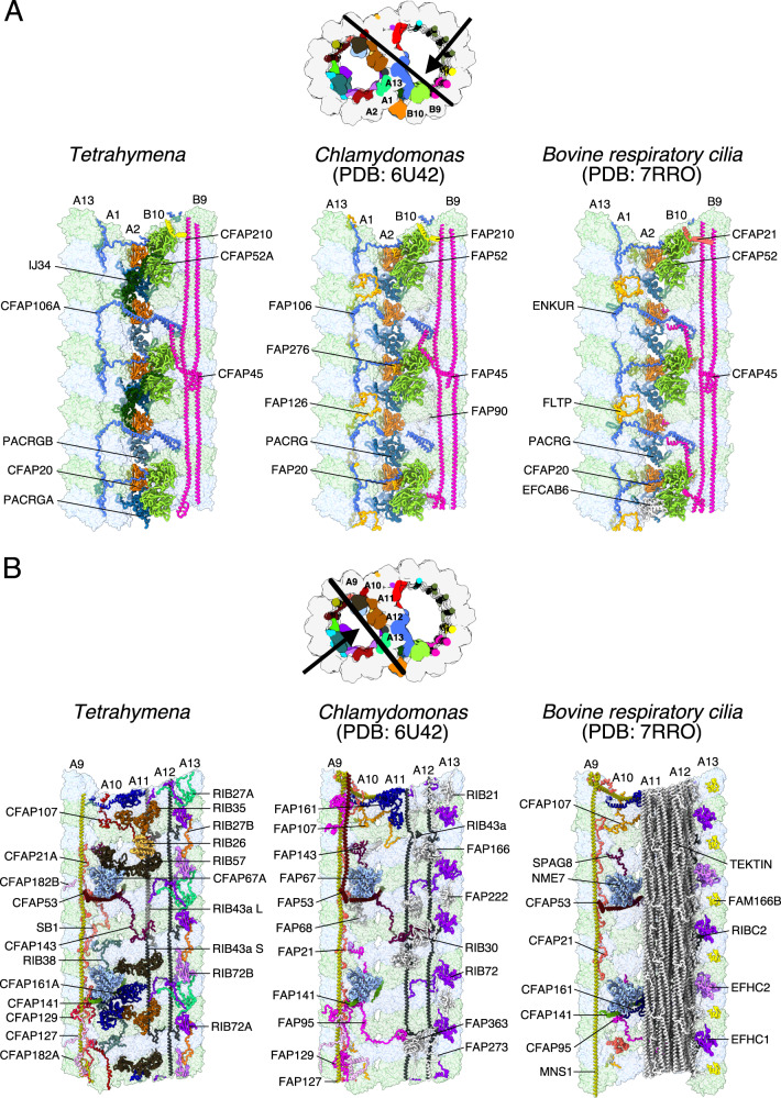
FAP52, FAP45, FAP106, PACRG, and FAP20 (Fig. 2A) are core MIPs of the inner junction region in Tetrahymena9, Chlamydomonas9,11, and bovine respiratory cilia12. Interestingly, Tetrahymena CFAP52 is stabilized by an ancillary protein IJ34 (homolog of CCDC81, this study) while in Chlamydomonas and bovine respiratory cilia FAP52 is stabilized by species-specific FAP2769,11 and EFCAB612 respectively. The two Tetrahymena paralogs of PACRG, TtPACRGA and TtPACRGB differ in their N-terminal region and are positioned alternately in the inner junction (Supplementary Fig. 3B). Consequently, the Tetrahymena inner junction has a 16-nm periodicity, which clearly defines the bimodality of the interdimer distance first reported in Tetrahymena4. The lack of the N-terminal region in TtPACRGB (Supplementary Fig. 3B) might explain why the inner junction of Tetrahymena is less stable upon salt treatment compared to Chlamydomonas9, pointing to the role of the N-terminal region of PACRG in inner junction stabilization. We did not see the unknown MIP densities previously reported in the subtomogram averaged map of the Tetrahymena axonemal doublet10. Perhaps, it is due to partial decoration of this unknown MIP.
Fig. 2. Comparison of the DMT structure from Tetrahymena, Chlamydomonas and bovine respiratory cilia.
A The inner junction, note that the architecture is well conserved. B The PF-ribbon region, note many species-specific MIPs.
The PF ribbon is the most divergent region in the DMT (Fig. 2B). RIB43a, the core filamentous MIP, is conserved across species and was reported previously to be crucial for the stability of the PF ribbon region4,17. In Tetrahymena, the PF ribbon region is composed of TtRIB72A, TtRIB72B, TtRIB43A_S, TtRIB43A_L, TtCFAP143, TtRIB27, TtRIB57, TtRIB35, and TtRIB26 (Fig. 2B). However, some of those proteins (TtRIB27, TtRIB57, TtRIB35, and TtRIB26) are not conserved in Chlamydomonas11 and bovine respiratory cilia12 (Supplementary Tables 1 and 2). Moreover, in bovine respiratory cilia, the PF ribbon is accompanied by tektin bundles (Fig. 2B)12. The divergence in the PF ribbon components between species demonstrates that the stability of the PF region is not dependent upon widely conserved proteins. Interestingly, three molecules of RIB57 displayed different conformations in the 48-nm doublet (Supplementary Fig. 3H). This conformational flexibility explains the observed length violation of intra-cross-links found in RIB57 in the Tetrahymena cilia when compared with the AlphaFold2 model31.
We observed similarities and differences in outer junction structures and protein composition between Tetrahymena, Chlamydomonas, and bovine respiratory cilia (discussed later). To summarize, each DMT region has core components while other MIPs are species-specific. These species-specific proteins possibly function to reinforce core components and contribute to species-specific motility. As such, species-specific MIPs may be essential for cilia stability and waveform regulation.
MIP distribution in Tetrahymena has a 96-nm periodicity
The DMT outer surface has a 96-nm periodicity regulated by the molecular ruler composed of CCDC39 and CCDC4032. In Chlamydomonas11 and bovine respiratory cilia12, the MIPs show a 48-nm periodicity. Interestingly, based on our observations, the Tetrahymena DMT has a 96-nm luminal repeat.
Tetrahymena TtCFAP115 is almost four times the size of the CrFAP115 (110kD vs. 26kD). The ColabFold29 prediction of TtCFAP115 shows that it contains four EF-hand pair domains (Interpro IPR011992) while CrFAP115 has only one EF-hand pair domain and repeats every 8 nm. We expected that TtCFAP115 should have a 32-nm periodicity. To accurately position TtCFAP115, we modeled it in a 96-nm map combining several datasets that were obtained at 3.7 Å resolution for Tetrahymena WT CU428 strain, and K40 acetylation-deficient strains with α-tubulin point mutation K40R33. As expected, TtCFAP115 shows a clear 32-nm repeat (Supplementary Fig. 3I). Since TtCFAP115 repeats at 32-nm while other MIPs such as TtRIB43a repeats at 48-nm, the Tetrahymena DMT must have a 96-nm luminal repeat consisting of three TtCFAP115 molecules per periodicity (Fig. 3A, B).
Fig. 3. MIPs in Tetrahymena exhibit a 96-nm periodicity.
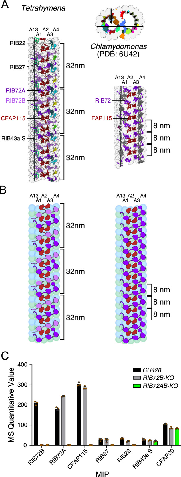
A TtCFAP115 shows 32-nm repeats leading to the true 96-nm periodicity of the MIPs of Tetrahymena. The FAP115 in Chlamydomonas only repeats with 8-nm periodicity, leading to the normal 48-nm repeat. B The cartoon of Tetrahymena and Chlamydomonas PFs A13-A4 showing the arrangement of FAP115 and RIB72. C Quantitative value of mass spectrometry (normal total spectra value) of WT, RIB72B-KO and RIB72A/B-KO cilia showing that TtCFAP115 is still intact after the knockout of RIB72B. n = 3 biological replicates for WT and each mutant. Data are presented as mean values +/− standard deviation. Values for each replicate are shown in brown circles. Source data are provided as a Source data file.
We observed that a single TtCFAP115 molecule interacts with two TtRIB72A and two TtRIB72B molecules (Fig. 3A). To support the prediction of interactions between TtRIB72 and TtCFAP115, we analyzed ciliary proteomes of RIB72B-KO and RIB72A/B-KO Tetrahymena knockout mutants21 by mass spectrometry (Fig. 3C and Supplementary Table 6). The top six proteins missing from the RIB27A/B-KO mutant are indeed MIPs, highlighting the accuracy of our proteomic analysis. Interestingly, TtCFAP115 was only missing from RIB72A/B double knockout mutant cilia. Our proteomic analysis showed that FAM166C, FAM166B, TtRIB22, and TtRIB27A are also missing only from the RIB72A/B double knockout mutant. In contrast, TtRIB27B is significantly reduced in RIB72B-KO and completely missing from RIB72A/B-KO cilia. Thus, similar to TtCFAP115, stable docking of FAM166B, FAM166C, TtRIB22 and TtRIB27A depends on TtRIB72A but not TtRIB72B (Figs. 1D and 3A), which reveals the role of TtRIB72A and TtRIB72B in the DMT assembly.
Currently no cryo-EM maps of the DMT have shown interactions that explain how the 48-nm repeat of MIPs and the 96-nm repeat of the outside proteins are established by the molecular ruler. Together with existing studies on MIPs11 and molecular rulers32, our findings on the 96-nm repeat organization of Tetrahymena MIPs suggest that the periodicities of the inside and outside proteins/complexes are self-regulated, probably through head-to-tail interactions of MIPs. Our finding showing that TtCFAP115 interacts with TtRIB72A/B is echoed by recent proteomics and low-resolution tomography studies10,20. Moreover, those studies demonstrated that knockout of TtCFAP115 leads to prolonged power stroke and decreased swimming speed10,20. Those findings further illustrate the important roles of TtCFAP115 in Tetrahymena cilia which are probably a by-product of DMT stabilization. Our structural analysis of TtCFAP115 highlights the importance of high-resolution reconstructions and the power of artificial intelligence prediction in the accurate validation of protein interactions. This strategy allowed us to structurally verify all MIPs that were lost in the mass spectrometry data of the RIB72A/B-KO mutants, namely FAM166B, FAM166C, TtRIB22, TtRIB27A, and TtRIB27B (Supplementary Table 6). Such validation is important as mass spectrometry studies often reveal a broad list of missing proteins, many of which are unspecific.
The outer surface of the intact DMT is associated with many filamentous proteins
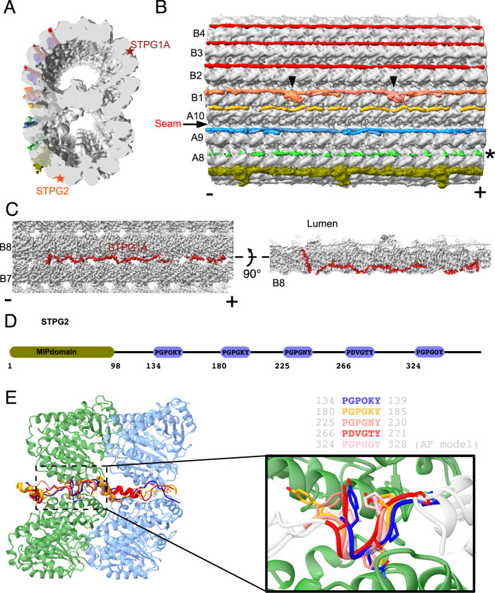
We observed numerous filamentous structures binding to the outer wedge between two PFs on the native DMT outer surface, associated with PFs B1 to B5, and A8 to A10 (Fig. 4A, B). These PFs were shown to serve as tracks for anterograde and retrograde intraflagellar transports (Supplementary Fig. 4A–D)5.
Fig. 4. The outer surface filaments on the native DMT.
A, B Cross-sectional (A) and longitudinal (B) views of the outer surface filaments on the native Tetrahymena DMT. The filaments are bound to adjacent pairs of PFs from A8 to B5. The filaments between PFs B2-B3, B3-B4, and B4-B5 appear similar and have a 48-nm periodicity. In contrast, the filaments between PFs A9-A10, A10-B1, and B1-B2 appear to have a 24-nm periodicity. These filaments have a clear head-to-tail periodic arrangement between PFs A9-A10 and B1-B2. The filament between PFs B1B2 has a globular domain (black arrowheads). The density of the filament between PFs A8-A9 is very weak, probably due to partial decoration. (−) and (+) signs indicate the minus and plus ends of the microtubule respectively. C STPG1A is a filamentous protein that is woven between PFs B7-B8, bound to the surface and the lumen. D Schematic representation of the protein topology of the filamentous MIP STPG2. E The PG-rich repeat motifs of STPG2 are structurally similar.
The low resolution of the outer surface filament suggests that they are either partially decorated or inherently flexible. At this resolution, the outer surface filaments of PFs A9A10, A10B1 and B1B2 still exhibit a clear 24-nm periodicity (Fig. 4B). Other filaments might have a 48-nm repeat since they appear similarly in the 96-nm map. While we could not identify most of the outer surface, we were able to trace the backbone of the filament between A10 and B1 (OJ3) since it binds tightly to the outer junction wedge and has better resolution. In bovine respiratory cilia and Chlamydomonas cilia, a similar density to OJ3 is observed but the identity of the protein remains unknown.
The outer surface filaments bind to the DMT in a similar fashion to MAP734. MAP7 is known to activate kinesin-1 even though its α-helical microtubule-binding domain recognizes a site on microtubules that partly overlaps with the kinesin-1 binding site. In contrast, dynein motor activity is not impacted by MAP7 binding to microtubules35. In Tetrahymena, both intraflagellar transport motors, kinesin-2 and dynein-2, must walk on the DMT decorated by these outer surface filaments. When we overlapped the kinesin and dynein microtubule-binding domains on our DMT structure, we noticed a clear steric clash (Supplementary Fig. 4B–D), similar to the case of MAP734. Thus, likely there is a conformational change upon molecular motor binding to facilitate intraflagellar transport in the cilia. High concentrations of MAP7 seem to inhibit kinesin-1 activity34. Therefore, it is reasonable to suggest that the low resolution of the outer surface filament is due to partial decoration and not inhibition of kinesin-2 activity.
In addition, we also observed two proteins binding tightly to the trench of the outer wedge of PF B8B9 and PF A6A7 (Fig. 4A, star labels). Since these two proteins bind tightly to the trench of the wedge, we were able to identify them as sperm tail PG-rich proteins STPG1A and STPG2 (Fig. 4C and Supplementary Fig. 4E, F). Interestingly, while most of STPG1A and STPG2 bind outside the DMT, both weave inside the DMT and contain a luminal domain (Fig. 4C). STPG1A contains five PG-rich motifs, each interspersed by about 40 amino acids (Fig. 4D). By superimposing the five STPG1A PG-rich motifs bound on the B-tubule, we discovered that the PG motif is a microtubule-binding motif and contains six residues with a P-G-P-G-x-Y pattern (Fig. 4E). The ~40 amino acids act as 8-nm spacers allowing each PG motif to bind to consecutive tubulins along the PF. That explains how STPG1A repeats and spans the 48-nm repeat without head-to-tail interactions. In our cilia proteome, there are five PG-rich proteins: STPG1A and its homolog STPG1B; STPG2; and two Outer Dense Fiber 3 Like (ODL3L) proteins (Supplementary Fig. 4E). Except for STPG2, all other proteins contain the P-G-P-G-x-Y motif and ~40 amino acid spacers. Both ODF3L proteins contain 12 P-G-P-G-x-Y motifs, suggesting that they bind to 12 tubulins or 96-nm. Interestingly, we found cross-links from both ODF3L proteins to the outer side of tubulins (Supplementary Table 5). For STPG2 the PG-rich motif has a five-residue pattern, consisting of P-G-P-x-Y, and forms a similar structure bound to microtubules (Supplementary Fig. 4F, G). In short, our structure of STPG1A allows us to conclude that the PG motif is a microtubule-binding motif and that ODF3L proteins are MAPs that bind the outer wedge of the microtubule.
CFAP77 stabilizes the outer junction
Among the identified MIPs are proteins that support the outer junction, which is believed to be the nucleation site for B-tubule formation. We observed three outer junction proteins: TtCFAP77 and OJ2, inside the B-tubule, and OJ3 in the wedge between A10 and B1 (Fig. 5A, B and Supplementary Movie 2). Bioinformatics analyses and examination of cryo-EM maps from Chlamydomonas (EMD-20631) and bovine respiratory cilia (EMD-24664) showed that OJ2 is Tetrahymena-specific. In contrast, CFAP77 is conserved in many species including humans and Chlamydomonas (Supplementary Fig. 5A, B).
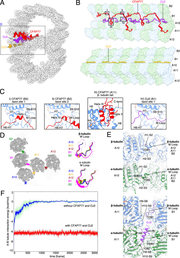
Fig. 5. CFAP77 stabilizes the outer junction.
A Cross-sectional view of the Tetrahymena outer junction highlighting proteins CFAP77 (red), OJ2 (purple) and OJ3 (yellow). B Architecture of the Tetrahymena outer junction including proteins CFAP77, OJ2 and OJ3. C Helices of CFAP77 occupy taxane-binding pockets of β-tubulin of PF B2. A loop of OJ2 occupies the taxane-binding pocket of β-tubulin of PF B1. The helix-turn-helix motif of CFAP77 is positioned near the C-terminus of β-tubulin of PF A11. D Cross-sectional view of the OJ. M-loops of tubulins are colored according to protofilament: B2 (green); B1 (purple); A10 (blue); A11 (gold); A12 (dark red). To the right, those same M Loops are superimposed. E The canonical (top) and unique (bottom) lateral interactions between tubulin subunits. PFs A11, A12, and B2 are superimposed and adopt the same overall conformation and lateral interactions (top). The lateral interactions between B1 and A11 are particularly unique in Tetrahymena because they involve OJ2 (bottom). F Molecular dynamics simulations of the outer junction with and without CFAP77 and OJ2.
Tetrahymena has two paralogs of CFAP77, CFAP77A and CFAP77B (Supplementary Fig. 5A and Supplementary Table 3). The mass spectrometry analyses revealed that CFAP77A is almost three times more abundant in cilia than CFAP77B. Because of the higher expression level, we decided to model CFAP77A.
The detailed analysis of the outer junction architecture reveals two MIPs, TtCFAP77 and OJ2, interacting with four outer junction PFs. TtCFAP77 contacts PFs A10, A11, B1, and B2, while OJ2 contacts PFs A11 and B1 (Fig. 5A, B). TtCFAP77 and OJ2 are 29 and 20 kDa acidic proteins, respectively, with short alpha helices interspersed with intrinsically disordered regions (Fig. 5B and Supplementary Fig. 5C, D). OJ3 is a filamentous MAP with four alpha helices broken by short intrinsically disordered regions, positioned between PFs A10 and B1. Unlike the 16 nm repeating unit shared by TtCFAP77 and OJ2 spanning two tubulin dimers, OJ3 repeats every 24 nm, spanning three tubulin dimers (Fig. 5B). Each of these three outer junction proteins acts as a thread that weaves across two or more PFs.
Remarkably, two short helices of TtCFAP77 occupy taxane-binding sites on β-tubulin subunits of PF B2 (Fig. 5C(i and ii)). The helix-turn-helix motif of TtCFAP77 is positioned on the β-tubulin C-terminus of PF A11 (Fig. 5C(iii)). OJ2 is positioned between PFs B1 and A11 and interacts with the taxane-binding site of a single β-tubulin along PF B1 (Fig. 5C(iv)).
We also examine the microtubule-binding loop (M-loop) critical for lateral interactions between PFs B1 and A-tubule and their interactions with the outer junction proteins. While the canonical lateral interactions between the M-loop and H1-S2 and H2-S3 loops of the adjacent PF36 are observed throughout the A-tubule and most of the B-tubule, the M-loop of α-tubulin of PF B1 adopts a unique conformation compared to all other M-loops of α-tubulin at the outer junction PFs (Fig. 5D, E). The M-loop of α-tubulin of PF B1 forms hydrogen bonds with both the α-tubulins of A11 and OJ2, providing structural evidence that OJ2 has a stabilizing role in the outer junction (Fig. 5E).
To investigate the significance of TtCFAP77 and OJ2 on the outer junction, we used molecular dynamics simulations to examine the behavior of protofilaments in the presence and absence of TtCFAP77 and OJ2. For the simulation, we used the PFs A10-A12 and B1-B2 as well as TtCFAP77 and OJ2 (see Methods). Our molecular dynamics simulation showed that tubulins from PFs B1-B2 dissociated from PFs A10-A12 in the absence of both TtCFAP77 and OJ2 (Fig. 5F). Molecular dynamics simulations indicate that mainly TtCFAP77 contributes to A- and B-tubule stability because the energy with and without OJ2 is almost the same (Supplementary Fig. 5E). In addition, further analysis shows that B-tubule bounding angle with A-tubule become unstable in the absence of both TtCFAP77 and OJ2 (Supplementary Fig. 5F). Therefore, TtCFAP77 is the key MIP for the stable binding of the B-tubule to the A-tubule.
The helix-turn-helix motif of TtCFAP77 and the wedge position between PFs A11 and B1 of OJ2 might suppress the intrinsically disordered C-terminal tails of β-tubulins during the assembly of the DMT based on its binding position to PF A11 (Supplementary Fig. 5C). Partial DMTs were previously reconstituted in vitro upon the addition of free intact tubulins to subtilisin-treated polymerized microtubules22. Formation of partial DMTs was observed only when microtubules lacked β-tubulin C-terminal tails. The pI of TtCFAP77 and OJ2 are 9.49 and 9.56 respectively, while the pI of β-tubulin is 4.79. Thus, under physiological conditions, these proteins carry positive and negative charges, respectively. It is possible that attractive electrostatic interactions between TtCFAP77, OJ2, and the C-terminal tails of β-tubulins are important for the DMT assembly (Supplementary Fig. 5C). In Tetrahymena, OJ2 might contribute more to the suppression of the C-terminal tails of β-tubulins due to its positive surface charge and proximity (Supplementary Fig. 5D). The cryo-EM maps of the 48-nm unit of the bovine respiratory cilia and Chlamydomonas DMTs have a weak but convincing density for CFAP77 but no equivalent density for OJ2.
Knockout of CFAP77 moderately reduces Tetrahymena cell motility
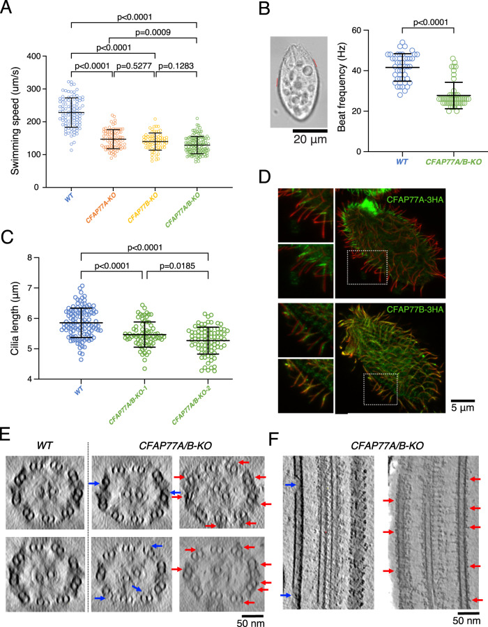
The structure and molecular dynamics simulation of TtCFAP77 suggested that it plays an important role in the stability of the DMT. Therefore, we investigated how deletion of TtCFAP77 will affect cell swimming, cilia beating, and DMT ultrastructure. Using a germ-line knock-out approach we engineered Tetrahymena cells and removed nearly the entire coding region of either CFAP77A (CFAP77A-KO) or CFAP77B (CFAP77B-KO), or both genes (CFAP77A/B-KO) (Supplementary Fig. 6A, B). Two independent clones were obtained for each mutant. The swimming trajectories of CFAP77 mutants, similar to WT cells, are straight but the cell swimming rate is reduced. Single CFAP77A-KO and CFAP77B-KO mutants covered approximately 66% and 63%, respectively of the WT distance while double CFAP77A/B-KO mutant traveled 58% of the WT distance (Fig. 6A). The analysis of the cilia beating using a high-speed camera revealed that in CFAP77A/B-KO mutant ciliary wave form and amplitude were similar to the one observed in the WT cells (Supplementary Fig. 6C) while the ciliary frequency was reduced (Fig. 6B and Supplementary Fig. 6D). Occasionally, in mutants, some asynchronously beating cilia were observed. The cilia length of two clones of CFAP77A/B-KO was about 90% that of WT cells, suggesting mild assembly defects (Fig. 6C).
Fig. 6. Knockout of CFAP77A/B caused mild defects in cilia.
A Knockout of CFAP77A or CFAP77B, or both led up to 40% swimming speed reduction (n = 99 for WT, n = 81 for CFAP77A-KO, n = 70 for CFAP77B-KO, n = 119 for CFAP77A/B-KO). Statistical analyses were done with two-sided Tukey’s multiple comparisons tests. B Tetrahymena cells with marked exemplary positions (red lines) where cilia beat was analyzed in recorded swimming cells. Graphical representation of measurements of cilia beating frequency in WT and CFAP77A/B-KO mutants. (n = 42 cilia from 12 cells for WT, n = 48 cilia from 12 cells for CFAP77A/B-KO). Statistical analyses were done with a two-sided Mann-Whitney comparisons test. C Cilia length measurements of WT and CFAP77A/B-KO mutants. On average cilia length was: WT = 5.85 µm (number of measured cilia, n = 115), CFAP77A/B-KO clone 1 = 5.46 µm (n = 74), CFAP77A/B-KO clone 2 = 5.27 µm (n = 83). Student’s t test WT/KO is 2E−08 and 6,8E−16, respectively. Data are presented as mean values +/− standard deviation in A–C. Source data are provided as a Source data file for A–C. D Differential localization of CFAP77A and CFAP77B in CFAP77A-3HA and CFAP77B-3HA knock-in mutants. Green—anti-HA; red—poly glycylated tubulin. E The tomographic cross-sections of WT and CFAP77A/B-KO mutants showing occasional damage in the outer junction of CFAP77A/B-KO mutants (blue arrows) and unknown densities near the outer junction region (red arrows). F Longitudinal sections from CFAP77A/B-KO tomogram showing outer junction damage (blue arrows) and unknown densities (red arrows). For (D–F), the experiments were done once.
Immunolocalization of TtCFAP77A and TtCFAP77B proteins expressed as fusions with C-terminal 3HA tag under the control of respective native promoters revealed that TtCFAP77A is present at the proximal end of the cilium while TtCFAP77B localizes along the entire cilium length except for the distal tip (Fig. 6D). However, how TtCFAP77A and TtCFAP77B are targeted for assembly is still unclear.
Using cryo-electron tomography and subtomogram averaging, we obtained structures of the WT and CFAP77A/B-KO mutant 96-nm axonemal repeat (Supplementary Fig. 7A, B). The averaged tomographic map of the CFAP77A/B-KO mutant axoneme looks similar to WT with no obvious defects. However, when we examined the raw tomograms of the CFAP77A/B-KO mutant, we observed gaps in outer junction regions in some of the tomograms (Fig. 6E, F, blue arrows). This suggests that the lack of CFAP77 likely destabilizes the outer junction mildly. We also observed some unknown and non-periodic densities binding to the outer junction from outside in some cilia in the CFAP77A/B-KO mutant (Fig. 6E, F, red arrows). Interestingly, the level of tubulin glutamylation in cilia was slightly increased in the CFAP77A/B-KO cells compared with the WT cells (Supplementary Fig. 7C–F).
Discussion
In this work, we used cryo-EM to determine the structure of native DMTs from the ciliate Tetrahymena thermophila and a combination of mass spectrometry and artificial intelligence approaches to identify and model proteins inside the DMT. Obtained structures show core MIPs and species-specific MIPs based on conservation with other species.
Since the DMT was obtained without harsh treatment or enzymatic digestion, its structure represents a close-to-native structure. It revealed a difference in periodicity between the outside and inside proteins of the Tetrahymena DMT without any clear connection between them. The inside periodicity is a multiple of 16 nm while the outside periodicity is a multiple of 24 nm. In the case of bovine respiratory cilia, it is proposed that Pierce 1 and 2 are the links between the outer 24-nm repeat of the outer dynein arm and the inner 48-nm repeat of MIPs12. In Tetrahymena, we observed STPG1A and STPG2 bind to the outer wedge and weave into the luminal space of the A- and B-tubule. However, there are many regions of the DMT having no contact between outside and inside proteins. Based on the observation of the outer surface filament, we speculate that the outside periodicity can also be achieved independently through a self-regulated head-to-tail arrangement as suggested for the molecular rulers CCDC39 and CCDD4032 and MIPs4,11. Perhaps initial templates of inside and outside proteins at the base of the cilia initiate the perfect registry between the proteins on the inside and outside surfaces. Interestingly, our structural analysis of the PG-rich motif indicates that ODF3L perhaps span 96-nm in the outer wedge of the microtubules. However, the role of ODF3L in the formation of the 96-nm periodicity of the outside is unknown.
The presence of the MAP7-like outer surface filaments in our structure is very interesting since it might have implications for intraflagellar transport on the DMT. As it was shown that MAP7 has biphasic regulation on kinesin-1 due to competitive binding to microtubules34, outer surface filaments might impose regulatory effects on dynein and kinesin, partitioning anterograde and retrograde transport tracks. Since there is no evidence that this is a conserved feature in the axoneme11,12, this might be a species-specific feature for intraflagellar transport in Tetrahymena. On the other hand, a sleeve-like density covering the DMT was observed in the Chlamydomonas in situ centriole structure37, which may prevent the continuous contact of intraflagellar transport motors with the DMT. While the mechanisms of intraflagellar transport to overcome the sleeves in Chlamydomonas and the outer surface filaments in Tetrahymena DMT are supposed to be different, a fundamental question of how the intraflagellar transport trains can accomplish that feat is an intriguing one.
Our study also identifies the conserved protein CFAP77 as an important outer junction protein. In human epithelial cilia CFAP77 localizes along the entire cilia length but is most abundant in their middle part38. Expression of human CFAP77 increases during ciliated epithelial cell differentiation38. In buffalo sperm, CFAP77 is both phosphorylated and ubiquitinated39. Hypomethylation of the CFAP77 gene has also been linked to opioid dependence40. It is still unclear how to link post-translation modifications of CFAP77 with its function in cilia.
Based on molecular dynamic simulation, knockout phenotype, and mass spectroscopy data we suggest that CFAP77 is important for the swimming of Tetrahymena probably through its stabilizing role. Interestingly, the phenotype of CFAP77A/B-KO is very similar to RIB72A/B-KO21 and CFAP115-KO mutants20 with lower ciliary beating frequency and coordination of beating between cilia and excessive curve or kink of some cilia. It is possible that MIPs are required for stabilization of the DMT as the force transducer for proper ciliary beating. Lacking certain MIPs reduces the tensile strength of the DMT, which leads to unexpected curves or kinks during ciliary bending and hence affects the beating coordination. Since the force during ciliary bending is stronger at the base, stability of the axoneme base is likely more important for ciliary function. Coincidently, more TtCFAP77A is present in the proximal half of the cilium (Fig. 6C); possibly the B-tubule is more stable in that region.
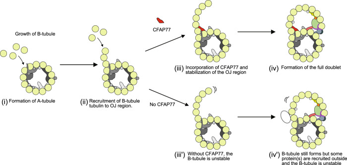
While TtCFAP77 structure and position suggest that it might be important for the suppression of the C-terminal tails of β-tubulins, our study shows that TtCFAP77 is not vital for the assembly of the DMT as the cilia still form with only occasional damages (Fig. 7). The mild phenotype might be because Tetrahymena has the species-specific protein OJ2, which might have a redundant role in ciliary assembly. Thus, the phenotype of a CFAP77-KO might be more severe in the case of other species and a potential ciliopathy gene in humans. Based on the increase in polyglutamylation in the CFAP77A/B-KO mutants and the presence of proteins outside the DMT, we speculate that the destabilization of the DMT might lead to a compensatory effect in post-translational modifications. Indeed, the increase in polyglutamylation of the DMT might compensate for the de-stabilization from the loss of CFAP77.
Fig. 7. A model of the role of CFAP77 on the formation of the B-tubule.
The A-tubule is formed (i), then free tubulins bind to form the hook at the outer junction (OJ) region (ii). With CFAP77, the newly formed B-tubule hook is stabilized (iii), leading to the final DMT formation (iv). Without CFAP77, the B-tubule is not properly stabilized (iii’) and there are additional protein(s) on the outer surface (iv’). The presence of additional protein(s) may or may not be related to elevated levels of tubulin polyglutamylation detected in CFAP77A/B-KO cilia.
Interestingly, CFAP77 was not found in the human primary cilia proteome41. Moreover, there is no orthologous protein encoded by the C. elegans genome. Yet the DMT exists in the human primary cilia and worm sensory cilia albeit in shorter segments. Therefore, it is likely that CFAP77 contributes more to the stability than the assembly of the outer junction. It was recently shown that Azami Green-fused Tau-derived peptide added to the microtubules induces formation of the more complex structures, including DMT42 similar to the doublet reconstitution of C-terminal tail truncated tubulins22. Therefore, it’s possible that other proteins in primary cilia bind to the outer surface of microtubules and interact with or suppress the C-terminal tails of tubulins.
In conclusion, using cryo-EM and improved cilia preparation methods, we have visualized the structure of the native DMT from Tetrahymena thermophila and identified 42 MIPs. Moreover, we have shown that an evolutionarily conserved protein, CFAP77, is an outer junction protein and its knockout affects cilia motility (Fig. 7). Our findings contribute to our understanding of MIP function but leave many questions regarding the identity of outer surface proteins and their implication in intraflagellar transport.
Methods
Cell culture
All Tetrahymena strains (CU428 WT, K40R, CFAP77A-3HA, CFAP77B-3HA CFAP77A-KO, CFAP77B-KO, CFAP77A/B-KO, RIB72B-KO, and RIB72A/B-KO mutants) used in this study were grown in 1 L of SPP media43 in a shaker incubator at 30 °C and 120 rpm. Cultures were harvested at an OD600 of approximately 0.744.
Cilia isolation using dibucaine treatment
Tetrahymena cells were harvested and deciliated44. Briefly, a 2 L cell culture was harvested by centrifugation at 2000 × g for 10 min at 23 °C. Whole cells were resuspended in fresh SPP and adjusted to a total volume of 24 mL. Dibucaine (25 mg in 1 mL SPP) was added, and the culture was gently swirled for 45 s. To stop the dibucaine treatment, 75 mL of ice-cold SPP supplemented with 1 mM EGTA was added and the dibucaine-treated cultures were centrifuged for 15 min at 2000 × g and 4 °C. The supernatant (cilia) was collected and centrifuged at 30,000 × g for 45 min at 4 °C. The pellet (cilia) was gently washed and resuspended in Cilia Wash Buffer (50 mM HEPES at pH 7.4, 3 mM MgSO4, 0.1 mM EGTA, 1 mM DTT, 250 mM sucrose). The resuspended cilia were flash-frozen with liquid nitrogen and stored at −80 °C.
Purification of the intact axoneme
The frozen cilia were thawed on ice and then centrifuged for 10 min at 8000 × g and 4 °C. The pellet (cilia) was washed and resuspended in Cilia Final Buffer (50 mM HEPES at pH 7.4, 3 mM MgSO4, 0.1 mM EGTA, 1 mM DTT, 0.5% trehalose). NP-40 Alternative (Millipore Sigma #492016) was added to a final concentration of 1.5% for 45 min on ice and then the de-membranated axonemes were centrifuged. Intact axonemes (membranes removed with detergent) can be used for tomography or further DMT preparation (see below). For tomography, the pelleted intact axonemes were resuspended in Cilia Final Buffer to a final concentration of 3.8 mg/mL. Cross-linking was done with glutaraldehyde at a final concentration of 0.015%. Bradford reagent (Bio-rad #5000201) was used to measure the total protein concentration.
Purification of DMTs
The intact axoneme pellet (see above) was resuspended with Cilia Final Buffer and ADP was added to a final concentration of 0.3 mM. The sample was incubated at room temperature for 10 min. ATP was added to a final concentration of 1 mM and the sample was again incubated at room temperature for 10 min. DMT samples were adjusted to 2.2 mg/mL using Cilia Final Buffer.
Cryo-EM sample preparation
C-Flat Holey thick carbon grids (Electron Microscopy Services #CFT312-100) were treated with chloroform overnight. For single-particle analysis, DMT sample (4 μL) was applied to the chloroform-treated, negatively glow-discharged (10 mA, 15 s) grids inside the Vitrobot Mk IV (Thermo Fisher) chamber. The sample was incubated on the grid for 15 s at 23 °C and 100% humidity then blotted with force 3 for 5 s then plunge frozen in liquid ethane.
Cryo-EM data acquisition
A total of 18,384 and 25,610 movies were collected for the WT and K40R mutant using the Titan Krios 300 keV FEG electron microscope (Thermo Fisher) equipped with direct electron detector K3 Summit (Gatan, Inc.) and the BioQuantum energy filter (Gatan, Inc.) using SerialEM45. The movies were collected with a beam shift pattern of four movies per hole and four holes per movement. The final pixel size is 1.370 Å/pixel. Each movie has a total dose of 45 electrons per Å2 over 40 frames. The defocus range was between −1.0 and −3.0 μm at an interval of 0.25 μm.
Image processing
Motion correction and dose-weighting of the movies were performed using MotionCor246 implemented in Relion 347, and the contrast transfer function parameters were estimated using Gctf48. Micrographs with apparent drift, ice contamination, and bad contrast transfer function estimation were discarded.
The particles were picked using a combination of manual and automatic pickings to speed up this process. The rare top views of the filaments were picked manually using e2helixboxer49. The side views of the filaments were picked automatically using Topaz by training with a set of side view manual picks50. The Topaz coordinates were converted into the filament coordinates using a customized clustering and line fitting Python scripts based on RANSAC algorithm.
Particles of 512 × 512 pixels were extracted with 8-nm overlapped, binned twice, and pre-aligned using a modified version of the Iterative Helical Real Space Reconstruction script51 in SPIDER52 to work with non-helical symmetry. The alignment parameters were then transferred to Frealign for aligning the particles for six iterations in Frealign53, and then converted into Relion 3.1. Iterative per-particle-defocus refinement and Bayesian polishing were done for the 8 nm particles.
Each particle was subtracted from its tubulin lattice signal and underwent 3D classification into two classes to obtain the 16-nm repeat particles. The 16-nm repeat particles were then subjected to 3D classification into 3 classes to obtain the 48-nm repeat particles. The 48-nm particles were then refined, resulting in a global resolution of 4.0 Å and 3.5 Å for WT and the K40R mutant (Table 1).
To improve the local resolution for modeling, we performed focused refinements of different regions by different masks to cover approximately two PFs. Next, the maps were merged into a composite map by Phenix combine_focused_maps tool54 and then enhanced by DeepEmhancer55 to improve visualization and interpretability.
To obtain the 96-nm map, we combined WT and K40R particles and performed a 3D classification into two classes with a mask focusing on the outside of the DMT and subsequent refinement of the 96-nm particles. We obtained a 3.75 Å resolution of the combined 96-nm map (Table 1).
Modeling
Modeling of tubulins was done based on PDB 6U0H. For MIP modeling, two strategies were carried out. For conserved MIPs with available models from Chlamydomonas reinhardtii (PDB 6U42), homologous models of Tetrahymena proteins were either constructed using Modeller56 or predicted using ColabFold29. The homologous models were fitted into the 48-nm density map of Tetrahymena based on the relative location in the Chlamydomonas map or by docking using UCSF ChimeraX function fitmap57. The final models were then modeled to the density using Coot58 and real-space refined in Phenix54 (Table 2). For unknown densities in the Tetrahymena map, connected density was first segmented manually using Chimera. The segmented density was then submitted to DeepTracer server27 to trace the C-alpha backbone. The C-alpha model was then searched for fold similarity in the library of ColabFold predicted models of all proteins in the proteome of Tetrahymena cilia using Pymol cealign function. The top candidate ColabFold predicted models were then fitted to the full Tetrahymena density map for evaluation. Suitable candidates were gone through modeling in Coot and real-space refinement in Phenix (Supplementary Fig. 1C). Alternatively, we searched for the identity of the proteins by using FindMySequence30 to search the C-alpha backbone trace within the database of the cilium proteome (Supplementary Fig. 1C). In certain cases, we used the same density from the Tetrahymena map of K40R mutant to facilitate modeling since the K40R map has a better resolution at 3.5 Å. For validation, we used both FindMySquence’s E-value as an indicator for a good identification and in situ cross-linking mass spectrometry of Tetrahymena cilia31 (Supplementary Fig. 1D, Supplementary Fig. 2, Supplementary Table 5). All of the maps and model visualization were done using ChimeraX57.
Cryo-ET sample preparation
To preserve the round shape of the axoneme, the axonemes for tomography were cross-linked by glutaraldehyde (final concentration 0.15%) for 40 min on ice and quenched by 1 M HEPES. Axonemes were approximately 3.6 mg/mL when mixed with 10 nm gold beads in a 1:1 ratio for a final axoneme concentration of 1.8 mg/mL. Cross-linked axoneme sample (4 μL) was applied to negatively glow-discharged (10 mA, 15 s) C-Flat Holey thick carbon grids (Electron Microscopy Services #CFT312-100) inside the Vitrobot Mk IV (Thermo Fisher) chamber. The sample was incubated on the grid for 15 s at 23 °C and 100% humidity then blotted with force 0 for 8 s then plunge frozen in liquid ethane.
Cryo-ET acquisition and reconstruction
Tilt series were collected using the dose-symmetric scheme from −60 to 60 degrees with an increment of 3 degrees. The defocus for each tilt series ranges from −2.5 to −3.5 μm. The total dose for each tilt series is 160 e− per Å2. For each view, a movie of 10–13 frames was collected. The pixel size for the tilt series is 2.12 Å. Tomograms were reconstructed using IMOD45. Frame alignment was done with Alignframes59. Aligned tilt series were manually inspected for quality and sufficient fiducial presence. Batch reconstruction was performed in IMOD. The cross-linked axonemes appear similar to non-cross-linked axoneme without any obvious artifacts.
Subtomogram averaging
Subtomogram averaging of the 4-times binned 96 nm repeating unit of WT and CFAP77A/B-KO mutants (2608 and 1702 subtomograms respectively) was done using the “axoneme align”60. CTF estimation was done with WARP61. Refinement of the 96 nm subtomogram averages was done with the Relion 4.0 pipeline62. The resolutions for the 96-nm repeating unit of WT and CFAP77A/B-KO DMT are 18 and 21 Å, respectively. For visualization, tomograms were CTF deconvolved and missing wedge corrected using IsoNet63.
Setting up for CFAP77 coarse-grained MD simulation
We used A10-12 and B1-2 PFs, OJ2 and TtCFAP77 to model Tetrahymena outer junction region. Based on the atomic model of the outer junction region, coarse-grained MD simulation was performed using CafeMol 2.164. Each amino acid was represented as a single bead located at its Cα position in the coarse-grained model. We used the energy functions AICG2+, excluded volume, and electrostatic interaction for predicting dynamics. In the AICG2+, the original reference structure was assumed as the most stable structure, and parameters were modified to represent the interactions in the all-atom reference structure65. Three residues (F133, G308, and E401) at the three A10 α-tubulins were anchored for the convenience of the analysis. It is known that the inter α-β dimer interaction is much weaker than intra α-β dimer interaction. Also, inter A-B tubule interaction is weaker than intra A- or B-tubule interaction. To replicate these features in our simulations, we set inter α-β dimer’s interacting force to 0.8 times the original value while the intra α-β dimer’s interacting force was left as the original value (1.0 times). Also, we set inter A-B tubule interacting force to 0.2 times the original value while that of intra A- or B-tubules PFs’ interacting force was set to 0.3 times the original value.
Then, we performed simulations of the outer junction region stability with and without OJ2 and CFAP77 molecules coarse-grained MD, 20 times for each setup. Each MD simulation took 3×10⁷ MD steps. Note, that one MD step roughly corresponds to approximately 1 ps. The MD simulations were conducted by the underdamped Langevin dynamics at 300 K temperature. We set the friction coefficient to 0.02 (CafeMol unit), and default values in CafeMol were used for other parameters.
To calculate angular elasticity between A- and B-tubules (Supplementary Fig. 5F), the two vectors used in the angle calculation were defined by the K401 of the central α-tubulin in A10 as a vector base point, and the center of mass of the central α-tubulin in A12 and B2 as an endpoint of the vector. Since K401 is a residue anchored in space for MD simulation efficiency, it serves as a base point for following the change in the angle between A and B tubules.
CFAP77 gene knock-ins and knock-outs
To reveal the localization of the CFAP77 protein paralogs, we amplified the entire open reading frame with the addition of MluI and BamHI restriction sites at 5’ and 3’ ends, respectively, and 3’UTR with the addition of PstI and XhoI restriction sites at 5’ and 3’ ends, respectively using Phusion HSII High Fidelity Polymerase (Thermo Fisher Scientific Baltics, Lithuania) and primers listed in Supplementary Table 7, and replaced fragments of the CFAP44 gene in CFAP44-3HA plasmid66. Such a construct enabled the expression of the C-terminally 3HA tagged CFAP77 paralogs under the control of their own promoters and selection of the positive clones based on the resistance to paromomycin (neo4 resistance cassette67). Approximately 10 μg of plasmids were digested with MluI and XhoI to separate a transgene from the plasmid backbone, precipitated onto DNAdel Gold Carrier Particles (Seashell Technology, La Jolla, CA, USA) according to the manufacturer’s instructions and used to biolistically transform CU428 cells. The positive clones were grown in SPP medium with the addition of an increasing concentration of paromomycin (up to 1 mg/mL) and decreasing concentration of CdCl2 (up to 0.2 µg/mL) to promote the transgene assortment.
To knock out Tetrahymena CFAP77 genes we employed the germline gene disruption approach68,69. Using primers listed in Supplementary Table 7 we amplified fragments of the targeted genes with the addition of selected restriction sites and cloned upstream and downstream of the neo4 resistance cassette67. In both loci, nearly the entire coding region was removed. To obtain strain with deletion of both CFAP77A and CFAP77B, we crossed CFAP77A and CFAP77B heterokaryons and followed with cell maturation and selection as described68. At least two independent clones were obtained for single and double mutants. The loci deletion was confirmed by PCR (primers in Supplementary Table 7).
Phenotypic and localization studies
Cells swimming rate and cilia beating were analyzed as previously described70. For protein localization studies, Tetrahymena cells were fixed either by adding equal volume of the mixture of 0.5% NP40 and 2% PFA in PHEM buffer or by permeabilization with 0.25% Triton-X-100 followed by fixation with 4% PFA (final concentrations) on coverslips, air-dried, blocked using 3%BSA/PBS and incubated overnight with anti-HA antibodies (Cell Signaling Technology, Danvers, MA, USA) diluted 1:200, and polyG antibodies71, 1:2000 at 4 °C. After washing, 3×5 min with PBS, samples were incubated with secondary antibodies (anti-mouse IgG conjugated with Alexa-488, and anti-rabbit IgG conjugated with Alexa-555 (Invitrogen, Eugene, OR, USA) both in concentration of 1:300. After washing, the coverslips were mounted in Fluoromount-G (Southern Biotech, Birmingham, AL, USA) and viewed using Zeiss LSM780 (Carl Zeiss Jena, Germany) or Leica TCS SP8 (Leica Microsystems, Wetzlar, Germany) confocal microscope. The expression of the tagged proteins was verified by Western blot as described before72.
Quantification of polyglutamylation
WT cells were incubated for 10 min in medium supplied with India Ink to form ink-filled dark food vacuoles. Next, WT and CFAP77A/B-KO cells were fixed side-by-side as described73 and stained with polyE anti-polyglutamic acid primary antibody (1:2000). WT and mutant cells position next to one another were recorded using a confocal microscope and the intensity of the polyE signal in cilia was measured using ImageJ program. For western blotting, 5 μg of cilia protein per lane were separated on a 10% SDS-PAGE and western blots were done as described74, with the primary antibodies at the following dilutions: GT335 anti-glutamylated tubulin mAb (1:1000), 12G10 anti-α-tubulin mAb (1:40,000) deposited to the DSHB by Frankel, J. / Nelsen, E.M. (DSHB Hybridoma Product 12G10 anti-alpha-tubulin), polyE antibodies (1:20,000). The intensity of detected bands was measured using ImageJ and presented as a graph showing a ratio of the glutamylated tubulin to α-tubulin.
Mass spectrometry
Samples prepared for cryo-EM were also analyzed by mass spectrometry. Approximately 25–30 μg proteins were loaded on the SDS-PAGE gel. Electrophoresis was performed, but the run was terminated before the proteins entered the resolving gel. The gel band was cut, reduced with DTT, alkylated with iodoacetic acid and digested with trypsin. Extracted peptides were re-solubilized in 0.1% aqueous formic acid and loaded onto a Thermo Acclaim Pepmap (Thermo, 75µM ID × 2 cm C18 3uM beads) precolumn and then onto an Acclaim Pepmap Easyspray (Thermo, 75µM × 15 cm with 2µM C18 beads) analytical column separation using a Dionex Ultimate 3000 uHPLC at 250 nL/min with a gradient of 2–35% organic (0.1% formic acid in acetonitrile) over 3 h. Peptides were analyzed using a Thermo Orbitrap Fusion mass spectrometer operating at 120,000 resolution (FWHM in MS1) with HCD sequencing (15,000 resolution) at top speed for all peptides with a charge of 2+ or greater. The raw data were converted into *.mgf format (Mascot generic format) for searching using the Mascot 2.6.2 search engine (Matrix Science) against Tetrahymena thermophila protein dataset from UniProt.
Mass spectrometry data were analyzed by Scaffold_4.8.4 (Proteome Software Inc.). Proteins with mean values of exclusive unique peptide count of 2 or more in the WT mass spectrometry results were used for analysis. Raw mass spectrometry data were normalized by total spectra. One-way ANOVA was applied to WT, RIB72A/B-KO and RIB72B-KO mass spectrometry results using biological triplicates. Proteins missing in RIB72A/B-KO and RIB72B-KO compared to WT were identified in Supplementary Table 6. We used the emPAI score calculation of ciliary proteins of specific sizes and periodicities as complementary information for protein identification. We compared the emPAI scores between salt-treated and non-salt-treated axonemes to filter out proteins that are on the surface of the microtubule, rather than the lumen.
Reporting summary
Further information on research design is available in the Nature Portfolio Reporting Summary linked to this article.
Supplementary information
Description of additional supplementary files
Acknowledgements
We thank Dr. Kelly Sears, Dr. Mike Strauss, Dr. Kaustuv Basu (Facility for Electron Microscopy Research at McGill University) for helping with data collection and Dr. Muneyoshi Ichikawa for critically reading of the manuscript. We acknowledge the RI-MUHC Proteomics and Molecular Analysis Platform for assistance with protein analysis. S.K. is supported by JSPS Overseas Research Fellowships. K.H.B. is supported by the grants from Canadian Institutes of Health Research (PJT-156354) and Natural Sciences and Engineering Research Council of Canada (RGPIN-2022-04774). E.M.M. acknowledges support from the Welch Foundation (F1515) and U.S. National Institutes of Health (R01 HD085901).
Source data
Author contributions
Conceptualization: K.H.B. Methodology: T.L., K.P., A.G., A.A.Z.K., C.D., M.V.-P., Z.F., P.H., C.L.M. Formal analysis: S.K., C.B., E.J., S.K.Y., C.L.M. Investigation: S.K., C.B., E.J., S.K.Y., T.L., K.P., A.G., A.A.Z.K., C.D., M.V.-P., Z.F., P.H., C.L.M., E.M.M. Resources: D.W., K.H.B. Writing—original draft: K.H.B., D.W., S.K., C.B. Writing—review and editing: K.H.B., D.W., S.K., C.B., E.J., K.P.
Peer review
Peer review information
Nature Communications thanks anonymous reviewer(s) for their contribution to the peer review of this work. Peer review reports are available.
Data availability
The data generated in this study are available in the following databases: 8G2Z (48-nm Tetra WT doublet), 8G3D (48-nm Tetra K40R doublet), EMD-29685 (48-nm Tetra WT doublet), EMD-29692 (48-nm Tetra K40R doublet), EMD-29693 (96-nm Tetra combined doublet), EMD-29667 (96-nm Tetra WT doublet subtomogram average), EMD-29666 (96-nm Tetra CFAP77A/B-KO doublet subtomogram average). All data used but not produced in this study are available in the following databases: 6U0H (48-nm Tetra WT doublet), 6U42 (48-nm Chlamydomonas WT doublet), 7RRO (48-nm Bovine WT doublet), EMD-20602 (48-nm Tetra WT doublet), EMD-20631 (48-nm Chlamydomonas WT doublet), EMD-24664 (48-nm Bovine WT doublet). The cilia length measurement, swimming speed, waveform and polyglutamylation quantification data, original gel and blot, and mass spectrometry of WT, RIB72B-KO, RIB72A/B-KO cilia generated in this study are provided in the Supplementary Information/Source Data file. Source data are provided with this paper.
Code availability
The Python code for filament fitting is available on Github.
Competing interests
The authors declare no competing interests.
Footnotes
Publisher’s note Springer Nature remains neutral with regard to jurisdictional claims in published maps and institutional affiliations.
These authors contributed equally: Shintaroh Kubo, Corbin S. Black, Ewa Joachimiak.
Contributor Information
Dorota Wloga, Email: d.wloga@nencki.edu.pl.
Khanh Huy Bui, Email: huy.bui@mcgill.ca.
Supplementary information
The online version contains supplementary material available at 10.1038/s41467-023-37868-0.
References
- 1.Reiter JF, Leroux MR. Genes and molecular pathways underpinning ciliopathies. Nat. Rev. Mol. Cell Biol. 2017;18:533–547. doi: 10.1038/nrm.2017.60. [DOI] [PMC free article] [PubMed] [Google Scholar]
- 2.Nicastro D, et al. The molecular architecture of axonemes revealed by cryoelectron tomography. Science. 2006;313:944–948. doi: 10.1126/science.1128618. [DOI] [PubMed] [Google Scholar]
- 3.Bui KH, Sakakibara H, Movassagh T, Oiwa K, Ishikawa T. Molecular architecture of inner dynein arms in situ in Chlamydomonas reinhardtii flagella. J. Cell Biol. 2008;183:923–932. doi: 10.1083/jcb.200808050. [DOI] [PMC free article] [PubMed] [Google Scholar]
- 4.Ichikawa M, et al. Tubulin lattice in cilia is in a stressed form regulated by microtubule inner proteins. Proc. Natl Acad. Sci. USA. 2019;116:19930–19938. doi: 10.1073/pnas.1911119116. [DOI] [PMC free article] [PubMed] [Google Scholar]
- 5.Stepanek L, Pigino G. Microtubule doublets are double-track railways for intraflagellar transport trains. Science. 2016;352:721–724. doi: 10.1126/science.aaf4594. [DOI] [PubMed] [Google Scholar]
- 6.Nicastro D, et al. Cryo-electron tomography reveals conserved features of doublet microtubules in flagella. Proc. Natl Acad. Sci. USA. 2011;108:E845–E853. doi: 10.1073/pnas.1106178108. [DOI] [PMC free article] [PubMed] [Google Scholar]
- 7.Maheshwari A, et al. alpha- and beta-Tubulin lattice of the axonemal microtubule doublet and binding proteins revealed by single particle cryo-electron microscopy and tomography. Structure. 2015;23:1584–1595. doi: 10.1016/j.str.2015.06.017. [DOI] [PubMed] [Google Scholar]
- 8.Ichikawa M, et al. Subnanometre-resolution structure of the doublet microtubule reveals new classes of microtubule-associated proteins. Nat. Commun. 2017;8:15035. doi: 10.1038/ncomms15035. [DOI] [PMC free article] [PubMed] [Google Scholar]
- 9.Khalifa AAZ, et al. The inner junction complex of the cilia is an interaction hub that involves tubulin post-translational modifications. Elife. 2020;9:e52760. doi: 10.7554/eLife.52760. [DOI] [PMC free article] [PubMed] [Google Scholar]
- 10.Li S, Fernandez JJ, Fabritius AS, Agard DA, Winey M. Electron cryo-tomography structure of axonemal doublet microtubule from Tetrahymena thermophila. Life Sci. Alliance. 2022;5:e202101225. doi: 10.26508/lsa.202101225. [DOI] [PMC free article] [PubMed] [Google Scholar]
- 11.Ma M, et al. Structure of the decorated ciliary doublet microtubule. Cell. 2019;179:909.e2–922.e2. doi: 10.1016/j.cell.2019.09.030. [DOI] [PMC free article] [PubMed] [Google Scholar]
- 12.Gui M, et al. De novo identification of mammalian ciliary motility proteins using cryo-EM. Cell. 2021;184:5791–5806.e5719. doi: 10.1016/j.cell.2021.10.007. [DOI] [PMC free article] [PubMed] [Google Scholar]
- 13.Yanagisawa HA, et al. FAP20 is an inner junction protein of doublet microtubules essential for both the planar asymmetrical waveform and stability of flagella in Chlamydomonas. Mol. Biol. Cell. 2014;25:1472–1483. doi: 10.1091/mbc.e13-08-0464. [DOI] [PMC free article] [PubMed] [Google Scholar]
- 14.Dymek EE, et al. PACRG and FAP20 form the inner junction of axonemal doublet microtubules and regulate ciliary motility. Mol. Biol. Cell. 2019;30:1805–1816. doi: 10.1091/mbc.E19-01-0063. [DOI] [PMC free article] [PubMed] [Google Scholar]
- 15.Owa M, et al. Inner lumen proteins stabilize doublet microtubules in cilia and flagella. Nat. Commun. 2019;10:1143. doi: 10.1038/s41467-019-09051-x. [DOI] [PMC free article] [PubMed] [Google Scholar]
- 16.Ikeda K, et al. Rib72, a conserved protein associated with the ribbon compartment of flagellar A-microtubules and potentially involved in the linkage between outer doublet microtubules. J. Biol. Chem. 2003;278:7725–7734. doi: 10.1074/jbc.M210751200. [DOI] [PubMed] [Google Scholar]
- 17.Norrander JM, deCathelineau AM, Brown JA, Porter ME, Linck RW. The Rib43a protein is associated with forming the specialized protofilament ribbons of flagellar microtubules in Chlamydomonas. Mol. Biol. Cell. 2000;11:201–215. doi: 10.1091/mbc.11.1.201. [DOI] [PMC free article] [PubMed] [Google Scholar]
- 18.King SM. Axonemal protofilament ribbons, DM10 domains, and the link to juvenile myoclonic epilepsy. Cell Motil. Cytoskeleton. 2006;63:245–253. doi: 10.1002/cm.20129. [DOI] [PubMed] [Google Scholar]
- 19.Suzuki T, Inoue I, Yamakawa K. Epilepsy protein Efhc1/myoclonin1 is expressed in cells with motile cilia but not in neurons or mitotic apparatuses in brain. Sci. Rep. 2020;10:22076. doi: 10.1038/s41598-020-79202-4. [DOI] [PMC free article] [PubMed] [Google Scholar]
- 20.Fabritius AS, et al. Proteomic analysis of microtubule inner proteins (MIPs) in Rib72 null Tetrahymena cells reveals functional MIPs. Mol. Biol. Cell. 2021;32:br8. doi: 10.1091/mbc.E20-12-0786. [DOI] [PMC free article] [PubMed] [Google Scholar]
- 21.Stoddard D, et al. Tetrahymena RIB72A and RIB72B are microtubule inner proteins in the ciliary doublet microtubules. Mol. Biol. Cell. 2018;29:2566–2577. doi: 10.1091/mbc.E18-06-0405. [DOI] [PMC free article] [PubMed] [Google Scholar]
- 22.Schmidt-Cernohorska M, et al. Flagellar microtubule doublet assembly in vitro reveals a regulatory role of tubulin C-terminal tails. Science. 2019;363:285–288. doi: 10.1126/science.aav2567. [DOI] [PubMed] [Google Scholar]
- 23.Vent J, et al. Direct involvement of the isotype-specific C-terminus of beta tubulin in ciliary beating. J. Cell Sci. 2005;118:4333–4341. doi: 10.1242/jcs.02550. [DOI] [PMC free article] [PubMed] [Google Scholar]
- 24.Bosch Grau M, et al. Tubulin glycylases and glutamylases have distinct functions in stabilization and motility of ependymal cilia. J. Cell Biol. 2013;202:441–451. doi: 10.1083/jcb.201305041. [DOI] [PMC free article] [PubMed] [Google Scholar]
- 25.Kubo S, et al. Remodeling and activation mechanisms of outer arm dyneins revealed by cryo-EM. EMBO Rep. 2021;22:e52911. doi: 10.15252/embr.202152911. [DOI] [PMC free article] [PubMed] [Google Scholar]
- 26.Akella JS, et al. MEC-17 is an alpha-tubulin acetyltransferase. Nature. 2010;467:218–222. doi: 10.1038/nature09324. [DOI] [PMC free article] [PubMed] [Google Scholar]
- 27.Pfab J, Phan NM, Si D. DeepTracer for fast de novo cryo-EM protein structure modeling and special studies on CoV-related complexes. Proc. Natl Acad. Sci. USA. 2021;118:e2017525118. doi: 10.1073/pnas.2017525118. [DOI] [PMC free article] [PubMed] [Google Scholar]
- 28.Jumper J, et al. Highly accurate protein structure prediction with AlphaFold. Nature. 2021;596:583–589. doi: 10.1038/s41586-021-03819-2. [DOI] [PMC free article] [PubMed] [Google Scholar]
- 29.Mirdita, M. et al. ColabFold: making protein folding accessible to all. Nat. Methods19, 679–682 (2022). [DOI] [PMC free article] [PubMed]
- 30.Chojnowski G, et al. findMySequence: a neural-network-based approach for identification of unknown proteins in X-ray crystallography and cryo-EM. IUCrJ. 2022;9:86–97. doi: 10.1107/S2052252521011088. [DOI] [PMC free article] [PubMed] [Google Scholar]
- 31.McCafferty, C. L., Pennington, E. L., Papoulas, O., Taylor, D. W. & Marcotte, E. M. Does AlphaFold2 model proteins’ intracellular conformations? An experimental test using cross-linking mass spectrometry of endogenous ciliary proteins. Preprint at bioRxiv10.1101/2022.08.25.505345 (2022). [DOI] [PMC free article] [PubMed]
- 32.Oda T, Yanagisawa H, Kamiya R, Kikkawa M. A molecular ruler determines the repeat length in eukaryotic cilia and flagella. Science. 2014;346:857–860. doi: 10.1126/science.1260214. [DOI] [PubMed] [Google Scholar]
- 33.Gaertig J, et al. Acetylation of lysine 40 in alpha-tubulin is not essential in Tetrahymena thermophila. J. Cell Biol. 1995;129:1301–1310. doi: 10.1083/jcb.129.5.1301. [DOI] [PMC free article] [PubMed] [Google Scholar]
- 34.Ferro LS, et al. Structural and functional insight into regulation of kinesin-1 by microtubule-associated protein MAP7. Science. 2022;375:326–331. doi: 10.1126/science.abf6154. [DOI] [PMC free article] [PubMed] [Google Scholar]
- 35.Monroy BY, et al. A combinatorial MAP code dictates polarized microtubule transport. Dev. Cell. 2020;53:60.e4–72.e4. doi: 10.1016/j.devcel.2020.01.029. [DOI] [PMC free article] [PubMed] [Google Scholar]
- 36.Nogales E, Whittaker M, Milligan RA, Downing KH. High-resolution model of the microtubule. Cell. 1999;96:79–88. doi: 10.1016/S0092-8674(00)80961-7. [DOI] [PubMed] [Google Scholar]
- 37.van den Hoek H, et al. In situ architecture of the ciliary base reveals the stepwise assembly of intraflagellar transport trains. Science. 2022;377:543–548. doi: 10.1126/science.abm6704. [DOI] [PubMed] [Google Scholar]
- 38.Blackburn K, Bustamante-Marin X, Yin W, Goshe MB, Ostrowski LE. Quantitative proteomic analysis of human airway cilia identifies previously uncharacterized proteins of high abundance. J. Proteome Res. 2017;16:1579–1592. doi: 10.1021/acs.jproteome.6b00972. [DOI] [PMC free article] [PubMed] [Google Scholar]
- 39.Zhang PF, et al. Integrated analysis of phosphoproteome and ubiquitylome in epididymal sperm of buffalo (Bubalus bubalis) Mol. Reprod. Dev. 2021;88:15–33. doi: 10.1002/mrd.23432. [DOI] [PMC free article] [PubMed] [Google Scholar]
- 40.Montalvo-Ortiz JL, Cheng Z, Kranzler HR, Zhang H, Gelernter J. Genomewide study of epigenetic biomarkers of opioid dependence in European-American Women. Sci. Rep. 2019;9:4660. doi: 10.1038/s41598-019-41110-7. [DOI] [PMC free article] [PubMed] [Google Scholar]
- 41.Mick DU, et al. Proteomics of primary cilia by proximity labeling. Dev. Cell. 2015;35:497–512. doi: 10.1016/j.devcel.2015.10.015. [DOI] [PMC free article] [PubMed] [Google Scholar]
- 42.Inaba H, et al. Generation of stable microtubule superstructures by binding of peptide-fused tetrameric proteins to inside and outside. Sci. Adv. 2022;8:eabq3817. doi: 10.1126/sciadv.abq3817. [DOI] [PMC free article] [PubMed] [Google Scholar]
- 43.Williams NE, Wolfe J, Bleyman LK. Long-term maintenance of Tetrahymena spp. J. Protozool. 1980;27:327. doi: 10.1111/j.1550-7408.1980.tb04270.x. [DOI] [PubMed] [Google Scholar]
- 44.Black C, Dai DC, Peri K, Ichikawa M, Bui KH. Preparation of doublet microtubule fraction for single particlecryo-electron microscopy. Bio Protoc. 2021;11:e4041. doi: 10.21769/BioProtoc.4041. [DOI] [PMC free article] [PubMed] [Google Scholar]
- 45.Mastronarde DN. Automated electron microscope tomography using robust prediction of specimen movements. J. Struct. Biol. 2005;152:36–51. doi: 10.1016/j.jsb.2005.07.007. [DOI] [PubMed] [Google Scholar]
- 46.Zheng SQ, et al. MotionCor2: anisotropic correction of beam-induced motion for improved cryo-electron microscopy. Nat. Methods. 2017;14:331–332. doi: 10.1038/nmeth.4193. [DOI] [PMC free article] [PubMed] [Google Scholar]
- 47.Zivanov J, et al. New tools for automated high-resolution cryo-EM structure determination in RELION-3. Elife. 2018;7:e42166. doi: 10.7554/eLife.42166. [DOI] [PMC free article] [PubMed] [Google Scholar]
- 48.Zhang K. Gctf: Real-time CTF determination and correction. J. Struct. Biol. 2016;193:1–12. doi: 10.1016/j.jsb.2015.11.003. [DOI] [PMC free article] [PubMed] [Google Scholar]
- 49.Tang G, et al. EMAN2: an extensible image processing suite for electron microscopy. J. Struct. Biol. 2007;157:38–46. doi: 10.1016/j.jsb.2006.05.009. [DOI] [PubMed] [Google Scholar]
- 50.Bepler T, et al. Positive-unlabeled convolutional neural networks for particle picking in cryo-electron micrographs. Nat. Methods. 2019;16:1153–1160. doi: 10.1038/s41592-019-0575-8. [DOI] [PMC free article] [PubMed] [Google Scholar]
- 51.Egelman EH. The iterative helical real space reconstruction method: surmounting the problems posed by real polymers. J. Struct. Biol. 2007;157:83–94. doi: 10.1016/j.jsb.2006.05.015. [DOI] [PubMed] [Google Scholar]
- 52.Frank J, et al. SPIDER and WEB: processing and visualization of images in 3D electron microscopy and related fields. J. Struct. Biol. 1996;116:190–199. doi: 10.1006/jsbi.1996.0030. [DOI] [PubMed] [Google Scholar]
- 53.Grigorieff N. FREALIGN: high-resolution refinement of single particle structures. J. Struct. Biol. 2007;157:117–125. doi: 10.1016/j.jsb.2006.05.004. [DOI] [PubMed] [Google Scholar]
- 54.Afonine PV, et al. Real-space refinement in PHENIX for cryo-EM and crystallography. Acta Crystallogr. D Struct. Biol. 2018;74:531–544. doi: 10.1107/S2059798318006551. [DOI] [PMC free article] [PubMed] [Google Scholar]
- 55.Sanchez-Garcia R, et al. DeepEMhancer: a deep learning solution for cryo-EM volume post-processing. Commun. Biol. 2021;4:874. doi: 10.1038/s42003-021-02399-1. [DOI] [PMC free article] [PubMed] [Google Scholar]
- 56.Webb B, Sali A. Protein structure modeling with MODELLER. Methods Mol. Biol. 2014;1137:1–15. doi: 10.1007/978-1-4939-0366-5_1. [DOI] [PubMed] [Google Scholar]
- 57.Pettersen EF, et al. UCSF ChimeraX: structure visualization for researchers, educators, and developers. Protein Sci. 2021;30:70–82. doi: 10.1002/pro.3943. [DOI] [PMC free article] [PubMed] [Google Scholar]
- 58.Emsley P, Lohkamp B, Scott WG, Cowtan K. Features and development of Coot. Acta Crystallogr. D Biol. Crystallogr. 2010;66:486–501. doi: 10.1107/S0907444910007493. [DOI] [PMC free article] [PubMed] [Google Scholar]
- 59.Mastronarde DN, Held SR. Automated tilt series alignment and tomographic reconstruction in IMOD. J. Struct. Biol. 2017;197:102–113. doi: 10.1016/j.jsb.2016.07.011. [DOI] [PMC free article] [PubMed] [Google Scholar]
- 60.Bui KH, Ishikawa T. 3D structural analysis of flagella/cilia by cryo-electron tomography. Methods Enzymol. 2013;524:305–323. doi: 10.1016/B978-0-12-397945-2.00017-2. [DOI] [PubMed] [Google Scholar]
- 61.Tegunov D, Cramer P. Real-time cryo-electron microscopy data preprocessing with Warp. Nat. Methods. 2019;16:1146–1152. doi: 10.1038/s41592-019-0580-y. [DOI] [PMC free article] [PubMed] [Google Scholar]
- 62.Zivanov J, et al. A Bayesian approach to single-particle electron cryo-tomography in RELION-4.0. Elife. 2022;11:e83724. doi: 10.7554/eLife.83724. [DOI] [PMC free article] [PubMed] [Google Scholar]
- 63.Liu Y-T, et al. Isotropic reconstruction of electron tomograms with deep learning. Nat. Commun. 2021;13:6482. doi: 10.1038/s41467-022-33957-8. [DOI] [PMC free article] [PubMed] [Google Scholar]
- 64.Kenzaki H, et al. CafeMol: a coarse-grained biomolecular simulator for simulating proteins at work. J. Chem. Theory Comput. 2011;7:1979–1989. doi: 10.1021/ct2001045. [DOI] [PubMed] [Google Scholar]
- 65.Li W, Wang W, Takada S. Energy landscape views for interplays among folding, binding, and allostery of calmodulin domains. Proc. Natl Acad. Sci. USA. 2014;111:10550–10555. doi: 10.1073/pnas.1402768111. [DOI] [PMC free article] [PubMed] [Google Scholar]
- 66.Urbanska P, et al. Ciliary proteins Fap43 and Fap44 interact with each other and are essential for proper cilia and flagella beating. Cell Mol. Life Sci. 2018;75:4479–4493. doi: 10.1007/s00018-018-2819-7. [DOI] [PMC free article] [PubMed] [Google Scholar]
- 67.Mochizuki K. High efficiency transformation of Tetrahymena using a codon-optimized neomycin resistance gene. Gene. 2008;425:79–83. doi: 10.1016/j.gene.2008.08.007. [DOI] [PubMed] [Google Scholar]
- 68.Dave D, Wloga D, Gaertig J. Manipulating ciliary protein-encoding genes in Tetrahymena thermophila. Methods Cell Biol. 2009;93:1–20. doi: 10.1016/S0091-679X(08)93001-6. [DOI] [PubMed] [Google Scholar]
- 69.Cassidy-Hanley D, et al. Germline and somatic transformation of mating Tetrahymena thermophila by particle bombardment. Genetics. 1997;146:135–147. doi: 10.1093/genetics/146.1.135. [DOI] [PMC free article] [PubMed] [Google Scholar]
- 70.Bazan R, et al. Ccdc113/Ccdc96 complex, a novel regulator of ciliary beating that connects radial spoke 3 to dynein g and the nexin link. PLoS Genet. 2021;17:e1009388. doi: 10.1371/journal.pgen.1009388. [DOI] [PMC free article] [PubMed] [Google Scholar]
- 71.Duan J, Gorovsky MA. Both carboxy-terminal tails of alpha- and beta-tubulin are essential, but either one will suffice. Curr. Biol. 2002;12:313–316. doi: 10.1016/S0960-9822(02)00651-6. [DOI] [PubMed] [Google Scholar]
- 72.Joachimiak E, et al. Composition and function of the C1b/C1f region in the ciliary central apparatus. Sci. Rep. 2021;11:11760. doi: 10.1038/s41598-021-90996-9. [DOI] [PMC free article] [PubMed] [Google Scholar]
- 73.Wloga D, et al. Members of the NIMA-related kinase family promote disassembly of cilia by multiple mechanisms. Mol. Biol. Cell. 2006;17:2799–2810. doi: 10.1091/mbc.e05-05-0450. [DOI] [PMC free article] [PubMed] [Google Scholar]
- 74.Janke C, et al. Tubulin polyglutamylase enzymes are members of the TTL domain protein family. Science. 2005;308:1758–1762. doi: 10.1126/science.1113010. [DOI] [PubMed] [Google Scholar]
Associated Data
This section collects any data citations, data availability statements, or supplementary materials included in this article.
Supplementary Materials
Description of additional supplementary files
Data Availability Statement
The data generated in this study are available in the following databases: 8G2Z (48-nm Tetra WT doublet), 8G3D (48-nm Tetra K40R doublet), EMD-29685 (48-nm Tetra WT doublet), EMD-29692 (48-nm Tetra K40R doublet), EMD-29693 (96-nm Tetra combined doublet), EMD-29667 (96-nm Tetra WT doublet subtomogram average), EMD-29666 (96-nm Tetra CFAP77A/B-KO doublet subtomogram average). All data used but not produced in this study are available in the following databases: 6U0H (48-nm Tetra WT doublet), 6U42 (48-nm Chlamydomonas WT doublet), 7RRO (48-nm Bovine WT doublet), EMD-20602 (48-nm Tetra WT doublet), EMD-20631 (48-nm Chlamydomonas WT doublet), EMD-24664 (48-nm Bovine WT doublet). The cilia length measurement, swimming speed, waveform and polyglutamylation quantification data, original gel and blot, and mass spectrometry of WT, RIB72B-KO, RIB72A/B-KO cilia generated in this study are provided in the Supplementary Information/Source Data file. Source data are provided with this paper.
The Python code for filament fitting is available on Github.


