Aortic thrombosis is a rare, often fatal condition that most commonly involves the abdominal aorta. Arterial embolic events in the setting of aortic disease are often due to thrombi associated with extensive underlying atherosclerosis. It is rare to find large, partially occlusive thrombi when there is minimal aortic atherosclerosis.
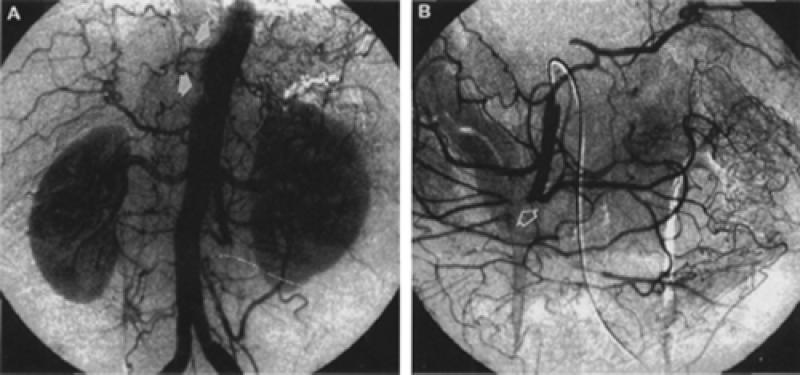
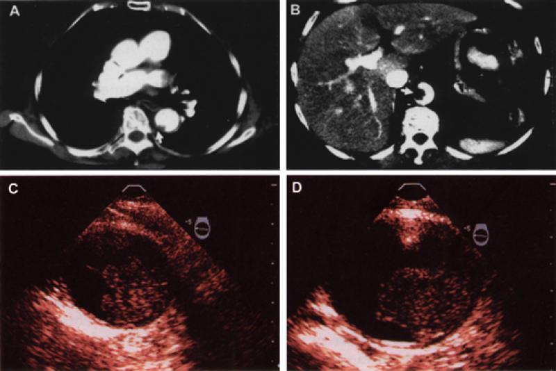
In May 1999, an 82-year-old woman presented with complaints of severe abdominal pain, vomiting, and bloody diarrhea. Despite the severe pain, physical examination showed only minimal abdominal tenderness. Mesenteric ischemia was suspected. Abdominal aortography demonstrated multiple filling defects along the right side of the aorta at the level of the superior celiac axis (Fig. 1A, arrows). Selective angiography of the superior mesenteric artery showed complete occlusion of the vessel just beyond the takeoff of the right colic branch (Fig. 1B, arrow). Contrast-enhanced computed tomography demonstrated filling defects within the lower thoracic and upper abdominal aorta (Figs. 2A and 2B, arrows).


The patient underwent surgical resection of the ischemic small intestine and embolectomy of the superior mesenteric artery. Transesophageal echocardiography showed no intracardiac mass or thrombus. However, examination of the descending thoracic and upper abdominal aorta showed that the aortic lumen was obstructed as much as 50% by large, protruding, heterogeneous masses that had sessile and mobile components (Figs. 2C and 2D). The patient was treated with anticoagulation.
Footnotes
Address for reprints: Jamshid Shirani, MD, The Jack D. Weiler Hospital of the Albert Einstein College of Medicine, 1825 Eastchester Road, Room W1-70K, Bronx, NY 10461-2373


