

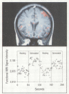
Abstract
A wide range of structural and functional techniques now exists to map the human brain in health and disease. These approaches span the gamut from external tomographic imaging devices (positron-emission tomography, single photon-emission computed tomography, magnetic resonance imaging, computed tomography), to surface detectors (electroencephalography, magnetoencephalography, transcranial magnetic stimulation), to measurements made directly on the brain's surface or beneath it (intrinsic signal imaging, electrocorticography). The noninvasive methods have been combined to provide unique and previously unavailable insights into the macroscopic organization of the functional neuroanatomy of human vision, sensation, hearing, movement, language, learning, and memory. All methods have been applied to patients with neurologic, neurosurgical, and psychiatric disease and have provided a rapidly expanding knowledge of the pathophysiology of diseases such as epilepsy, cerebrovascular disease, neoplasms, neurodegenerative diseases, mental illness, and addiction states. In addition, these new methods have become a mainstay of preoperative surgical planning and the monitoring of pharmacologic or surgical (transplantation) interventions. Most recently, the ability to observe the reorganization of the human nervous system after acute injury, such as occurs with cerebral infarction or head trauma, or in the course of a progressive degenerative process such as Alzheimer's or Parkinson's disease, may provide new insights and methods in the rapidly expanding field of neurorehabilitation. Our newfound ability to generate maps and databases of human brain development, maturation, skill acquisition, aging, and disease states is both an exciting and formidable task.
Full text
PDF


Images in this article


Selected References
These references are in PubMed. This may not be the complete list of references from this article.
- Belliveau J. W., Kennedy D. N., Jr, McKinstry R. C., Buchbinder B. R., Weisskoff R. M., Cohen M. S., Vevea J. M., Brady T. J., Rosen B. R. Functional mapping of the human visual cortex by magnetic resonance imaging. Science. 1991 Nov 1;254(5032):716–719. doi: 10.1126/science.1948051. [DOI] [PubMed] [Google Scholar]
- Bradley W. G., Jr, Waluch V. Blood flow: magnetic resonance imaging. Radiology. 1985 Feb;154(2):443–450. doi: 10.1148/radiology.154.2.3966131. [DOI] [PubMed] [Google Scholar]
- Burt C. T., Koutcher J. A. Multinuclear NMR studies of naturally occurring nuclei. J Nucl Med. 1984 Feb;25(2):237–248. [PubMed] [Google Scholar]
- Chollet F., DiPiero V., Wise R. J., Brooks D. J., Dolan R. J., Frackowiak R. S. The functional anatomy of motor recovery after stroke in humans: a study with positron emission tomography. Ann Neurol. 1991 Jan;29(1):63–71. doi: 10.1002/ana.410290112. [DOI] [PubMed] [Google Scholar]
- Cohen D., Cuffin B. N., Yunokuchi K., Maniewski R., Purcell C., Cosgrove G. R., Ives J., Kennedy J. G., Schomer D. L. MEG versus EEG localization test using implanted sources in the human brain. Ann Neurol. 1990 Dec;28(6):811–817. doi: 10.1002/ana.410280613. [DOI] [PubMed] [Google Scholar]
- Cohen L. B., Lesher S. Optical monitoring of membrane potential: methods of multisite optical measurement. Soc Gen Physiol Ser. 1986;40:71–99. [PubMed] [Google Scholar]
- Cohen M. S., Weisskoff R. M. Ultra-fast imaging. Magn Reson Imaging. 1991;9(1):1–37. doi: 10.1016/0730-725x(91)90094-3. [DOI] [PubMed] [Google Scholar]
- Demaerel P., Johannik K., Van Hecke P., Van Ongeval C., Verellen S., Marchal G., Wilms G., Plets C., Goffin J., Van Calenbergh F. Localized 1H NMR spectroscopy in fifty cases of newly diagnosed intracranial tumors. J Comput Assist Tomogr. 1991 Jan-Feb;15(1):67–76. doi: 10.1097/00004728-199101000-00009. [DOI] [PubMed] [Google Scholar]
- Frackowiak R. S. Functional mapping of verbal memory and language. Trends Neurosci. 1994 Mar;17(3):109–115. doi: 10.1016/0166-2236(94)90119-8. [DOI] [PubMed] [Google Scholar]
- Freed C. R., Breeze R. E., Rosenberg N. L., Schneck S. A., Kriek E., Qi J. X., Lone T., Zhang Y. B., Snyder J. A., Wells T. H. Survival of implanted fetal dopamine cells and neurologic improvement 12 to 46 months after transplantation for Parkinson's disease. N Engl J Med. 1992 Nov 26;327(22):1549–1555. doi: 10.1056/NEJM199211263272202. [DOI] [PubMed] [Google Scholar]
- Frostig R. D., Lieke E. E., Ts'o D. Y., Grinvald A. Cortical functional architecture and local coupling between neuronal activity and the microcirculation revealed by in vivo high-resolution optical imaging of intrinsic signals. Proc Natl Acad Sci U S A. 1990 Aug;87(16):6082–6086. doi: 10.1073/pnas.87.16.6082. [DOI] [PMC free article] [PubMed] [Google Scholar]
- Gadian D. G., Frackowiak R. S., Crockard H. A., Proctor E., Allen K., Williams S. R., Russell R. W. Acute cerebral ischaemia: concurrent changes in cerebral blood flow, energy metabolites, pH, and lactate measured with hydrogen clearance and 31P and 1H nuclear magnetic resonance spectroscopy. I. Methodology. J Cereb Blood Flow Metab. 1987 Apr;7(2):199–206. doi: 10.1038/jcbfm.1987.45. [DOI] [PubMed] [Google Scholar]
- Grafton S. T., Martin N. A., Mazziotta J. C., Woods R. P., Vinuela F., Phelps M. E. Localization of motor areas adjacent to arteriovenous malformations. A positron emission tomographic study. J Neuroimaging. 1994 Apr;4(2):97–103. doi: 10.1111/jon19944297. [DOI] [PubMed] [Google Scholar]
- Kauer J. S. Real-time imaging of evoked activity in local circuits of the salamander olfactory bulb. Nature. 1988 Jan 14;331(6152):166–168. doi: 10.1038/331166a0. [DOI] [PubMed] [Google Scholar]
- Kaufman L., Crooks L. E., Sheldon P. E., Rowan W., Miller T. Evaluation of NMR imaging for detection and quantification of obstructions in vessels. Invest Radiol. 1982 Nov-Dec;17(6):554–560. doi: 10.1097/00004424-198211000-00006. [DOI] [PubMed] [Google Scholar]
- Lindvall O., Sawle G., Widner H., Rothwell J. C., Björklund A., Brooks D., Brundin P., Frackowiak R., Marsden C. D., Odin P. Evidence for long-term survival and function of dopaminergic grafts in progressive Parkinson's disease. Ann Neurol. 1994 Feb;35(2):172–180. doi: 10.1002/ana.410350208. [DOI] [PubMed] [Google Scholar]
- Mazziotta J. C., Frackowiak R. S., Phelps M. E. The use of positron emission tomography in the clinical assessment of dementia. Semin Nucl Med. 1992 Oct;22(4):233–246. doi: 10.1016/s0001-2998(05)80118-7. [DOI] [PubMed] [Google Scholar]
- Ogawa S., Tank D. W., Menon R., Ellermann J. M., Kim S. G., Merkle H., Ugurbil K. Intrinsic signal changes accompanying sensory stimulation: functional brain mapping with magnetic resonance imaging. Proc Natl Acad Sci U S A. 1992 Jul 1;89(13):5951–5955. doi: 10.1073/pnas.89.13.5951. [DOI] [PMC free article] [PubMed] [Google Scholar]
- Petroff O. A., Prichard J. W., Behar K. L., Alger J. R., den Hollander J. A., Shulman R. G. Cerebral intracellular pH by 31P nuclear magnetic resonance spectroscopy. Neurology. 1985 Jun;35(6):781–788. doi: 10.1212/wnl.35.6.781. [DOI] [PubMed] [Google Scholar]
- Petroff O. A., Prichard J. W., Behar K. L., Rothman D. L., Alger J. R., Shulman R. G. Cerebral metabolism in hyper- and hypocarbia: 31P and 1H nuclear magnetic resonance studies. Neurology. 1985 Dec;35(12):1681–1688. doi: 10.1212/wnl.35.12.1681. [DOI] [PubMed] [Google Scholar]
- Prichard J. W., Rosen B. R. Functional study of the brain by NMR. J Cereb Blood Flow Metab. 1994 May;14(3):365–372. doi: 10.1038/jcbfm.1994.47. [DOI] [PubMed] [Google Scholar]
- Prichard J., Rothman D., Novotny E., Petroff O., Kuwabara T., Avison M., Howseman A., Hanstock C., Shulman R. Lactate rise detected by 1H NMR in human visual cortex during physiologic stimulation. Proc Natl Acad Sci U S A. 1991 Jul 1;88(13):5829–5831. doi: 10.1073/pnas.88.13.5829. [DOI] [PMC free article] [PubMed] [Google Scholar]
- Ross J. S., Masaryk T. J., Modic M. T., Harik S. I., Wiznitzer M., Selman W. R. Magnetic resonance angiography of the extracranial carotid arteries and intracranial vessels: a review. Neurology. 1989 Oct;39(10):1369–1376. doi: 10.1212/wnl.39.10.1369. [DOI] [PubMed] [Google Scholar]
- Stehling M. K., Turner R., Mansfield P. Echo-planar imaging: magnetic resonance imaging in a fraction of a second. Science. 1991 Oct 4;254(5028):43–50. doi: 10.1126/science.1925560. [DOI] [PubMed] [Google Scholar]
- Ts'o D. Y., Frostig R. D., Lieke E. E., Grinvald A. Functional organization of primate visual cortex revealed by high resolution optical imaging. Science. 1990 Jul 27;249(4967):417–420. doi: 10.1126/science.2165630. [DOI] [PubMed] [Google Scholar]
- Watson J. D., Myers R., Frackowiak R. S., Hajnal J. V., Woods R. P., Mazziotta J. C., Shipp S., Zeki S. Area V5 of the human brain: evidence from a combined study using positron emission tomography and magnetic resonance imaging. Cereb Cortex. 1993 Mar-Apr;3(2):79–94. doi: 10.1093/cercor/3.2.79. [DOI] [PubMed] [Google Scholar]
- Woods R. P., Cherry S. R., Mazziotta J. C. Rapid automated algorithm for aligning and reslicing PET images. J Comput Assist Tomogr. 1992 Jul-Aug;16(4):620–633. doi: 10.1097/00004728-199207000-00024. [DOI] [PubMed] [Google Scholar]
- Woods R. P., Mazziotta J. C., Cherry S. R. MRI-PET registration with automated algorithm. J Comput Assist Tomogr. 1993 Jul-Aug;17(4):536–546. doi: 10.1097/00004728-199307000-00004. [DOI] [PubMed] [Google Scholar]