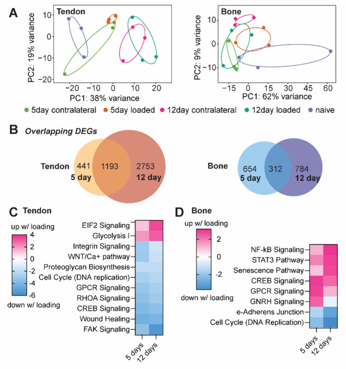
Figure 4.
(A) Principal component analysis plots of differential gene expression for naïve, contralateral, and loaded tendons and bones after 5day and 12 days of loading. (B) In tendon and bone 1,193 and 312 genes, respectively, were differentially expressed after 5 and 12 days of loading. (C) In tendon, optogenetic loading induced upregulation of EIF2 signaling and Glycolysis I pathways, and downregulation of FAK and RHOA signaling pathways. (D) In bone, optogenetic loading led to activation of inflammatory pathways such as NF-B and STAT3 signaling, senescence, and CREB signaling, as well as downregulation of cell cycle (DNA replication) and epithelial adherens junction signaling.

