To the Editor:
It was interesting to read the report of giant left atrium by Panayotis Fasseas and colleagues, 1 in a recent issue of the Texas Heart Institute Journal. In this context, it may be worth sharing with readers the appearance of a 100% cardiothoracic ratio on a chest radiograph. Such cardiomegaly due to left atrial (LA) enlargement is a rare entity in the current era.
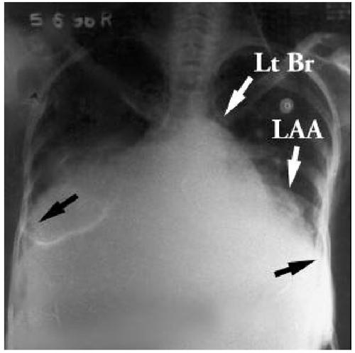
A 30-year-old woman presented at Sri Sathya Sai Institute of Higher Medical Sciences (Puttaparthi, India) with severe rheumatic mitral regurgitation and atrial fibrillation. The posteroanterior view of the chest radiograph showed 100% cardiothoracic ratio (Fig. 1). Left atrial enlargement was evident because of elevation of the left main bronchus, which had led to splaying of the carina. A double shadow was visible through the right side of the cardiac shadow. The enlarged LA appendage was seen as a bulge on the left border of the heart. Such an irregular border with bulges differentiates LA enlargement from a pericardial effusion, which has a smooth border. Enlargement of the LA causes the chamber to bulge to the right side, pushing the right lung. Further enlargement causes it to approach the right lateral chest wall. When the LA comes within an inch of the lateral chest wall on either side, aneurysmal enlargement is said to exist.

Fig. 1 Chest radiograph in the posteroanterior view shows a 100% cardiothoracic shadow. Black arrows demarcate the cardiac borders.
LAA = left atrial appendage; Lt Br = elevated left main bronchus
Footnotes
Letters to the Editor should be no longer than 2 double-spaced typewritten pages and should contain no more than 5 references. They should be signed, with the expectation that the letters will be published if appropriate. The right to edit all correspondence in accordance with Journal style is reserved by the editors.
References
- 1.Fasseas P, Lee-Dorn R, Sokil AB, VanDecker W. Giant left atrium. Tex Heart Inst J 2001;28:158–9. [PMC free article] [PubMed]


