Abstract
Background
Acidosis is one of the most common rumen diseases characterized by changes in the rumen environment and the circulatory system. Recent alternative trends in rearing small ruminants have led to the use of probiotics, rumenotorics and prebiotics to treat acidosis in animals.
Purpose
This study aimed to evaluate the efficacy of probiotics and the combination of probiotics with prebiotics and probiotics with rumenotorics for the treatment of acidosis in sheep.
Methods
This experimental study was conducted from September 2018 to May 2019. For the therapeutic study, 25 sheep were randomly divided into 5 equal groups. Acidosis was induced by an oral dose of 50 g/kg with wheat flour after a 24 hour fast. Four regimens of therapy were employed: PT probiotics, PPT probiotics with prebiotics; PRT probiotics with rumenotorics and standard ST treatment were adopted. Before and after therapy, laboratory analyses on rumen fluid, serum analysis, physical signs, and hematological changes were conducted.
Results
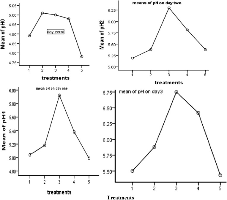
When probiotics were combined with rumenotorics (PRT), the mean standard deviation of rumen pH at day zero was 4.96±0.837 (PRT). Rumen pH improved from day one today three to 5.92±0.54, 6.30±041 and 6.75±0.34, respectively. The change in rumen pH was statistically significant after treatment on day 3 (p=0.002). The therapeutic regimens of PRT had improved heart rate and respiratory rate after treatment and the change was statistically significant (p=0.006 and p=0.000) compared to the control group. The PCV of the PRT treated sheep was also improved.
Conclusion
Probiotics with rumenotorics were the most successful therapeutic regimen for the treatment of ruminal acidosis in sheep. Therefore, the use of probiotics with rumenotorics is the promising alternative for the treatment of acidosis.
Keywords: experiment, hematology, therapeutic, rumen, pH
Introduction
Ruminants can potentially affect the financial and social part of most African rural communities. A 53% of the total ruminants, small ruminants such as sheep and goats in the developing world are found in Asia, particularly India and Pakistan.1 Ethiopia has around 28 million sheep in different local sheep breeds and they can be divided into around 14 traditional sheep populations mainly based on their location.2,3
Sheep make significant contributions to the agricultural economy and play an important role in the living hood security of marginalized and landless pastoralists.4 They provide household food security and family income through meat, wool, hide, milk and manure with little or no supplementation.5 Nevertheless, the performance of sheep farming in Ethiopia compares poorly to other African countries due to inadequate feed and nutrition, widespread diseases and other health-related problems, poor management and marketing system.3 Among the various health problems faced by ruminants, rumen disorders cause significant economic losses to producers in the form of feed wastage, delayed marketing and discarding of tripe and liver or whole carcasses, reduced nutritional value, reduced water-binding capacity of meat and several organoleptic defects and death of animals.6
Rumen acidosis is a mismanagement disease and sometimes a man-made disorder of ruminants; particularly fattening animals.7 The most common causes of acidosis are accumulations of apples, corn, wheat, sugar beets, and concentrated sucrose solutions. In clinically diseased ruminants, the morbidity rate varies between 10 and 50%, and death from lactic acidosis can reach up to 90% in untreated cases, whereas it can reach 30 to 40% in properly treated or treated cases.8 The clinical and path-physiological consequences of a Acidosis is heme-concentration, dehydration, diarrhea, cardiovascular collapse, renal failure, muscle fatigue, sudden depression associated with a drop in blood pressure, and death. Surviving animals induce mycosis, ruminants for a long time, hepatic necrobacillosis due to migration of the microorganism from the rumen to the liver, or chronic laminitis due to septicemia, and signs of rumen scars.9,10
This problem is particularly related to the sudden change in ration, since the type of diet affects the number and type of bacteria and protozoa in the rumen and a change requires a period of microbial adaptation. When rumen pH falls, the amplitude and frequency of rumen contractions decrease, and at around pH 5, rumen atony grain stasis occurs in ruminants. Hematological and biochemical changes in ruminal acidosis are important in assessing disease severity, and severe dehydration and cardiovascular involvement are common.11
An understanding of the effective and efficient therapeutic regimen for the ruminal disorder is very essential for the successful management of ruminal acidosis. Recent trends in sheep rearing have led to the use of prebiotics, rumenotorics (substances and formulations that optimize stomach function) and probiotics as feed additives or treatment to promote growth by increasing feed efficiency, improving rumen motility and manipulating rumen microbial flora.12 Probiotics are beneficially affecting the host upon ingestion of acidic nutrients by improving the balance of the gastrointestinal micro flora.13 Lactic acid producing bacteria (Lactobacilli and Enterococci) provide a constant lactic acid supply in the rumen, stimulate lactate utilizing bacteria and stabilize the ruminal pH.14,15
Probiotics can contain prebiotics as substrates and are then referred to as symbiotic responsible for improving animal performance, and probiotics are one of the best antibiotic alternatives.16,17 Prebiotics reach the large intestine as non-digestible food components that escape digestion by mammalian enzymes intact and metabolized by beneficial members of the native microbiota.18 Prebiotics (fructo-oligosaccharides) promote the growth of beneficial bacteria in the gastrointestinal tract of ruminants.19 Rumenotics (potassium antimony tartrate, ferrous sulfate, copper and cobalt) act as cofactors required for vitamin B12 synthesis and serve as a substrate for rumen microbial growth, restoration of impaired rumen function and subsequent appetite recovery.20 Rumenotoris (bolus rumenotars) is also used to restore rumen motility and appetite.21 Sheep populations are raised in various agro-ecologies for various purposes such as meat production, income generation, and as a source of fur and skin in Ethiopia.3 The production system, however, will be affected by several limitations. Fore-stomach diseases result in large losses for producers in terms of deaths, wasted feed, delayed marketing, uneconomical animals recovered and additional labor costs for preventive and therapeutic measures.22 Large amount of carbohydrate-rich food and causes proliferation of rumen lactic acid-producing bacteria, especially Streptococcus, resulting in high mortality.12 During acute rumen acidosis, the higher concentration of protons is particularly high amount of lactate in the rumen affects the integrity of the rumen epithelial cells with small lesions and parakeratosis, which can directly affect the metabolic system of ruminants.23 In addition, the acidic environment, high osmotic pressure and high lactate concentration in the rumen can cause death and lysis of the gram-negative bacteria. It causes relevant metabolic disorders such as laminitis, abomasal displacement; fatty liver and sudden death syndrome.23–25 Chemical buffers, ionophores, and probiotics are emerging among strategies to prevent lactic acidosis. Chemical buffers, magnesium oxide and sodium bicarbonate are the most common alkalizing agents used to treat acidosis in both rumen and systemic acidosis in ruminants. Currently, the use of probiotic and prebiotic additives is evolving as an alternative to ameliorate rumen disorders.9 Recent evidence suggests that yeast and bacterial probiotic (BP) products are an effective (not just acid neutralized) and economical alternative to using traditional buffers for moderation of rumen pH.26 Probiotic yeasts like Saccharomyces cerevisiae and bacterial strains like bifidobacteria have been utilized to maintain rumen pH and boost animal productivity. Probiotics that produce or utilize lactate are thought to help prevent sub-acute rumen acidosis in ruminants by diminishing lactate levels.
The effect of probiotics and the physicochemical conditions of rumen contents on the survival of pathogenic strains could have important implications for farm management and food safety, as well as reducing the risk of foodborne diseases.27 Antimicrobial peptide synthesis by probiotics, such as bacteriocins, can inhibit the growth of pathogenic bacteria or the production of enzymes capable of hydrolyzing bacterial toxins can be impeded.28 However, the application of probiotics and the connection with the rumen microflora has not been given enough attention in our country. Therefore, they play a key role in reducing the risk of rumen acidosis. Lactate-consuming bacteria have also been proposed and successfully used as probiotics to lower lactate levels, promote acidosis-damaged microbiota, and maintain a rumen pH.29
Case Definition
Rumenotorics are agents and mixtures that promote fore-stomach function (fermentation and motility) are known as ruminotorics. Formulations that contain glycogenic substrates, minerals, cofactors, and bitters (eg, nux vomica) have limited application in current therapy of ruminoreticular indigestion.
Probiotics are defined as “live microorganisms which when administered in adequate amount confer health benefits to the host” (FAO/WHO).
Prebiotics are a special form of dietary fiber that acts as a fertilizer for the good bacteria in the gut.
Materials and Methods
Study Animals
Local breed sheep, Northwest part of Ethiopia, Gondar district 727 km away from Capital city of Addis Ababa, were bought from the same origin and as much as possible equivalent age, body condition and similar physical size for experimentally induced acidosis intervention. Ethical clearance was obtained from Research and Publication Directorate of the Institutional Ethical Review Board of the University of Gondar ethical committee (Re.VP/RTT/05/2925/2018).
Study Area
The study was performed in University of Gondar at College of veterinary medicine and animal science, on the farm in the laboratory animal’s room.
Experimental Design, Period and Sampling Procedures
Experiment was conducted from September 2018 to May 2019 to evaluate and compare the therapeutic effectiveness of probiotics, combination of probiotics with prebiotics and probiotics with rumenotorics in the treatment of acidosis in sheep. Completely randomized experimental design was assigned for different treatments to the study animals after induction of acidosis. There were 5 experimental groups, namely, negative control group (CG), probiotics (PT), probiotics plus prebiotics (PPT), probiotics plus rumenotorics (PRT) and sodium bicarbonate (ST). In each group 5 sheep of equivalent age, body condition and physically similar size were randomly assigned. The total number of animals used for the study was 25 sheep. The experimental procedure was divided into five phases (Table 1) and (Supplementary Information (ppt)-different phases of the experiment with images).
Table 1.
Different Phases of the Experiment
| Phases | Activities |
|---|---|
| Phase-1 | Pre-induction phase: Evaluation of general health status of the animals |
| Phase-2 | Fasting phase: Prohibited sheep from feeding for 24 hour by keeping in the laboratory animal room |
| Phase-3 | Induction phase: Oral administration of 50g/kg wheat flour; clinical signs and change of vital parameters were indication of induction of acidosis |
| Phase-4 | Treatment phase: There was random administration of different treatments for three consecutive days (day1, day2 and day 3). |
| Phase-5 | Recovery phase: In the last day of the experiment there was better improvement of vital parameters (HR, RT and T°C). |
Initially, the animals were housed in separate cages with wooden floors and free access to water, an environmentally controlled space. During the experimental periods, animals were fed 50 g/kg of wheat flour orally dosed according to the experimental design after a 24 hour fasting period to induce rumen acidosis.23 Then, we had a follow-up until the clinical signs and other parameters appeared for 24 hours. In this experiment, six veterinarians were divided based on their interest to collect the sample in each sampling type. For physical parameters, rumen fluctuation and hematological sampling, pre- and post-treatment samples were taken by two veterinarians in each type. The first procedure consisted of physical parameter measurements such as temperature, heart rate and respiratory rate, followed by blood sampling and rumen fluid extraction. The collection of rumen fluid was performed for the last time in the sampling procedures as the rumen microflora was to be evaluated shortly after collection. Finally, the intervention was carried out according to the type of treatment. In this case, each sheep has its own identification ID, we have marked the animals in five groups in relation to the type of treatment/intervention. A random sampling procedure was used to select the type of treatment for each group of animals.
Four different types of treatments were allocated randomly to ascertain the comparative efficacy of various regimens by comparing with the negative control group and within the group. Randomized control design was adopted for assessing the ameliorative potential of probiotics and the combinations of probiotics with rumenotorics, probiotics with sodium bicarbonate in those animals suffering from the ruminal disorder. Four therapeutic regimens were employed (PT-probiotics, PPT-probiotics with prebiotics, PRT-probiotics with rumenotorics, ST-standard treatment/sodium bicarbonate and negative control group were adopted for the study as per protocol depicted as (Table 2)). Sampling was performed before treatment and thereafter on 1st, 2nd, and 3rd-day of post-treatment.30
Table 2.
Probiotics: Composition, Dose, Route and Production Company
| Treatments Groups | Composition | N | Dose | Company | Country | Route | Duration |
|---|---|---|---|---|---|---|---|
| Probiotic alone | Saccharomyces spp Bifidobacteriumspp |
5 | 1gram | IndiaMART | India | Oral | 3 days |
| Probiotic and Prebiotic | Saccharomyces sppBifidobacteriumspp | 5 | 1gram | IndiaMART | India | Oral | 3 days |
| FructoOligoSacchride | 100 miligarm | ||||||
| Probiotic and Rumenotoric | Saccharomyces sppBifidobacteriumspp | 5 | 1gram | IndiaMART | India | Oral | 3 days |
| AntimonyPotassiumtartarate ferrous sulphate, Copper sulphate, cobalt chloride | 5 | 1 bolib.i.d | |||||
| Sodium bicarbonate | NaHco3 | 5 | 1g/kg | – | China | Oral | 3 day |
Notes: N= number of sheep in each group, Saccharomyces spp=Saccharomyces Boulardii. Bifidobacterium spp= BifidobacteriumBifidium and longum.
Experimental Data Evaluation
General Physical Examination
General physical examination including clinical signs, body temperature, heart rate, and respiratory rate were taken from animals before and after treatment. Heart rate expressed in a number of beats per minute and was measured using a stethoscope and a stopwatch for 60 seconds and multiplying the result by two to obtain this record in minutes. The respiration rate was measured by a stethoscope expressed in a number of breaths per minute was measured using a stethoscope and stopwatch upon auscultation of respiratory movements for 30 seconds and the value obtained multiplied by two to obtain this result in minutes.31 Body temperature was measured with a clinical thermometer. Body temperatures of each sheep were recorded by using the digital thermometer. It was kept in the rectum for two minutes with care in close contact with the mucous membrane of the rectum.32
Evaluation of Hematological Parameters
Blood sample was collected from the jugular vein into 5 mL out of which 2 mL of blood was taken in EDTA (1.5 mg/mL) containing vials for the estimation of hemoglobin (Hgb), blood pH and packed cell volume (PCV) using automatic hemo-analyzer (Model: AD0824HB-A70GL, brand ADDA, China). And it was kept at 4°Cas per method described by33 and rest 3 mL of blood was kept in deep refrigerator (Model NJ 50TD, class: T, E.U) at −20°C until the serum analysis. Serum analysis was performed to quantify albumin, total proteins and enzymes (AST, ALP) using a visible light spectrophotometer (CE2031, 2000 series, manufacturer Buck Scientific Inc, ENGLAND).
Determination of blood pH was performed by collecting 5 mL whole blood into a test tube from the jugular vein using 19 gauge needles and allowed to clot at room temperature for 1hr to obtain serum. Serum pH was measured with the use of a wide range pH indicator paper and pH meter. The pH indicator paper was sinked into serum and the color of the strip was compared with the standard colors. The reading of the strip was taken immediately after dipping to avoid the change in color by exposure to air. The reading of the pH meter was recorded and the mean value of both the readings was calculated.32
Physical and Micro-Biochemical Examination of Rumen Fluid
Physical parameters of ruminal liquor (color, thickness, odor and SAT) were evaluated. Also, Micro-biochemical changes including protozoa motility, density, methylene blue reduction tests (MBRT), and pH of rumen fluid was examined. The power of hydrogen (pH) in ruminal fluid was measured with a portable pH meter (Model: CG 840, Ag/AGCL, Schott Gerate, Hofheim, Germany) pre- and post-treatment.
One day before the trial, the site prepared aseptically for the start of the trials; the sheep’s wool was cut off in the left flank area. Then, the area was scrubbed with tincture of iodine (2%). The next day of sampling, the site was disinfected with 70% alcohol and punctured with a 16 gauge needle. One needle was used for each animal at each sampling point. Five milliliters (5 mL) of rumen fluids were collected aseptically by rumenocentesis in the left par lumbar fossa using 16-gauge needles with a disposable syringe (in some sheep) and (15 mL) through the use of gastric tube (Ascitechcampany, Hangzhou, China) based on measured body size (length from mouth to rumen). Then, about 10 mL of rumen fluid collected by the nasogastric tube was discarded to avoid contamination with saliva, and the fluid was immediately transferred to a pre-warmed (390°C) anaerobic flask until such time as the sample was collected from all animals, then taken back to the laboratory (Clinical Pathology Laboratory, UoG) for assessment of rumen pH and protozoa activity.34
The ruminal fluid was put in beaker and pH meter was inserted, then pH value was recorded.35 The pH sensor was adjusted with pH 6.67 buffer solutions and was inserted in the sampling bottle which contained ruminal fluid. Ruminal pH was recorded continuously after the collection of the ruminal fluid throughout the experimental period. Then, measured each sample, the pH sensor was washed by tap water for every trial.
Few drops of ruminal fluid were placed on a glass slide with a toothpick and cover slip is put on it to see the motility of ruminal protozoa. Herein, protozoan motility was graded in four categories: ++++ highly motile and very crowded (good): >10 mobile protozoa per field; +++ motile and crowded (fair): 6–9 mobile protozoa per field; ++ sluggish motility and low number (subnormal): 3–5 mobile protozoa per field; + no or sporadic alive fauna (very low): <3 mobile protozoa per field.34,35
Methylene blue reduction test (MBRT) was performed to check the anaerobic fermentative metabolism of the bacterial population. To perform this activity, 6 mL ruminal fluid was taken and mixed with 0.03% Methylene blue in the test tube. Then, it was incubated in 37 °C for six minutes (Incubator). Finally, the time was measured needed for the color of the mixture to be changed before and after treatment. Sedimentation activity test (SAT) was done at the induction phase of the acidosis and after therapeutic procedures to check the sedimentation features of the particles. Sample of rumen fluid was put in a test tube and stand properly. The time was measured needed for completion of sedimentation of fine particles and flotation of coarse solid particles.
Data Management and Statistical Analysis
The data collected were coded and entered into an MS Excel spreadsheet. After validation, it was transferred and processed using SPSS version 25 computer software. Both descriptive (mean, standard deviation and minimum and maximum values) and analytical analysis were performed. The data were statistically analyzed using the technique of multivariate analysis of variance (MANOVA). The difference in hematological, biochemical and physical parameters (considered as dependent variables) before and after treatment for each treatment group (PT, PPT, PRT and ST) as a fixed effect were analyzed by Scheffe multiple comparison as a post hoc test. For statistical conclusions, a 5% level of significance was considered statistically significant. When treatments were statistically significant, comparison between negative control group (GC) vs (PT, PPT, PRT) or standard treatment (ST) vs (PT, PPT, PRT) showed a significant effect.
Results
Induction of Acidosis and Clinical Investigations
Acidosis was successfully induced in all test animals in the study, and almost all sheep showed nervous depression and symptoms including watery, teeth-grinding (bruxism), yellowish and sour-smelling diarrhea, cessation of feeding and severe symptoms of foot pain and difficulty walking after induction of acidosis. All the sheep were able to stand, but they move slowly with their heads down, their eyes dull and sunken.
Recovery Rate
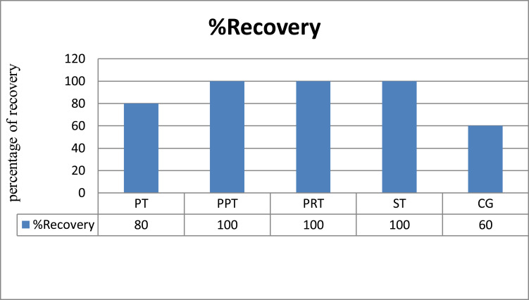
The results of the present study indicated that most of the therapeutic regimens tested in the experiment were found to be effective in eliciting a beneficial response in sheep with acidosis. Aside from one sheep (20%) dying from treatment of PT, there was no death due to acidosis in the treatment groups compared to the death of two sheep (40%) in the control group, possibly related to better treatment Effect of probiotics with prebiotics or rumenotorics on rumen acidosis versus probiotics alone.
Improvements of Vital Parameters
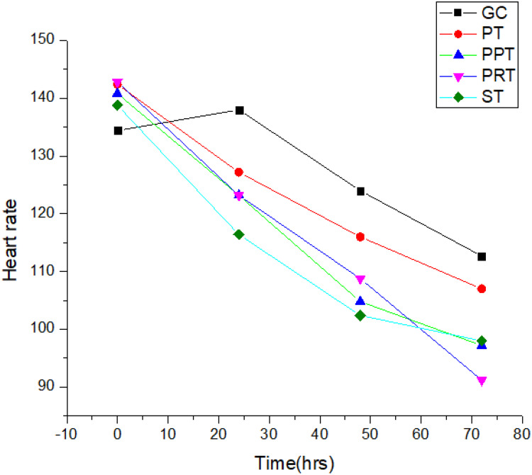
At the beginning of the experimental period (before treatment), the mean total heart rate was 139.84±9.290 in all animal groups, while changing to 127±0.20, 109±9.540 and 98.27±8.49 on days 1, 2 and 3, respectively. After treatment, this parameter was improved so that the progress is shown in (Figure 1). Three treatment groups, such as PPT, PRT and ST treatment group, showed a statistically significant difference compared to the control group on the last day of treatment. The PRT treatment group was most therapeutically effective in correcting heart rate on day two, which was statistically significant (p<0.05, p=0.006). The standard treatment group (ST) also had a highly significant difference between the control groups in improvement in heart rate (p<0.05, P=0.00). The combination of probiotics and prebiotics (PPT) also produced a statistically significant difference (P=0.001) for heart rate stabilization. The mean rectal temperature in all groups before treatment was 37.9 ± 0.612 °C, while the temperatures on days 1, 2 and 3 after treatment were 38, 360.97, 39.00 ± 0.617 and 400.0 (see Table 3) was. Analysis using four multivariate tests (Pillais Trace, P=0.038, Wilks Lambda = P=0.009, Hotelling’s Trace, p=0.02, and Roy’s Largest Root, p=0.00) also showed statistical significance (p<0, 05).
Figure 1.
The average change value of heart rate in acidotic sheep after treatment, with probiotics treatment alone (PT), probiotics plus prebiotics (PPT), probiotics plus rumenotorics (PRT), standard treatment (ST) and without treatment (control Group).
Table 3.
Effect of Various Treatment Regimens on Physical Parameters for Induced Acidosis
| Parameter | Time | GC | PT | PPT | PRT | ST |
|---|---|---|---|---|---|---|
| Heart rate | Day0 | 134.40±9.21 | 142.40±10.88 | 140.80±14.45 | 142.80±8.67 | 138.8±7.16 |
| Day1 | 138.00±8.72 | 127.20±7.16 | 123.20±8.67 | 123.20±126 | 116.40±4.6 | |
| Day2 | 124.00±6.93 | 116.00±8.64 | 104.80±5.23# | 108.8±7.64# | 102.40±4.6# | |
| Day3 | 112.67±3.05 | 107.00±8.87 | 97.20±6.88 | 91.20±7.69# | 98.00±8.00 | |
| Temperature | Day0 | 38.40±8.764 | 38.00±0.707 | 37.60±548 | 37.40±0.548 | 38.20±0.447 |
| Day1 | 38.60±0.89 | 38.20±0.837 | 38.00±0.707 | 38.80±0.447 | 39.20±1.05 | |
| Day2 | 38.60±0.000 | 39.00±0.000 | 38.40±548 | 39.40±0.548 | 39.20±0.837 | |
| Day3 | 38.80±000 | 39.00±39.00 | 39.00±0.000 | 40.00±0.000 | 39.60±0.55 | |
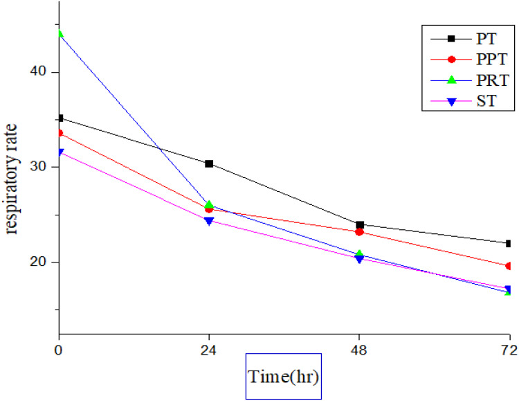
| Respiratory rate((min) | Day0 | 40.40±8.764 | 35.2±3.347 | 33.60±8.295 | 44.00±0.5.66 | 31.60±8.52 |
| Day1 | 33.60±7.266 | 30.40±4.561 | 25.60±4.561 | 26.00±4.472# | 24.40±2.966# | |
| Day2 | 28.67±1.16 | 24.00±3.266 | 23.20±3.347# | 20.80±3.37# | 20.40±2.96 | |
| Day3 | 22.67±5.03 | 22.00±2.309 | 19.60±2.966# | 16.80±1.05# | 17.20±2.63# |
Notes: Values expressed as #Superscript are significant between control groups at p<0.05. Values were expressed by means ±SD, by SPSS version 25, MANOVA followed by Scheffe post hoc test was used to compare between groups.
Abbreviations: GC, control group; PT, probiotics treatment alone; PPT, probiotics plus prebiotics; PRT, probiotics plus rumenotorics; ST, standard treatment.
The mean±SD respiratory rate (breaths/minute) was 36.968±0.85 before administration of treatments in all animal groups in the induction phase. As shown in Figure 2 and Table 3, the post-treatment (1st, 2nd and 3rd day) was changed to 285 ±0.745, 26.00 ±018, 19.27±3.47. Analysis of the multivariate tests (Pillais Trace, p=0.025, Wilks Lambda, p=0.024, Hotelling’s Trace, p=0.028, and Roy’s Largest Root, p=0.003) also showed statistical significance (p<0.05).
Figure 2.
The average changes of the value respiratory rate in acidotic sheep after treatment with probiotics treatment alone (PT), probiotics plus prebiotics (PPT), probiotics plus rumenotorics (PRT), standard treatment (ST) and without treatment (control Group).
Hematological Results
The hemoglobin concentration (standard mean deviation) of the test animals before the treatment group PT, PPT, PRT, ST and the control group were 14.00±2.00, 15.80±0.447, and 12.80 ±1.924, 14.40±1.517, 15.40 ±1.673, and after the treatment on the first day it changed to 13.20±2.168, 14.5±0.707, 12.10 ± 2.19, 13.80±1.78 and 14.80±2.168 concentrates the blood components such as Hematocrit and hemoglobin. Similarly, all treatment groups showed statistically significant after treatment on day two and day three (p<0.05), an apparent change shown as (Table 4). This can possibly have a positive effect due to treatments to neutralize the amount of lactate in acidotic sheep, and then the rumen osmotic pressure is reduced. Eventually, the concentration of blood components including hemoglobin was reduced.
Table 4.
Effect of Various Treatment Regimens on Hematological Parameters on Acidotic Sheep (Mean±SD)
| Parameter | Time | GC | PT | PPT | PRT | ST |
|---|---|---|---|---|---|---|
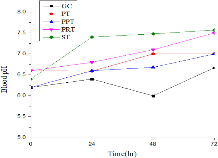
| Blood pH | Day 0 | 6.20±0.447 | 6.60±0.548 | 6.20±0.447 | 6.60±0.548 | 6.40±0.48 |
| Day 1 | 6.40±0.548 | 6.59±0.648 | 6.60±0.548 | 6.80±0.447 | 7.40±0.548 | |
| Day 2 | 6.00±0.00 | 7.00±0.00 | 6.68±0.547 | 7.100±0.100# | 7.480±0.447# | |
| Day 3 | 6.67±0.577 | 7.00±00 | 7.00±00 | 7.50±00# | 7.57±0.577# | |
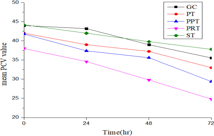
| PCV | Day 0 | 44.00±4.183 | 42.00±5.7 | 41.80±3.768 | 38.00±6.819 | 44.20±2.387 |
| Day 1 | 43.20±3.70 | 39.00±3.742 | 37.40±2.88# | 34.60±5.367# | 42.00±2.345 | |
| Day 2 | 39.00±1.00 | 37.25±4.272 | 35.60±1.881 | 29.80±6.261# | 39.80±1.483# | |
| Day 3 | 35.55±7.12 | 33.00±3.559 | 29.40± 2.99# | 24.80±10.134# | 37.80±1.789# | |
| Hemoglobin | Day 0 | 15.40±1.67 | 14.00±2.000 | 15.80±0.447 | 12.80±1.92 | 14.40±1.517 |
| Day 1 | 14.80±2.168 | 13.20±2.168 | 14.5±0.707 | 12.10 ±2.19 | 13.80±1.78 | |
| Day 2 | 13.00±1.000 | 12.00±2.168 | 11.40±0.548 | 11.40±1.817# | 13.20±1.095 | |
| Day 3 | 12.67±0.577 | 11.50±1.732 | 11.10±0.89# | 11.00±2.121# | 13.00±1.000 |
Notes: Values expressed as #Superscript are significant between control groups at p<0.05. Values were expressed by means ±SD, by SPSS version 25MANOVA followed by Scheffe post hoc test was used to compare between groups.
Abbreviations: GC, control group; PT, probiotics treatment alone; PPT, probiotics plus prebiotics; PRT, probiotics plus rumenotorics; ST, standard treatment.
Animals with experimentally induced acidosis had increased packed cell volume (PCV) initially, however there were a decreased PCV level after treatment regimens, were found to be statistically significant (p<0.05) between groups. The treatment Group PRT (Probiotics with Rumenotorics) and Standard Treatment group (ST), the mean ± SD of PCV revealed a statistically significant difference (p=0.047, p=0.017) from day two-day three, respectively. Treatment Group PRT blood pH value was significantly (p= 0.024) improved in the first day of treatment. Therefore, this result was shown that treatment Group PRT had a significant effect on the value of PCV and blood pH (see Figures 3 and 4) on day two and day three after treatment.
Figure 3.
The average change of blood pH in three consecutive days post treatment with probiotics treatment alone (PT), probiotics plus prebiotics (PPT), probiotics plus rumenotorics (PRT), standard treatment (ST) and without treatment (control Group).
Figure 4.
The average change of value packed cell volume (PCV) in acidotic sheep after treatment with probiotics treatment alone (PT), probiotics plus prebiotics (PPT), probiotics plus rumenotorics (PRT), standard treatment (ST) and without treatment (control Group).
Qualitative and Quantitative Ruminal Fluid Analysis
Chemical and physical examination of ruminal fluid were examined in the present experimental trials. During the induction phase of ruminal acidosis, the ruminal pH, protozo account and motility were reduced. The cellulose digestion and glucose fermentation activity were completely stopped in almost all experimental animals as indicated in their respective tests in a laboratory in day zero or before treatment.
The analysis of ruminal fluid pH was significantly increased (P<0.05) in acidotic sheep after treatment (Figure 5). A comparison of each treatment groups with the control group (GC), in the combination of probiotics with rumenotorics treatment group (PRT), the mean±SD 4.96±0.837, 5.92±0.54, 6.30±0.41 and 6.75±0.34 were found a statistically significant difference (p=0.07, 0.04, 0.002), respectively, from day one to day three. The standard treatment Group ST (sodium bicarbonate) also was revealed a statistically significant difference (p<0.05).
Figure 5.
The means ruminal pH between treatment groups for three consecutive day, pH0= ruminal pH on day zero; pH1=ruminal pH on day one; pH2=ruminal pH on day two; pH3=ruminal pH on day three. The x-axis is a list of treatments; treatment PT –probiotics; Treatment PPT- probiotics with prebiotics; Treatment PRT- probiotics withrumenotorics; Treatment ST-standard treatment (sodium-bicarbonate).
Sedimentation activity time and methylene blue reduction times were significantly different (P<0.05) in all groups after employed the treatments for three successive days. From treatment groups, probiotics with rumenotorics were found to be significantly (P<0.05) decreased. The mean value of the methylene blue reduction time (min) before and after treatment was 9.40±2.302 and 4.60±3.507, respectively (see Table 5). Also, sedimentation activity time was 1.60±0.894 and 12.00±3.082 significantly (P<0.05) increased after treatment. The analysis of qualitative ruminal fluid after treatment such as protozoan motility and concentration was found a statistically significant difference (p <0.05).
Table 5.
Effect of Various Treatment Regimens of Rumen Liquor Experimental Induced Acidosis in Sheep
| Parameter | Time | GC | PT | PPT | PRT | ST |
|---|---|---|---|---|---|---|
| Ruminal (pH) | Day 0 | 4.40±0.54 | 4.80±0.46188 | 5.00. ±447 | 4.96±0.837 | 4.97±0.637 |
| Day1 | 4.96±0.43 | 5.04±0.27 | 5.18±0.31 | 5.92±0.54# | 5.38±0.540 | |
| Day2 | 5.31±0.53 | 5.19±0.33 | 5.380±0.23 | 6.30±0.41# | 5.85±0.540# | |
| Day3 | 5.4333±0.4934 | 5.50±0.216 | 5.880±0.19)# | 6.75±0.34# | 6.42±0.570# | |
| PM | Day 0 | 0.00*(0.0) | 0.00*(0–00) | 0.00*(0.00) | 0.00*(0–00) | 0.0*(0 −00) |
| Day 1 | 0.00*(0 1.00 | 0.00*(0 −2.00) | 1.00*(0–2.00) | 2.0*(0–2.00) | 1.00*(0 −1.00) | |
| Day 2 | 1.00(1.0–1.00) | 1.00*(0–1.00) | 1.00*(1–2.00) | 2.0*(2–3.00)# | 1.00*(1–1.00) | |
| Day 3 | 1.00*(1 −2.00) | 1.50*(1.00–3.00) | 1.00*(1 −300)# | 3.0*(2–3.0) # | 2.00*(2–3.00)# | |
| MBRT | Day 0 | 10.33±1.528 | 9.25±1.708 | 9.40±2.966 | 9.40±2.302 | 10.20±1.643 |
| (min) | After (RX) | 9.67±1.528 | 8.25±1.708 | 7.60±1.51 | 4.60±3.507# | 6.40±1.673# |
| SAT | Before (RX) | 1.33±0.577 | 1.50±0.577 | 1.40±548 | 1.60±0.894) | 1.20±0.447 |
| (Min) | After (RX) | 4.00±1.000 | 3.25±0.500 | 6.60±0.219# | 12.00±3.08# | 4.00±1.00 |
Notes: Values expressed as #Superscript are significantly significant between control groups at p<0.05. *Indicates significantly different from between group. Values are Medin ± S.D, the values of protozoan motility was median (Q0 - Q3) and Kruskal–Wallis equality-of-populations rank test.
Abbreviations: PM, protozoan motility; MBRT, methylene blue reduction time; SAT, sedimentation activity time of the ruminal fluid before and after treatment between groups; GC, control Group; PT, probiotics treatment alone; PPT, probiotics plus prebiotics; PRT, probiotics plus rumenotorics; ST, standard treatment.
Serum Analysis Results
The treatment regimens were revealed statistically significant difference between groups to improve the abnormal value of enzymatic and protein disorders in serum analysis results. All the treatment Groups PPT, PRT and ST were showed clinical improvement for total protein, albumin, and AST. The mean values of total protein (g/dl), albumen (g/dl) and AST (u/L) before treatments (ST, PRT and PPT) were 5.5±0.24, 2.08±0.36, 254±59, then these were altered to 7±0.80, 3.3±0.4, 109±3, respectively, after treatment (see Table 6). However, the mean total protein (g/dl) and albumen concentration of experimental animals for treatment Group PRT were found a statistically significant difference (p<0.05). The average value of total protein (g/dl) and albumen concentration were 5.2±0.54, 2.08±0.36 before treatment then changed into 6.8±0.40, 2.47±0.27 after treatment, respectively.
Table 6.
Various Treatment Effects on Serum Proteins and Liver Enzyme Profile in Acidosis Sheep
| Parameter | Time | GC | PT | PPT | PRT | ST |
|---|---|---|---|---|---|---|
| TPn (g/dl) | Before | 5.32±0.43 | 5.70±0.46 | 5.75±0.54 | 5.2±0.54 | 5.5±0.24 |
| After (RX) | 5.7±0.32 | 5.9±0.35 | 6.00±0.63* | 6.8±0.40 | 7±0.80 | |
| Albumin (g/dl) | Before | 2.17±0.7 | 1.75±0.86# | 1.71±0.36# | 2.08±0.36* | 2.4±0.38#* |
| After (RX) | 2.08±0.8 | 2.52±0.32 | 2.3±0.10 | 2.47±0.27# | 3.3±0.4* | |
| AST | Before (R) | 219±39 | 241.6±44 | 202.4±0.36 | 181±91* | 254±59* |
| (U/l) | After (RX) | 232±28 | 176±61.8 | 173.4±67 | 111±27 | 109±34 |
| ALP (U/l) | Before | 285±86 | 404.3±46 | 366±12 | 415±31. | 434±42 |
| After (RX) | 222±52 | 336.0±78 | 346.7±99 | 121.8±87 | 99±43 |
Notes: Values were expressed by means ±SD, by SPSS version 25, MANOVA followed by scheffe, post hoc test was used to compare between groups *Indicates significantly different from the control group; #Indicates significantly different between groups; *#Indicates significantly different between groups and control group.
Abbreviations: GC, control Group; PT, probiotics treatment alone; PPT, probiotics plus prebiotics; PRT, probiotics plus rumenotorics; ST, standard treatment; TPn, total protein; AST, Aspartate transaminase; ALP, Alkaline phosphatase.
Discussion
A total of 25 sheep were used in this experimental study. Rumen acidosis induced by wheat flour intake after a 24-hour fast. Likewise,23 showed that in malnourished sheep (24 hours fasting) 50 g wheat/kg body weight of the sheep was used to induce acute rumen acidosis. Induction was effective in inducing rumen acidosis, which was observed through the manifestation of classic clinical signs of acidosis, such as decreased feed intake and rumination, depression, teeth grinding and diarrhea, after acid induction diet. The clinical signs were not concurrent and the nature of the signs was different in each group of animals. Over 20 (80%) animals showed the clinical signs after 24 hours and about 22 (88%) sheep showed similar clinical signs like diarrhea and teeth grinding. After induction, significant differences in rumen, blood pH and physical parameters between the treatment and control groups were observed and persisted until treatment was applied. It is unlikely23,36 to induce acute acidosis in adult sheep fasted for 24 hours by feeding 90 g of soaked wheat per kg body weight.
Heart rate and respiratory rate were increased to 152/min and 52/min, respectively, in the whole group before treatment. Similarly, a study was reported by6 in acidotic sheep in which heart rate (120–140/min) and respiratory rate (60–190) could be increased. Increased heart rate could be due to toxic effects of lactic acid and reduced plasma volume and circulatory failure. Increased respiration could be due to stimulation of the respiratory center by the increased blood carbon dioxide concentration and decreased blood pH.6,9 Improvements were found in the temperature, heart rate, and respiratory rate scores of the groups before and after treatment. An increased heart rate was observed in sheep with lactic acidosis, for which similar findings were reported by Hajikolaei et al.37 Reduced body temperature expressed in the present study was consistent with,38 which may be due to rumen acidosis leading to diarrhea and dehydration.
The present investigation showed that acute acidosis was successfully induced by a marked reduction in ruminal pH to 4.82±0.62. Similar results were observed in previous studies on induction of acute acidosis reported by Minuti et al.23 At this low pH, almost entire rumen protozoa activity ceases and no live protozoa population was observed under low power magnification due to lack of nutrients and optimal pH. Reduced ruminal pH is may be related to VFA production and lactic acid accumulation after feeding a grain diet.13 The result of this study coincide with findings reported by21 in acute ruminant acidosis in small ruminants and conversely, induction of acidosis was studied in sheep by sucrose at a dose of 18 g kg-1 bodyweight.39
In this study, sheep suffering from induced lactic acidosis showed reduced protozoa motility, glucose fermentation activity and cellulose indigestion and protozoa count. There was an increase in SAT, MBRT and gram-positive bacteria. Furthermore, increased sedimentation activity and reduced methylene reduction indicate reduced microbial activity and suppressed microbial fermentation activity. Mean sedimentation activity time before treatment was 1.41± 0.59, then after treatments it changed to 6.91±3.99 as well as methylene blue reduction time was 9.68±2.317, then after treatment it reduced to 7.05 ±2.34. Therefore, there was an improvement in both SAT and MBRT after treatment. This may be due to increased microbial activity in the anaerobic rumen. The hematological parameters (blood pH, hemoglobin and PCV) were influenced by the experimentally induced acidosis in such a way that the mean PCV value and the hemoglobin concentration (g/dl) were increased in all animal groups. A similar result was also reported by 40. This may be due to hem-concentration and increased osmolality of the rumen contents, thereby withdrawing fluid from the intravascular compartments.41 And the normal value that closely agrees with.32 Decreased pH could be due to rumen distension, which impedes venous blood return to the heart. This condition decreased hepatic blood flow and reduced lactic acid utilization, which in turn leads to systemic lactic acidosis, which is reflected in decreased blood pH.
In the present study, after exhaustive therapeutic treatment, sheep were treated with four treatment regimens, and all animals recovered and were clinically normal after treatment, except that one of them died, even after treatment group PT (probiotics only). Treatment group PPT (probiotics with prebiotics) had a better therapeutic effect than treatment group PT (probiotics alone). This result is consistent with20 treating simple digestive disorders due to acidosis. This, in the context of prebiotics (fructo-oligo-saccharide), can promote the growth of the beneficial bacteria in the gastrointestinal tract of ruminants, and the combination of probiotics and prebiotics has been considered more effective, and it can be pointed out that the combination has a synergistic effect on the growth of beneficial bacterial species in the rumen, the stabilization of rumen pH conditions and the utilization of feed components by the rumen flora.
Feeding FOS helps in the proliferation of these probiotics bacteria which inhibit the growth of more harmful bacteria.19 It can be postulated that prebiotics provides a substrate for the growth of bacterial probiotics.42
The mortality rate of each treatment group was compared to the control group. The mortality rate of the control group, as shown in Figure 6, was highest, 2 out of 5 sheep (40%), whereas the treatment group that treated only probiotics, 1 out of 5 sheep (20%) died on the second day after treatment. In contrast, previous findings reported that the case fatality rate could be as high as 90% in untreated cases, while it could be as high as 30–40% in treated cases.43 Experimental animals/sheep in other treatment groups such as group PPT (probiotics with prebiotics), PRT (probiotics with rumenotorics) and ST (NaHCO3) did not die after treatment. However, their activity and recovery time differed in the different treatment groups. In this study, the treatment group PT treated with probiotics showed no statistically significant effect compared to a control group treated acidotic sheep, while the combination of probiotics and rumenotorics showed the most therapeutically effective effect of acidotic sheep on day one, which is shown in agreement stands with findings reported by20 This therapeutic combination had a better effect on physiological parameters; in particular, heart rate and respiratory rate improved early after treatment. This may be due to the synergistic effect of probiotics and rumenotorics and increased the growth of the probiotics as well as the normal beneficial rumen microbes (cellulolytic bacteria) even better than the prebiotics with probiotics combination. In ruminants, since some of the prebiotics are digested as such by the action of rumen bacteria, some of them remained unavailable to the probiotics or beneficial microorganisms in the rumen, while rumenotorics are metallic elements and are therefore absolutely indigestible by the microbes in the rumen.
Figure 6.
The recovery rate of acidotic sheep treat with probiotics (PT), probiotic with prebiotics (PPT), probiotic with rumenotorics (PRT) and standard treatment (ST).
In this experimental trial, the combination of bacterial probiotics, yeast probiotics and rumenotorics (PRT) was more successful to improve the ruminal pH. Other studies showed that the beneficial effects on ruminal pH were observed for acidosis treatments associated with a bacterial probiotics with yeast probiotics only.44 Conversely, other reports showed that the effects of administering a single bacterial probiotic strain in ruminal acidosis have no profound effect on ruminal pH.45 While treating acidosis in sheep, a combination of bacterial probiotics with yeast can amend ruminal pH compared over the control group in short-time period.46 However, the treatment of acute acidosis by yeast only had no favorable effect on ruminal pH in sheep.47 Because yeast probiotics jointly with bacterial probiotic have a high buffering outcome in the rumen, by intervening the immediate drops in rumen pH and encourage numbers of rumen cellulolytic (cellulose processing) microorganisms and enhancements within fiber digest.48 Alternatively, the supplement of single strain of bacterial probiotic such as Megasphaera elsdeniistrain NCIMB 41125 in ruminants can control and prevent ruminal acidosis during the transition period from forage to grain since the bacterium could able to utilize the ruminal lactic acid and keeps it constant.49
The presence of yeast reduced the adverse effects of a drop in pH on the digestibility of a 70% concentrate feed ration; because there is an interaction between yeast supplementation and concentrate content for fiber digestibility.50 In contrast,51 found no effect of S. cerevisiae culture containing only yeast fermentation metabolites on rumen fermentation. The present result suggests that repeated administration of a multi-strain BP reduces the risk of acidosis to some extent followed by treatment in sufficient dose of BP containing yeast probiotics, while bacterial probiotics with yeast probiotics together with rumenotorics improve the absolute pH in the rumen. This may be due to the increase in activity of lactate-consuming bacteria and rumenotorics acting as co-factors required for vitamin B12 synthesis, as well as a substrate for rumen microbial growth, restoration of impaired rumen function and subsequent revival of the appetite.52 In the current study, the standard treatment group ST (sodium bicarbonate) also improved rumen pH after treatments.
All treatments were able to correct the acidotic state of blood parameters after their administration, as evidenced by increased blood pH, decreased PCV and hemoglobin concentration. In contrast, the results contradict the findings of,53 which showed that the use of probiotics as a dietary supplement leads to a statistically significant increase in hemoglobin concentration and Hematocrit. Other studies also reported that probiotics did not affect the blood components that comprise hemoglobin concentrations.54 This experimental study, which provided probiotics with rumenotorics and probiotics with prebiotics, was most successful in correcting the concentration of hemoglobin. Since probiotic yeasts and bacteria have different mechanisms of action in combination with prebiotics or rumenotorics, synergetic effect and higher viability could be expected when both types of probiotics are mixed.55 Meanwhile, PCV and blood pH improved early by the PRT (probiotics with rumenotorics) treatment group as well as the ST (sodium bicarbonate) treatment group. Cumulatively, sodium bicarbonate corrected blood pH earlier than probiotics with rumenotorics.
Blood pH change was higher on the first day after treatment with sodium bicarbonate. This is possibly due to the fact that sodium bicarbonate can multiply osmolality, water utilization, and salivation and increase the dilution rate of rumen volatile fatty acids and the pathway rate of the CSF phase.56 The strong ion difference theory suggests that any orally administered Na compound in which the corresponding anion of the Na salt is either metabolized or not retained could be effective in raising blood pH. Consequently, sodium bicarbonate acts in two ways: as a foundation of sodium to set the stage and help provide a definitive balance in the cation–anion relationship.57
Conclusion
In conclusion, the adopted model successfully induced acute rumen acidosis in sheep as indicated by clinical signs, vital physical parameters, rumen fluid changes and hematological parameters. The repeated therapeutic combination of probiotics was the optimal solution for the microbial composition and functional rumen disorder as well as the correction of hematological disorders due to an experimentally induced rumen acidosis. Therefore, the results of the present experiment indicate that probiotics with rumenotorics and a standard treatment (sodium bicarbonate) represented a comparatively better therapeutic modality to treat experimentally induced ruminal acidosis in sheep. In addition, probiotics, along with rumenotorics treatment, offer superior efficacy over other treatment groups. Therefore, the present work could provide guidance for veterinary professionals to improve rumen acidosis and can be used by clinicians for early recovery of animals affected by rumen acidosis.
Acknowledgments
First, we would like to praise the almighty God with his mother St. Mary for his endless mercy. We would like to acknowledge University of Gondar for partial financial support.
Funding Statement
The partial funding was provided by University of Gondar for experimental animals and some laboratory materials with budget code VPRCS 6232,2018-2019/20.
Data Share Statement
The datasets generated during and/or analyzed the current study are available from the corresponding author on reasonable request.
Ethical Approval
The experimental trial has been carried out as per the guideline of the Institutional Animal Ethics Committee, College of Veterinary Medicine and Animal Sciences, University of Gondar. The study was approved by the Institutional Review Board of the University of Gondar (RE.VP/RTT/05/2925/2018). The guide is entitled as “Guideline for Ethics in research and Teaching involving animals”.
Consent to Publication
People in the images in the Supplementary Information have provided informed consent for their images to be published.
Author Contributions
All authors made a significant contribution for this experiment, whether that is in the conception, experimental design, execution, collection of data, analysis and interpretation, took part in drafting, revising or critically reviewing the article; gave final approval of the version to be published; have agreed on the journal to which the articles has been submitted; and agree to be accountable for all aspects of the work.
Disclosure
The authors declare that they have no competing interest.
References
- 1.Asche F, Bellemare MF, Roheim C, Smith MD, Tveteras S. Fair enough? Food security and the international trade of seafood. World Dev. 2015;26:151–160. doi: 10.1016/j.worlddev.2014.10.013 [DOI] [Google Scholar]
- 2.Mengesha M, Tsega W. Indigenous sheep production in Ethiopia: a review. Iran J Appl Anim Sci. 2012;2:311–318. [Google Scholar]
- 3.Gizaw S, Komen H, Hanotte O, Van Arendonk JA. Indigenous sheep resources of Ethiopia: types, production systems and farmers preferences. Anim Genet Resour. 2008;43:25–39. doi: 10.1017/S1014233900002704 [DOI] [Google Scholar]
- 4.Shad F, Tufanil N, Ganie A, Ahmed H. Flushing in ewes for higher fecundity and fertility. Livest Int. 2011;15:10–11. [Google Scholar]
- 5.Hassen A, Ebro A, Kurtu M, Treydte A. Livestock feed resources utilization and management as influenced by altitude in the central highlands of Ethiopia. Livest Res Rural Dev. 2010;22:229. [Google Scholar]
- 6.Radostits O, Gay C, Hinchclitt K, Constable P. Veterinary Medicine, a Text Book of the Disease of Cattle, Horses, Sheep, Goats, and Pigs. New York: Elsevier; 2010. [Google Scholar]
- 7.Sargison N, Scott P. The implementation and value of diagnostic procedures in sheep health management. Small Rumin Res. 2010;92(1–3):2–9. doi: 10.1016/j.smallrumres.2010.04.019 [DOI] [Google Scholar]
- 8.Ragfar N. Ruminal acidosis prevention and treatment. J Anim Sci. 2007;4(10):20–56. [Google Scholar]
- 9.Allen HK, Levine UY, Looft T, Bandrick M, Casey TA. Treatment, promotion, commotion: antibiotic alternatives in food-producing animals. Trends Microbiol. 2013;21:114–119. doi: 10.1016/j.tim.2012.11.001 [DOI] [PubMed] [Google Scholar]
- 10.Dhanapalan P. Anatomy of the ruminant stomach and its application in ruminant medicine. Training Manual Ruminant Med . 2001;5:150–220. [Google Scholar]
- 11.Singh D. Clinical Study on Bovine Ruminal Acidosis with Reference to Its Impact on Liver and Kidney Functions and Therapeutic Management. Sher-e-Kashmir University of Agricultural Sciences and Technology of Jammu; 2017. [Google Scholar]
- 12.Roberfroid M, Gibson GR, Hoyles L, et al. Prebiotic effects: metabolic and health benefits. Br J Nutr. 2010;104:S1–S63. doi: 10.1017/S0007114510003363 [DOI] [PubMed] [Google Scholar]
- 13.Aschenbach J, Penner G, Stumpff F, Gäbel G. Ruminant nutrition symposium: role of fermentation acid absorption in the regulation of ruminal pH. J Anim Sci. 2011;2011(89):1092–1107. doi: 10.2527/jas.2010-3301 [DOI] [PubMed] [Google Scholar]
- 14.Kenney N, Vanzant E, Harmon D, Mcleod K. Direct-fed microbials containing lactate-producing bacteria influence ruminal fermentation but not lactate utilization in steers fed a high-concentrate diet. J Anim Sci. 2015;93:2336–2348. doi: 10.2527/jas.2014-8570 [DOI] [PubMed] [Google Scholar]
- 15.Seo JK, Kim S-W, Kim MH, Upadhaya SD, Kam DK, Jong K. Ha directfed microbials for ruminant animals Asian-Aust. J Anim Sci. 2010;23:1657–1667. [Google Scholar]
- 16.Kays SJ, Nottingham SF. Biology and Chemistry of Jerusalem Artichoke: Helianthus Tuberosus L. CRC press; 2007. [Google Scholar]
- 17.Callaway T, Edrington T, Anderson R, et al. Probiotics, prebiotics and competitive exclusion for prophylaxis against bacterial disease. Anim Health Res Rev. 2008;9(2):217–225. doi: 10.1017/S1466252308001540 [DOI] [PubMed] [Google Scholar]
- 18.Adesogan A. Using dietary additives to manipulate rumen fermentation and improve nutrient utilization and animal performance. Proceedings: 20th Florida Ruminant Nutrition Symposium. Gainesville. 2009:13–37. [Google Scholar]
- 19.Simon O, Jadamus A, Vahjen W. Probiotic feed additives-effectiveness and expected modes of action. J Anim Feed Sci. 2001;10:51–68. doi: 10.22358/jafs/70012/2001 [DOI] [Google Scholar]
- 20.Shah OS, Hussain T, Amin U, et al. Efficacy of different therapeutic regimens for simple indigestion in sheep. J Entomol Zool Stud. 2017;5(6):1502–1504. [Google Scholar]
- 21.Tufani N, Makhdoomi D, Hafiz A. Rumen acidosis in small ruminants and its therapeutic management. Iran J Appl Anim Sci. 2013;3:19–24. [Google Scholar]
- 22.Walker B. Grain poisoning of cattle and sheep. J Anim Health. 2006;330(3):87–120. [Google Scholar]
- 23.Minuti A, Ahmed S, Trevisi E, et al. Experimental acute rumen acidosis in sheep: consequences on clinical, rumen, and gastrointestinal permeability conditions and blood chemistry. J Anim Sci. 2014;92(9):3966–3977. doi: 10.2527/jas.2014-7594 [DOI] [PubMed] [Google Scholar]
- 24.Zebeli Q, Aschenbach J, Tafaj M, Boguhn J, Ametaj B, Drochner W. Invited review: role of physically effective fiber and estimation of dietary fiber adequacy in high-producing dairy cattle. J Dairy Sci. 2012;95(3):1041–1056. doi: 10.3168/jds.2011-4421 [DOI] [PubMed] [Google Scholar]
- 25.Ametaj BN, Koenig KM, Dunn SM, Yang WZ, Zebeli Q, Beauchemin KA. Backgrounding and finishing diets are associated with inflammatory responses in feedlot steers. J Anim Sci. 2009;87(4):1314–1320. doi: 10.2527/jas.2008-1196 [DOI] [PubMed] [Google Scholar]
- 26.Der Bedrosian M. The Effect of Sodium Bicarbonate or Live Yeast Culture (Saccharomyces Cerevisiae) on the Metabolism and Production of Lactating Dairy Cows. University of Delaware; 2009. [Google Scholar]
- 27.Rigobelo EC, De Ávila FA. Protective Effect of Probiotics Strains in Ruminants. Probiotic in Animals. IntechOpen; 2012. [Google Scholar]
- 28.Heng NC, Wescombe PA, Burton JP, Jack RW, Tagg JR. The diversity of bacteriocins in gram-positive bacteria. In: bacteriocins. Berlin, Heidelberg: Springer; 2007:45–92. [Google Scholar]
- 29.Campbell AA. Optimizing the Adaptation Rate of Feedlot Steers Dosed with Megasphaera elsdenii NCIMB 41125 and Fed High Starch Diets. University of Pretoria; 2010. [Google Scholar]
- 30.Markowiak P, Śliżewska K. Effects of probiotics, prebiotics, and synbiotics on animal health. Nutrients. 2017;9(9):1021. doi: 10.3390/nu9091021 [DOI] [PMC free article] [PubMed] [Google Scholar]
- 31.Dalcin VC, Fischer V, Daltro DDS, et al. Physiological parameters for thermal stress in dairy cattle. Rev Bras Zootec. 2016;45:458–465. doi: 10.1590/S1806-92902016000800006 [DOI] [Google Scholar]
- 32.Ullah H, Khan J, Khan M, et al. Clinico-therapeutical trials of lactic acidosis in small ruminants. J Anim Plant Sci. 2013;23:80–83. [Google Scholar]
- 33.Marzouk MS, Moustafa MM, Mohamed NM. Evaluation of immunomodulatory effects of some probiotics on cultured Oreochromis niloticus. In 8th International Symposium on Tilapia in Aquaculture; 2008. [Google Scholar]
- 34.Gentile A, Sconza S, Lorenz I, et al. D‐lactic acidosis in calves as a consequence of experimentally induced ruminal acidosis. J Vet Med Ser A. 2004;51:64–70. doi: 10.1111/j.1439-0442.2004.00600.x [DOI] [PubMed] [Google Scholar]
- 35.Mohamed AEA. Studies on ruminal disorders in sheep. Assiut Vet Med. 2004;41:1393. [Google Scholar]
- 36.Lettat A, Nozière P, Silberberg M, Morgavi DP, Berger C, Martin C. Rumen microbial and fermentation characteristics are affected differently by bacterial probiotic supplementation during induced lactic and subacute acidosis in sheep. BMC Microbiol. 2012;12(1):142. doi: 10.1186/1471-2180-12-142 [DOI] [PMC free article] [PubMed] [Google Scholar]
- 37.Hajikolaei MH, Nouri M, Afshar FS, Dehkordi A. Effects of experimentally induced ruminal lactic acidosis on blood pH, bicarbonate and pCO2 in the sheep. Pak J Biol Sci. 2006;9:2003–2005. doi: 10.3923/pjbs.2006.2003.2005 [DOI] [Google Scholar]
- 38.Khafipour E, Krause DO, Plaizier JC. A grain-based subacute ruminal acidosis challenge causes translocation of lipopolysaccharide and triggers inflammation. J Dairy Sci. 2009;92(3):1060–1070. doi: 10.3168/jds.2008-1389 [DOI] [PubMed] [Google Scholar]
- 39.Jafari-Dehkordi A, Haji-Hajikolaei MR, Karimi-Dehkordi Z. ECG changes in acute experimental ruminal lactic acidosis in sheep. Vet Res Forum. 2011;2:203–208. [Google Scholar]
- 40.Mohan GC, Naik BR, Kumar AC. Therapeutic efficacy of poly-herbal formulations and heterologous cud transplantation with probiotics in ameliorating simple indigestion in buffaloes. Int J Pharm Bio Sci. 2015;6:3846. [Google Scholar]
- 41.Nithin B. Evaluation of Essential Oils as an Adjunct Therapy in the Treatment of Lactoacidosis in Goats (Capra Hircus). Nagpur: College of Veterinary & Animal Sciences, Parbhani, MAFSU; 2017. [Google Scholar]
- 42.Losada MA, Olleros T. Towards a healthier diet for the colon: the influence of fructooligosaccharides and lactobacilli on intestinal health. Nutr Res. 2002;22(1–2):71–84. doi: 10.1016/S0271-5317(01)00395-5 [DOI] [Google Scholar]
- 43.Bramley E, Lean I, Fulkerson W, Costa N. Feeding management and feeds on dairy farms in New South Wales and Victoria. Anim Prod Sci. 2012;52(1):20–29. doi: 10.1071/AN11214 [DOI] [Google Scholar]
- 44.Nocek JE, Kautz WP. Direct-fed microbial supplementation on ruminal digestion, health, and performance of pre- and postpartum dairy cattle. J Dairy Sci. 2006;89:260. doi: 10.3168/jds.S0022-0302(06)72090-2 [DOI] [PubMed] [Google Scholar]
- 45.Chiquette J, Allison M, Rasmussen M. Use of prevotella bryantii 25A and a commercial probiotic during subacute acidosis challenge in midlactation dairy cows. J Dairy Sci. 2012;95:5985–5995. doi: 10.3168/jds.2012-5511 [DOI] [PubMed] [Google Scholar]
- 46.Bach A, Iglesias D, Devant M. Daily rumen pH pattern of loosehoused dairy cattle as affected by feeding pattern and live yeast supplementation. Anim Feed Sci Technol. 2007;136:146–153. doi: 10.1016/j.anifeedsci.2006.09.011 [DOI] [Google Scholar]
- 47.Vyas D, Uwsvizeye A, Mohammed R, et al. The effects of active dried, killed dried yeast on subacute ruminal acidosis, ruminal fermentation and nutrient digestibility in beef heifers. J Anim Sci. 2014;92:724–732. [DOI] [PubMed] [Google Scholar]
- 48.Habeeb AAM. Current view of the significance of yeast for ruminants a review 1-role of yeast and modes of action. Am J Inf Sci Technol. 2017;1(1):8–14. [Google Scholar]
- 49.Henning P, Horn C, Stein D, Meissner H, Hagg F. The potential of Megasphaeraelsdeniiisolates to control ruminal acidosis. Anim Feed Sci Technol. 2010;157:13–19. [Google Scholar]
- 50.Carro MD, Lebzien P, Rohr K. Influence of yeast culture on the in vitro fermentation (Rusitec) of diets containing variable portions of concentrate. Anim Feed Sci Technol. 2014;37:219–229. [Google Scholar]
- 51.Hristov AN, Varga G, Cassidy T, et al. Effect of Saccharomyces cerevisiae fermentation product on ruminal fermentation and nutrient utilization in dairy cows. J Dairy Sci. 2010;93(2):682–692. doi: 10.3168/jds.2009-2379 [DOI] [PubMed] [Google Scholar]
- 52.Rowley CA, Kendall MM, Leong JM. To B12 or not to B12: five questions on the role of cobalamin in host-microbial interactions. PLoS Pathog. 2019;15(1):107–479. doi: 10.1371/journal.ppat.1007479 [DOI] [PMC free article] [PubMed] [Google Scholar]
- 53.Cetin N, Gu C, Lu BK, Cetin E. The effects of probiotics and mannanosachride on some haematological and immunological parameters. J Vet Med Ser A. 2005;52:263–267. doi: 10.1111/j.1439-0442.2005.00736.x [DOI] [PubMed] [Google Scholar]
- 54.Dimcho D, Svetlana B, Tsvetomira S, Tatiana V. Effect of feeding Lactina probiotic on performance, some blood parameters and caecal microflora of mule ducklings. Traki J Sci. 2005;3:22. [Google Scholar]
- 55.Rima H, Steve L, Ismail F. Antimicrobial and probiotic properties of yeasts: from fundamental to novel applications. Front Microbiol. 2012;3:421. doi: 10.3389/fmicb.2012.00421 [DOI] [PMC free article] [PubMed] [Google Scholar]
- 56.Gastaldello A, Pires A, Susin I, et al. Limestone with different particle size and sodium bicarbonate to feedlot lambs fed high grain diets with or without monensin. Small Rumin Res. 2013;114:80–85. doi: 10.1016/j.smallrumres.2013.05.009 [DOI] [Google Scholar]
- 57.Lean IJ, Golder HM, Hall MB. Feeding, evaluating, and controlling rumen function. Vet Clin. 2014;30:539–575. [DOI] [PubMed] [Google Scholar]


