Key Clinical Message
Trichuris trichiura parasitizes only humans through fecal‐oral transmission. In non‐endemic areas, the frequency of endoscopic identification has been increasing due to the increasing number of immigrants from endemic countries. To prevent infection, it is important to pay attention to sanitary conditions such as soil and water sources.
Keywords: endoscopy, parasite, Trichuris trichiura, whipworm
Short abstract
Colonoscopy revealed infection with male and female Trichuris trichiura.
1. EXPLANATION
A Burmese man in his 20s underwent colonoscopy at our hospital in Japan because of abdominal discomfort. He had come to Japan from Myanmar 2 years ago and had worked on a pig farm. He had had diarrhea for 5 months. Blood samples showed elevated fractions of eosinophils (white blood cells 7900/μL, eosinophils 15.6%). Colonoscopy (PCF‐H290ZI; Olympus, Tokyo, Japan) showed that there were four whipworms including one brown whipworm and three white whipworms in the cecum and ascending colon. The white whipworm was attached to the cecum mucosa (Figures 1 and 2). The brown one was detected at the ascending colon. Magnified endoscopy and narrow band imaging showed that it had a stripe pattern and that its whip‐like anterior end was burrowing into the colonic mucosa. (Figure 3). The whipworms coiled themselves up and wound slowly in response to a stimulus. We removed all of them by using biopsy forceps. Histopathological examination revealed that the brown one was a female whipworm (Trichuris trichiura) and the three white worms were male (Figures 4 and 5). The female had a uterus with worm eggs. A stool examination for parasites' eggs was negative. After oral administration of Mebendazole, the patient reported resolution of abdominal discomfort.
FIGURE 1.
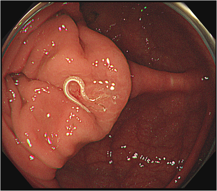
White whipworm in the cecum. Histopathological examination revealed that it was a male worm. The other two white whipworms were in the ascending colon.
FIGURE 2.
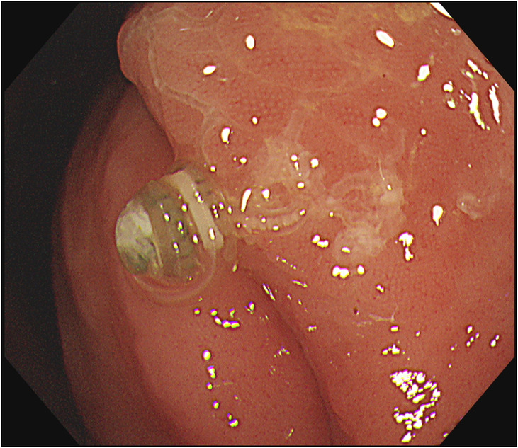
Brown whipworm in the ascending colon. Histopathological examination revealed that it was a female worm.
FIGURE 3.
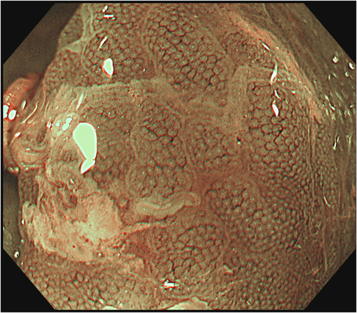
Picture of the female worm obtained by using narrow band imaging showed that its cranium was burrowing under the colonic mucosa.
FIGURE 4.
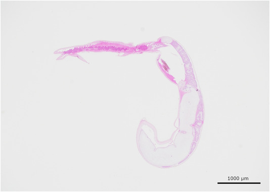
Histopathological image of a male whipworm.
FIGURE 5.
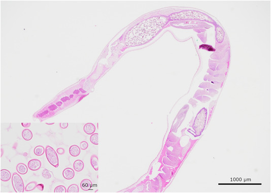
Histopathological image of the female whipworm.
T. trichiura infection is prevalent in tropical regions and is non‐endemic in Japan. T. trichiura is thought to live for one to 8 years as an adult. Therefore, it is likely that the patient acquired the infection while in Myanmar prior to arriving in Japan. T. trichiura parasitizes only humans through fecal‐oral transmission. In non‐endemic areas, the frequency of endoscopic identification has been increasing due to the increasing number of immigrants from endemic countries. 1 To prevent infection, it is important to pay attention to sanitary conditions such as soil and water sources. 2
AUTHOR CONTRIBUTIONS
Masaki Inoue: Data curation; writing – original draft. Marin Ishikawa: Supervision; writing – review and editing. Sho Tanaka: Data curation. Xinhan Zhang: Writing – review and editing. Hiromi Okada: Data curation; investigation. Takuto Miyagishima: Writing – review and editing.
FUNDING INFORMATION
None.
CONFLICT OF INTEREST STATEMENT
We have no financial relationships to disclose.
CONSENT
Written informed consent was obtained from the patient to publish this report in accordance with the journal's patient consent policy.
Inoue M, Ishikawa M, Tanaka S, Zhang X, Okada H, Miyagishima T. Infection with male and female Trichuris trichiura diagnosed in a non‐endemic area. Clin Case Rep. 2023;11:e7250. doi: 10.1002/ccr3.7250
DATA AVAILABILITY STATEMENT
Data available on request due to privacy/ethical restrictions.
REFERENCES
- 1. Lorenzetti R, Campo SM, Stella F, Hassan C, Zullo A, Morini S. An unusual endoscopic finding: Trichuris trichiura. Case report and review of the literature. Dig Liver Dis. 2003;35(11):811‐813. doi: 10.1016/s1590-8658(03)00455-9 [DOI] [PubMed] [Google Scholar]
- 2. Kattula D, Sarkar R, Rao Ajjampur SS, et al. Prevalence & risk factors for soil transmitted helminth infection among school children in South India. Indian J Med Res. 2014;139(1):76‐82. [PMC free article] [PubMed] [Google Scholar]
Associated Data
This section collects any data citations, data availability statements, or supplementary materials included in this article.
Data Availability Statement
Data available on request due to privacy/ethical restrictions.


