PLOS ONE logoLink to PLOS ONE
. 2023 Apr 24;18(4):e0284201. doi: 10.1371/journal.pone.0284201

Mycobacterium ulcerans culture results according to duration of prior antibiotic treatment: A cohort study

Brodie Tweedale 1,2, Fiona Collier 1, Nilakshi T Waidyatillake 2,3, Eugene Athan 1,2, Daniel P O’Brien 2,4,*
Editor: Nitin Gupta5
PMCID: PMC10124831  PMID: 37093836

Abstract

Mycobacterium ulcerans disease is a necrotising disease of the skin and subcutaneous tissue and is effectively treated with eight-weeks antibiotic therapy. Significant toxicities, however, are experienced under this prolonged regimen. Here, we investigated the length of antibiotic duration required to achieve negative cultures of M. ulcerans disease lesions and evaluated the influence of patient characteristics on this outcome. M. ulcerans cases from an observational cohort that underwent antibiotic treatment prior to surgery and had post-excision culture assessment at Barwon Health, Victoria, from May 25 1998 to June 30 2019, were included. Antibiotic duration before surgery was grouped as <2 weeks, ≥2-<4 weeks, ≥4-<6 weeks, ≥6-<8 weeks, ≥8-<10 weeks and ≥10–20 weeks. Cox regression analyses were performed to assess the association between variables and culture positive results. Ninety-two patients fitted the inclusion criteria. The median age was 60 years (IQR 28–74.5) and 51 (55.4%) were male. Rifampicin-based regimens were predominantly used in combination with clarithromycin (47.8%) and ciprofloxacin (46.7%), and the median duration of antibiotic treatment before surgery was 23 days (IQR, 8.0–45.5). There were no culture positive results after 19 days of antibiotic treatment and there was a significant association between antibiotic duration before surgery and a culture positive outcome (p<0.001). The World Health Organisation category of the lesion and the antibiotic regimen used had no association with the culture outcome. Antibiotics appear to be effective at achieving negative cultures of M. ulcerans disease lesions in less than the currently recommended eight-week duration.

Introduction

Mycobacterium ulcerans disease, known as the Buruli ulcer (BU), is a necrotising infection of the skin and subcutaneous tissue and can cause large ulcerative lesions [1]. BU has been reported in 33 countries worldwide, primarily in tropical and subtropical countries including Côte D’Ivoire, Ghana and Bénin where the largest burden of disease is observed [2, 3]. It also occurs in temperate regions such as south-eastern Australia where an increase in incidence is being observed with for example the number of cases managed at a tertiary referral centre in Victoria doubling between 2005–2010 and 2011–2017 [4]. It is the third most common mycobacterial disease amongst immunocompetent people and can result in significant morbidities and chronic deformities [3, 5]. The World Health Organisation (WHO) categorises lesions according to severity determined by size, number and involvement of critical sites [6]. It is important to understand optimum treatment therapies that minimise complications and maximise cure of the disease.

Eight-weeks of rifampicin-based combination antibiotic therapy is the currently recommended treatment for M. ulcerans disease [6, 7], and surgery may be used in addition to antibiotics [8]. Despite the effectiveness of these antibiotic regimens [9, 10], severe toxicities including gastrointestinal intolerance, hepatitis and rash are experienced in over 20% of patients in Australian populations and almost 15% of those require hospitalisation to manage adverse effects [11]. The risk of experiencing treatment side-effects exists throughout the whole treatment course [11], with the median time to develop antibiotic complications being 28 days (IQR 17–45 days). A shortened antibiotic duration therefore has the potential to reduce the incidence of antibiotic complications.

Initial evidence to support reduced treatment duration for M. ulcerans disease has come from studies in murine models. Ji, et al. reported the sterilisation of M. ulcerans infected mice footpads after only four weeks of antibiotic therapy [12]; and a more recent study by Chauffour, et al. has demonstrated comparable results [13]. Previous work by Cowan, et al. reported the effectiveness of a reduced antibiotic duration in humans after achieving cure in all patients receiving between four and six weeks antibiotic therapy combined with surgery [14]. Additionally, all of five excised lesions from which tissue was cultured for M. ulcerans following 28–35 days of antibiotics were negative, suggesting that only 4–6 weeks of antibiotics may be required to cure lesions [14], noting that the sensitivity of M. ulcerans culture is estimated at about 50% [15]. More recently, O’Brien et al. investigated the efficacy of six weeks of antibiotic therapy against the recommended eight weeks for the least severe, WHO category 1 lesions [16]. Cure was reported in 100% of 53 patients in the six-week treatment group, further supporting the potential for reduced antibiotic durations for the treatment of M. ulcerans disease [1214].

To better understand the minimum duration of antibiotics required to achieve cure of M. ulcerans disease lesions, we investigated the length of antibiotic duration required to achieve culture negativity in a group of patients that underwent both surgery and post-excision culture assessment; with the consideration of the influence of patient characteristics on this outcome.

Materials and methods

Data collection

Cohort identification

Analysis was performed on a subset of prospectively collected data of all M. ulcerans cases treated from May 25 1998 to June 30 2019 by Barwon Health staff; a tertiary hospital located in Geelong, Australia. Cases encompassed patients from endemic regions of South-eastern Victoria, including the Bellarine Peninsula, Mornington Peninsula and surrounds. Data was prospectively collected using Epi-Info 6 (CDC, Atlanta) [17].

Inclusion criteria

For inclusion in this analysis, patients with diagnosed M. ulcerans must have (i) had surgery, (ii) received rifampicin based antibiotic treatment prior to surgery, and (iii) had Mycobacterium cultures performed on the excised lesions. The cohort included 20 cases published in an earlier study from our group [14]. The type of surgery performed included wide excision, conservative excision, curette and debridement as previously described [8]. A M. ulcerans case was defined as the presence of a lesion clinically suggestive of M. ulcerans, plus any of:

  1. a culture of M. ulcerans from the lesion

  2. a positive IS2404 polymerase chain reaction from a swab or biopsy of the lesion

  3. histopathology of an excised lesion showing a necrotic granulomatous ulcer with the presence of acid-fast bacilli consistent with acute M. ulcerans infection

Mycobacterial cultures from surgical specimens were performed at Barwon Health microbiological laboratories using Lowenstein-Jensen media and incubated at 30°C for 12 weeks. Specimens were sent immediately from surgery to the laboratory which was on-site at Barwon Health. Here they were kept at room temperature (20°C) and within 1–6 hours of arrival were finely diced with a sterile scalpel blade, without decontamination or dilution, prior to plating a tissue aliquot of approximately 2 mm in diameter.

Definitions used during the analysis

Duration of symptoms was defined as the time between symptom onset and diagnosis; site of lesion was defined as upper limb, lower limb and trunk; antibiotic duration was grouped as <2 weeks, ≥2-<4 weeks, ≥4-<6 weeks, ≥6-<8 weeks, ≥8-<10 weeks and ≥10–20 weeks.

Data analysis

Statistical analyses were performed using StataIC 16 (StataCorp, Texas, USA) [18]. Summary statistics were tabulated to describe the cohort characteristics. If antibiotics were changed due to adverse effects, only initial antibiotic combinations were examined when analysing the association with antibiotic regimens in the analysis. A Pearson’s chi-squared test was used to assess the relationship between antibiotic duration and culture positive outcome, and a Kaplan-Meier curve was used to demonstrate the cumulative incidence of positive M. ulcerans cultures according to the days of antibiotic treatment. Rates of positive M. ulcerans cultures associated with identified variables were described in 100-person days of antibiotics. A Cox regression model was then used to assess the crude hazard ratios of variables with positive M. ulcerans cultures. Then a multivariate Cox regression analysis was performed including the variables sex and age a priori and the variable immune suppression as the only other variable to show evidence of an association with positive cultures in the crude analysis (assessed by p ≤ 0.20). The p-values for assessing the strength of the association of each variable with positive M. ulcerans cultures, controlled for all the other variables in the model, were determined by the likelihood ratio test.

Ethics approval and consent to participate

This study was approved by the Barwon Health Human Research and Ethics Committee (HREC No. 04/60). All previously gathered human medical data were analysed in a de-identified fashion.

Results

Cohort characteristics

From May 25th 1998 to June 30th 2019, lesions excised from 92 patients following antibiotic therapy were cultured and included in the study. This represented 13.5% of 679 patients who were treated with antibiotics and 40.2% of 229 patients who had surgery in addition to antibiotics in the Barwon Health cohort during this time. Baseline cohort characteristics are presented in Table 1. The median age of patients included in this study was 60 years (IQR, 28–74.5), and 51 (55.4%) were male. Nine (9.8%) patients had diabetes mellitus and 12 (13%) were immune suppressed due to disease or medication. None were known to be HIV positive. BCG status was not collected noting that since the 1980s it has not been included in routine vaccine schedules in the local population. The majority of lesions were classified as WHO category 1 lesions (58.7%, n = 54), followed by WHO category 3 (23.9%, n = 22) and WHO category 2 lesions (17.4%, n = 16). There were no cases associated with osteomyelitis. Forty-seven days (IQR, 35–76) was the median time from onset of symptoms to commencement of treatment. The first-line antibiotic regimens were rifampicin and clarithromycin in 44 (47.8%) cases, rifampicin and ciprofloxacin in 43 (46.7%) cases, and other regimens in 5 (5.4%) cases. The median duration of antibiotic treatment before surgery was 23 days (IQR, 8–45.5). Thirty-three (35.9%) required a split-skin graft and 8 (8.7%) a vascularised flap following surgery. Thirty-six (39.1%) of cases experienced paradoxical reactions. All patients were cured following treatment with no disease relapses.

Table 1. Baseline cohort characteristics.

Treatment Cohort (n = 92)
n (%) Mean ± SD (Range) and Median (IQR)
Gender
    • Male
    • Female

51 (55.4)
41 (44.6)
Age (years)
    • 0–19
    • 20–59
    • ≥ 60

19 (20.7)
26 (28.3)
47 (51.1)
Median = 60 (IQR 28–74.5)
WHO Category of Lesions
    • 1
    • 2
    • 3

54 (58.7)
16 (17.4)
22 (23.9)
Lesion Type
    • Nodule
    • Oedematous
    • Plaque
    • Ulcer

4 (4.4)
24 (26.1)
4 (4.4)
60 (65.2)
Antibiotic duration (days) Median = 23 (IQR 8–45.5)
Lesion Site
    • Upper limb
    • Lower limb
    • Head/Trunk

29 (31.5%)
61 (66.3%)
2 (2.2%)
Antibiotic regimen
    • RCla *
    • RCp **
    • Other

44 (47.8%)
43 (46.7%)
5 (5.4%)

*Rifampicin + Clarithromycin

**Rifampicin + Ciprofloxacin

Number of weeks of antibiotic treatment and M. ulcerans culture result

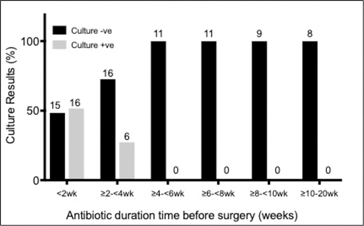
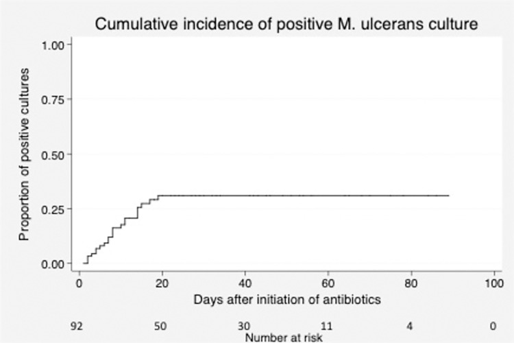
The proportion of patients with a positive M. ulcerans culture following less than two weeks of antibiotics was 51.6% (n = 31). After two to four weeks of treatment only 27.3% of patients had a positive culture, and there were no culture positive results evident after 2.7 weeks (19 days) (Figs 1 & 2). A chi-square test of goodness-of-fit confirmed a significant association between antibiotic duration before surgery in weeks and a culture positive outcome (p < 0.001) (Fig 1).

Fig 1. Relationship between antibiotic duration before surgery (weeks) and culture results.

Fig 1

Figure labels on columns represent actual number of cases.

Fig 2. Kaplan-Meier curve showing the cumulative incidence of positive M. ulcerans cultures according to days of antibiotic treatment.

Fig 2

Association between patient characteristics and M. ulcerans culture results

The association between patient characteristics and the outcome of the post-excision M. ulcerans culture was examined (Table 2). Using multivariable Cox regression model analysis adjusting for age, gender and immune suppression, there was a trend that older age (p = 0.07) and lack of immune suppression (p = 0.06) were associated with a positive culture. However, there was no association between culture positivity and gender, WHO category, lesion type or site, diabetes, antibiotic regimen, weight or duration of symptoms prior to diagnosis.

Table 2. Cox regression model showing adjusted and unadjusted associations between identified variable and rates of positive M. ulcerans culture.

Variable Failures (Positive culture) (%) Follow-up (days) Rate per 100-person days (95% CI) Crude hazard ratio (95% CI) p-value Adjusted hazard ratio (95% CI) p-value
Gender
Female 10 (24.4) 1115 9.0 (4.83,16.67) 1 0.97 1 0.86
Male 12 (23.5) 1587 7.6 (4.5,14.1) 1.0 (0.4,2.3) 0.9 (0.4,2.2)
Age (years)
0–15 1 (7.1) 603 1.7 (0.2–11.8) 1 0.14 1 0.07
16–64 9(23.1) 995 9.0 (4.7,17.4) 4.1 (0.5,32.6) 4.3 (0.5,34.5)
≥65 12(30.8) 1104 10.9 (6.2,19.1) 5.1 (0.7,39.3) 6.6 (0.8,50.8)
WHO category of lesion
1 14 (25.9) 1160 12.1 (7.1,20.4) 1 0.29 - -
2 2 (12.5) 703 2.8 (0.7,11.4) 0.4 (0.1,1.6) -
3 6 (27.3) 839 7.2 (3.2,15.9) 0.9 (0.4,2.5) -
Lesion type
Ulcer 15(25.0) 1396 2.7 (2.1,3.4) 0.7 (0.1–5.6) 0.44 - -
Nodule 1 (25.0) 113 3.6 (1.6,8.0) 1 - -
Oedema 6 (25.0) 993 3.0 (1.6,5.8) 0.7 (0.1,5.5) - -
Plaque 0 (0.0) 200 0.0 - - -
Lesion site
Upper limb 9 (30.0) 755 11.9 (6.2,22.9) 1 0.38 - -
Lower Limb 13 (21.3) 1913 7.0 (3.9,11.7) 0.6 (0.3,1.4) - -
Head/Trunk 0 (0.0) 34 0.0 - - -
Immune suppression
No 21 (26.3) 2351 8.9 (5.8,13.7) 1 0.16 - 0.06
Yes 1 (8.3) 351 2.8 (0.4,20.2) 0.3 (0.0,2.3) 0.2 (0,1.6)
Diabetes
No 22 (27.2) 2271 9.7 (6.4,14.7) - - - -
Yes 0 (0.0) 238 0 - -
Antibiotic regimen
RCla* 9(20.5) 1517 5.9 (3.1,11.4) 1 0.48 - -
RCp** 12 (27.9) 1026 11.7 (6.6,20.6) 1.7 (0.7,4.0) -
other 1 (20.0) 159 6.3 (0.9,44.6) 1.0 (0.1,7.7) -
Weight (kg)
0–90 4 (19.1) 707 5.7 (2.1,15.1) 1 0.71 - -
≥ 90 2 (25.0) 251 8.0 (2.0,31.9) 1.3 (0.2,7.3) - -
Missing 16 (25.4) 1744 9.2 (5.6,15.0) 1.6 (0.5,4.7) - -
Duration of symptoms prior to diagnosis
0–42 15 (27.8) 1593 9.4 (5.7,15.6) 1 0.50 - -
≥42 6 (16.7) 1030 5.8 (2.6,12.0) 0.6 (0.2,1.6) - -
Missing 1 (50) 79 12.7 (1.8, 89.9) 1.7 (0.2,12.8) - -

*Rifampicin + Clarithromycin

**Rifampicin + Ciprofloxacin

Discussion

Our results suggest BU lesions can be culture negative following antibiotic therapy significantly earlier than the recommended eight-weeks of treatment with no lesions culture positive after 19 days of treatment; a finding that supports the potential to reduce antibiotic treatment duration. Our study utilised data from a group of BU patients that had both rifampicin-based antibiotic treatment prior to surgical excision and M. ulcerans culture testing of the excised tissue. This enabled assessment of the antibiotic treatment duration needed for achieving culture negativity of lesions as well as assessing the influence of a number of patient covariates. We highlight the relationship between duration of antibiotic therapy and culture outcome, whereby an increased length of antibiotic therapy before surgery coincides with a decrease in the proportion of culture positive results.

Comparable results were yielded in an early study in Ghana in 2005, albeit in small numbers, where the cultures of 11 patients were negative after four-weeks of rifampicin-based therapy [19]. Additionally, an Australian study (n = 4) also demonstrated the inability to culture M. ulcerans from excised lesions following up to six weeks of antibiotic therapy [20]. Further, we showed in an earlier analysis of the Barwon Health cohort that 100% of lesions excised after 28–38 days of antibiotics were culture negative [14]. Importantly, four years on and with more than a four-fold increase in the number of patients in this specific cohort subpopulation (n = 92 compared to n = 20), the findings between these studies remain consistent, lending support to a reduced antibiotic duration for the treatment of M. ulcerans disease. Also, we recently reported that the cure of WHO category 1 lesions is achievable with just six-weeks of antibiotic therapy–affording further evidence for a reduced antibiotic duration [16]. Notably, the current study included nearly 40% of lesions which were WHO category 2 and 3, suggesting a shorter duration of antibiotic therapy may also be possible for more severe lesions.

Contrasting results have also been reported. In a study of patients treated with two weeks of rifampicin and streptomycin followed by six-weeks oral rifampicin and clarithromycin [21], three lesions (42.9%) were culture positive after 12-weeks from commencement of treatment. Additionally, in a randomised trial in Ghana, positive cultures were reported from three lesions (60%) after eight-weeks of antibiotic therapy [10]. Despite this, all lesions healed without any additional surgical intervention and no recurrence was reported after 12-month follow-up. This is suggestive that a reduced bacterial load, and not necessarily complete lesion sterilisation, may be adequate to permit an immune response strong enough to overcome mycolactone production and achieve wound healing. This was highlighted recently by O’Brien, et al. reporting a case series of five adults whose small ulcerative lesions spontaneously healed without specific treatment, emphasizing that a vigorous host immune response can overcome the suppressive effects of mycolactone and achieve cure [22].

A reduced antibiotic duration required to achieve cure will immediately benefit patients by reducing the daily inconvenience associated with antibiotic consumption. More importantly, the chance of experiencing drug-toxicities [11], the cost of treatment, and the impact on the microbiome will decrease [23]. This current study not only supports our previous proposition of a 25% reduction in the currently recommended eight-week treatment time for the sterilisation of selected small WHO category I BU lesions [16]; but also raises the possibility of an even further reduced antibiotic treatment time. This is significant as the median time for emergence of severe antibiotic complications is four weeks (IQR 17–45 days) [11]. Theoretically, over 50% of severe antibiotic complications could be prevented by reducing treatment time from eight-weeks to four-weeks. However, culture results of excised specimens don’t necessarily correlate with clinical outcomes, especially as the sensitivity of M. ulcerans culture is estimated at about 50% [15]. Therefore, prospective randomised control trials comparing the reduced duration against the recommended eight-week therapy should be conducted to assess the clinical effectiveness of shortened antibiotic regimens.

Other potential benefits of shortened antibiotic treatment regimens include a reduction in hospital and treatment fees associated with side-effects, as well as the decrease to the cost of antibiotics themselves; saving money for both patients and the health care system [24]. The potential effect on a patient’s microbiome due to prolonged antibiotic use will also be reduced, therefore minimising disruptions to patient health [23, 25]. Additionally, the risk of antibiotic resistance will be reduced with a shortened treatment duration [26].

We found that there was some evidence that older age of the patient was associated with culture positivity (Table 2). We have recently reported that age greater than 65 years is a risk factor for experiencing more severe disease (Category 2 and 3); potentially due to reduced immunity and control of the bacteria [4, 27]. Thus it is also possible that a reduced immunity in older age slows the sterilising rate of lesions by antibiotics. Conversely, young individuals may be able to sterilise wounds more quickly through a greater penetration of wounds with antibiotics via improved tissue circulation. A possible association of increased culture positive results with no immune suppression is unexpected and the reasons for this are unclear.

We acknowledge that our study is limited by its observational nature, the fact that not all surgical specimens from our cohort were cultured and the relatively small sample size. Furthermore, postulations that M. ulcerans strains in Africa are more virulent due to production of increased quantities and more potent forms of mycolactone may limit the extrapolation of findings more globally [28]. However, we feel that the findings offer sufficient information to stimulate more research into the effectiveness of shortened antibiotic treatment regimens.

Conclusions

We have shown in an Australian cohort that BU lesions can be culture negative following antibiotic therapy significantly earlier than the recommended eight-weeks of treatment. This provides the potential to significantly reduce toxicities, inconvenience and cost experienced by patients by reducing the duration of antibiotic treatment.

Data Availability

As the data may contain potentially identifiable information, the authors are not authorized by the Barwon Health Human Research Ethics Committee to publicly share a de-identified data set. Data is available upon request from the Barwon Health Human Research Ethics Committee via email (regi@barwonhealth.org.au) for researchers who meet the criteria for access to confidential data.

Funding Statement

The authors received no specific funding for this work.

References

  • 1.Sizaire V, Nackers F, Comte E, Portaels F: Mycobacterium ulcerans infection: control, diagnosis, and treatment. The Lancet infectious diseases 2006, 6(5):288–296. doi: 10.1016/S1473-3099(06)70464-9 [DOI] [PubMed] [Google Scholar]
  • 2.O’Brien DP, Jeanne I, Blasdell K, Avumegah M, Athan E: The changing epidemiology worldwide of Mycobacterium ulcerans. Epidemiology and Infection 2018, 14719. doi: 10.1017/S0950268818002662 [DOI] [PMC free article] [PubMed] [Google Scholar]
  • 3.World Health Organization: Working to overcome the global impact of neglected tropical diseases: first WHO report on neglected tropical diseases. In.: Geneva: World Health Organization; 2010. [Google Scholar]
  • 4.Tai AYC, Athan E, Friedman ND, Hughes A, Walton A, O’Brien DP: Increased Severity and Spread of Mycobacterium ulcerans, Southeastern Australia. Emerging infectious diseases 2018, 24(1):58–64. doi: 10.3201/eid2401.171070 [DOI] [PMC free article] [PubMed] [Google Scholar]
  • 5.Portaels F, Silva MT, Meyers WM: Buruli ulcer. Clinics In Dermatology 2009, 27(3):291–305. doi: 10.1016/j.clindermatol.2008.09.021 [DOI] [PubMed] [Google Scholar]
  • 6.World Health Organization: Treatment of Mycobacterium ulcerans disease (Buruli ulcer): guidance for health workers. 2012. [Google Scholar]
  • 7.O’Brien DP, Jenkin G, Buntine J, Steffen CM, McDonald A, Horne S, et al. Treatment and prevention of Mycobacterium ulcerans infection (Buruli ulcer) in Australia: guideline update. Medical Journal of Australia 2014, 200(5):267–270. doi: 10.5694/mja13.11331 [DOI] [PubMed] [Google Scholar]
  • 8.O’Brien DP, Callan P, Friedman ND, Athan E, Hughes A, McDonald A: Mycobacterium ulcerans disease management in Australian patients: the re-emergence of surgery as an important treatment modality. ANZ Journal of Surgery 2019, 89(6):653–658. doi: 10.1111/ans.14829 [DOI] [PubMed] [Google Scholar]
  • 9.Friedman ND, Athan E, Walton AL, O’Brien DP: Increasing experience with primary oral medical therapy for Mycobacterium ulcerans disease in an Australian cohort. Antimicrobial agents and chemotherapy 2016, 60(5):2692–2695. doi: 10.1128/AAC.02853-15 [DOI] [PMC free article] [PubMed] [Google Scholar]
  • 10.Nienhuis WA, Stienstra Y, Thompson WA, Awuah PC, Abass KM, Tuah W, et al. Antimicrobial treatment for early, limited Mycobacterium ulcerans infection: a randomised controlled trial. The Lancet 2010, 375(9715):664–672. doi: 10.1016/S0140-6736(09)61962-0 [DOI] [PubMed] [Google Scholar]
  • 11.O’Brien DP, Friedman D, Hughes A, Walton A, Athan E: Antibiotic complications during the treatment of Mycobacterium ulcerans disease in Australian patients. Internal medicine journal 2017, 47(9):1011–1019. doi: 10.1111/imj.13511 [DOI] [PubMed] [Google Scholar]
  • 12.Ji B, Chauffour A, Robert J, Lefrançois S, Jarlier V: Orally administered combined regimens for treatment of Mycobacterium ulcerans infection in mice. Antimicrobial agents and chemotherapy 2007, 51(10):3737–3739. doi: 10.1128/AAC.00730-07 [DOI] [PMC free article] [PubMed] [Google Scholar]
  • 13.Chauffour A, Robert J, Veziris N, Aubry A, Jarlier V: Sterilizing activity of fully oral intermittent regimens against Mycobacterium ulcerans infection in mice. PLoS neglected tropical diseases 2016, 10(10)0005066. doi: 10.1371/journal.pntd.0005066 [DOI] [PMC free article] [PubMed] [Google Scholar]
  • 14.Cowan R, Athan E, Friedman ND, Hughes AJ, McDonald A, Callan P, et al. Mycobacterium ulcerans treatment–can antibiotic duration be reduced in selected patients? PLoS neglected tropical diseases 2015, 9(2)0003503. doi: 10.1371/journal.pntd.0003503 [DOI] [PMC free article] [PubMed] [Google Scholar]
  • 15.Herbinger K-H, Adjei O, Awua-Boateng N-Y, Nienhuis WA, Kunaa L, Siegmund V, et al. Comparative Study of the Sensitivity of Different Diagnostic Methods for the Laboratory Diagnosis of Buruli Ulcer Disease. Clinical Infectious Diseases 2009, 48(8):1055–1064. doi: 10.1086/597398 [DOI] [PubMed] [Google Scholar]
  • 16.O’Brien D, Friedman ND, Cowan R, Walton A, Athan E.: Six versus eight weeks of antibiotics for small Mycobacterium ulcerans lesions in Australian Patients. Clinical Infectious Diseases 2019, 70(9):1993–1997. [DOI] [PubMed] [Google Scholar]
  • 17.Dean AG AT, Sunki GG, Friedman R, Lantinga M, Sangam S, Zubieta JC, et al. Epi Info™, a database and statistics program for public health professionals. In. Atlanta, GA, USA: CDC; 2011. [Google Scholar]
  • 18.StataCorp: Stata Statistical Software: Release 16. In. TX: StataCorp LLC: College Station; 2019. [Google Scholar]
  • 19.Etuaful S, Carbonnelle B, Grosset J, Lucas S, Horsfield C, Phillips R, et al. Efficacy of the combination rifampin-streptomycin in preventing growth of Mycobacterium ulcerans in early lesions of Buruli ulcer in humans. Antimicrobial agents and chemotherapy 2005, 49(8):3182–3186. doi: 10.1128/AAC.49.8.3182-3186.2005 [DOI] [PMC free article] [PubMed] [Google Scholar]
  • 20.Gordon CL, Buntine JA, Hayman JA, Lavender CJ, Fyfe JA, Hosking P, et al. All-oral antibiotic treatment for buruli ulcer: a report of four patients. PLoS neglected tropical diseases 2010, 4(11)770. doi: 10.1371/journal.pntd.0000770 [DOI] [PMC free article] [PubMed] [Google Scholar]
  • 21.Phillips RO, Sarfo FS, Abass MK, Abotsi J, Wilson T, Forson M, et al. Clinical and bacteriological efficacy of rifampin-streptomycin combination for two weeks followed by rifampin and clarithromycin for six weeks for treatment of Mycobacterium ulcerans disease. Antimicrobial agents and chemotherapy 2014, 58(2):1161–1166. doi: 10.1128/AAC.02165-13 [DOI] [PMC free article] [PubMed] [Google Scholar]
  • 22.O’Brien DP, Murrie A, Meggyesy P, Priestley J, Rajcoomar A, Athan E: Spontaneous healing of Mycobacterium ulcerans disease in Australian patients. PLoS neglected tropical diseases 2019, 13(2)0007178–e0007178. doi: 10.1371/journal.pntd.0007178 [DOI] [PMC free article] [PubMed] [Google Scholar]
  • 23.Ferrer M, Méndez-García C, Rojo D, Barbas C, Moya A: Antibiotic use and microbiome function. Biochemical Pharmacology 2017, 134:114–126. doi: 10.1016/j.bcp.2016.09.007 [DOI] [PubMed] [Google Scholar]
  • 24.Pak J O ’Brien DP, Quek T, Athan E: Treatment costs of Mycobacterium ulcerans in the antibiotic era. International health 2012, 4(2):123–127. doi: 10.1016/j.inhe.2011.12.005 [DOI] [PubMed] [Google Scholar]
  • 25.Blaser MJ: Antibiotic use and its consequences for the normal microbiome. Science 2016, 352(6285):544. doi: 10.1126/science.aad9358 [DOI] [PMC free article] [PubMed] [Google Scholar]
  • 26.Wilson HL, Daveson K, Del Mar CB: Optimal antimicrobial duration for common bacterial infections. Aust Prescr 2019, 42(1):5–9. doi: 10.18773/austprescr.2019.001 [DOI] [PMC free article] [PubMed] [Google Scholar]
  • 27.O’Brien DP, Friedman ND, Cowan R, Pollard J, McDonald A, Callan P, et al. Mycobacterium ulcerans in the Elderly: More Severe Disease and Suboptimal Outcomes. Plos Neglected Tropical Diseases 2015, 9(12)0004253–e0004253. doi: 10.1371/journal.pntd.0004253 [DOI] [PMC free article] [PubMed] [Google Scholar]
  • 28.Mve-Obiang A, Lee RE, Portaels F, Small P: Heterogeneity of mycolactones produced by clinical isolates of Mycobacterium ulcerans: implications for virulence. Infection and immunity 2003, 71(2):774–783. doi: 10.1128/IAI.71.2.774-783.2003 [DOI] [PMC free article] [PubMed] [Google Scholar]

Decision Letter 0

Nitin Gupta

19 Dec 2022

PONE-D-22-30052Mycobacterium ulcerans culture results according to duration of prior antibiotic treatment: a cohort studyPLOS ONE

Dear Dr. O'Brien,

Thank you for submitting your manuscript to PLOS ONE. After careful consideration, we feel that it has merit but does not fully meet PLOS ONE’s publication criteria as it currently stands. Therefore, we invite you to submit a revised version of the manuscript that addresses the points raised during the review process.

ACADEMIC EDITOR: This is an interesting 20-year cohort study on duration of antimicrobial therapy for patients with Buruli ulcer. While the premise is intriguing and the data seems robust, the reviewers have raised pertinent questions here that needs to be addressed. This article can be considered for publication after the aricle has been revised appropriately.  

Please submit your revised manuscript by Feb 02 2023 11:59PM. If you will need more time than this to complete your revisions, please reply to this message or contact the journal office at plosone@plos.org. When you're ready to submit your revision, log on to https://www.editorialmanager.com/pone/ and select the 'Submissions Needing Revision' folder to locate your manuscript file.

Please include the following items when submitting your revised manuscript:

  • A rebuttal letter that responds to each point raised by the academic editor and reviewer(s). You should upload this letter as a separate file labeled 'Response to Reviewers'.

  • A marked-up copy of your manuscript that highlights changes made to the original version. You should upload this as a separate file labeled 'Revised Manuscript with Track Changes'.

  • An unmarked version of your revised paper without tracked changes. You should upload this as a separate file labeled 'Manuscript'.

If you would like to make changes to your financial disclosure, please include your updated statement in your cover letter. Guidelines for resubmitting your figure files are available below the reviewer comments at the end of this letter.

If applicable, we recommend that you deposit your laboratory protocols in protocols.io to enhance the reproducibility of your results. Protocols.io assigns your protocol its own identifier (DOI) so that it can be cited independently in the future. For instructions see: https://journals.plos.org/plosone/s/submission-guidelines#loc-laboratory-protocols. Additionally, PLOS ONE offers an option for publishing peer-reviewed Lab Protocol articles, which describe protocols hosted on protocols.io. Read more information on sharing protocols at https://plos.org/protocols?utm_medium=editorial-email&utm_source=authorletters&utm_campaign=protocols.

We look forward to receiving your revised manuscript.

Kind regards,

Nitin Gupta, MBBS, MD, DM, AAHIVS, DTM&H

Academic Editor

PLOS ONE

Journal Requirements:

When submitting your revision, we need you to address these additional requirements.

1. Please ensure that your manuscript meets PLOS ONE's style requirements, including those for file naming. The PLOS ONE style templates can be found at 

https://journals.plos.org/plosone/s/file?id=wjVg/PLOSOne_formatting_sample_main_body.pdf and 

https://journals.plos.org/plosone/s/file?id=ba62/PLOSOne_formatting_sample_title_authors_affiliations.pdf

2. Please amend your current ethics statement to address the following concerns:

a) Did participants provide their written or verbal informed consent to participate in this study?

b) If consent was verbal, please explain i) why written consent was not obtained, ii) how you documented participant consent, and iii) whether the ethics committees/IRB approved this consent procedure.

3. In your Data Availability statement, you have not specified where the minimal data set underlying the results described in your manuscript can be found. PLOS defines a study's minimal data set as the underlying data used to reach the conclusions drawn in the manuscript and any additional data required to replicate the reported study findings in their entirety. All PLOS journals require that the minimal data set be made fully available. For more information about our data policy, please see http://journals.plos.org/plosone/s/data-availability.

Upon re-submitting your revised manuscript, please upload your study’s minimal underlying data set as either Supporting Information files or to a stable, public repository and include the relevant URLs, DOIs, or accession numbers within your revised cover letter. For a list of acceptable repositories, please see http://journals.plos.org/plosone/s/data-availability#loc-recommended-repositories. Any potentially identifying patient information must be fully anonymized.

Important: If there are ethical or legal restrictions to sharing your data publicly, please explain these restrictions in detail. Please see our guidelines for more information on what we consider unacceptable restrictions to publicly sharing data: http://journals.plos.org/plosone/s/data-availability#loc-unacceptable-data-access-restrictions. Note that it is not acceptable for the authors to be the sole named individuals responsible for ensuring data access.

We will update your Data Availability statement to reflect the information you provide in your cover letter.

4. Your ethics statement should only appear in the Methods section of your manuscript. If your ethics statement is written in any section besides the Methods, please move it to the Methods section and delete it from any other section. Please ensure that your ethics statement is included in your manuscript, as the ethics statement entered into the online submission form will not be published alongside your manuscript. 

Additional Editor Comments:

This is an interesting 20-year cohort study on duration of antimicrobial therapy for patients with Buruli ulcer. While the premise is intriguing and the data seems robust, the reviewers have raised pertinent questions here that needs to be addressed. This article can be considered for publication after the aricle has been revised appropriately.

[Note: HTML markup is below. Please do not edit.]

Reviewers' comments:

Reviewer's Responses to Questions

Comments to the Author

1. Is the manuscript technically sound, and do the data support the conclusions?

The manuscript must describe a technically sound piece of scientific research with data that supports the conclusions. Experiments must have been conducted rigorously, with appropriate controls, replication, and sample sizes. The conclusions must be drawn appropriately based on the data presented.

Reviewer #1: Yes

Reviewer #2: Yes

**********

2. Has the statistical analysis been performed appropriately and rigorously?

Reviewer #1: Yes

Reviewer #2: Yes

**********

3. Have the authors made all data underlying the findings in their manuscript fully available?

The PLOS Data policy requires authors to make all data underlying the findings described in their manuscript fully available without restriction, with rare exception (please refer to the Data Availability Statement in the manuscript PDF file). The data should be provided as part of the manuscript or its supporting information, or deposited to a public repository. For example, in addition to summary statistics, the data points behind means, medians and variance measures should be available. If there are restrictions on publicly sharing data—e.g. participant privacy or use of data from a third party—those must be specified.

Reviewer #1: Yes

Reviewer #2: Yes

**********

4. Is the manuscript presented in an intelligible fashion and written in standard English?

PLOS ONE does not copyedit accepted manuscripts, so the language in submitted articles must be clear, correct, and unambiguous. Any typographical or grammatical errors should be corrected at revision, so please note any specific errors here.

Reviewer #1: Yes

Reviewer #2: Yes

**********

5. Review Comments to the Author

Please use the space provided to explain your answers to the questions above. You may also include additional comments for the author, including concerns about dual publication, research ethics, or publication ethics. (Please upload your review as an attachment if it exceeds 20,000 characters)

Reviewer #1: Comments

Summary: the objective of the study is to investigate the length of antibiotic treatment duration required to achieve sterilization of M.ulcerans disease lesions. To reach such a target, the authors studied a cohort of 92 patients diagnosed with M.ulcerans disease who, from May 25, 1998 to June 30, 2019, had antibiotic treatment (mainly daily rifampicin + clarithromycin or ciprofloxacin) before surgery at Barton Health, Victoria, Australia and had culture of the excision lesions on Loewenstein Jensen medium.

Comment to the authors

The manuscript is well presented but, while the results of study are totally based on the sterilization or not of the excised lesions, almost no information is given on the technical means of the culture of excised specimens. It is only written “Mycobacterial cultures from surgical specimens were performed at Barwon Health microbiological laboratories using Lowenstein-Jensen media and incubated for 12 weeks (page 5, lines 129-130)”. More details are needed: time between lesion excision surgery and culture, temperature to which the excised lesions are kept, the interval of time between surgical excision and culture, mincing of the specimen, decontamination procedures if any, dilutions of the excision specimen, amount seeded on Loewenstein Jensen medium and culture temperature. All precisions on the culture procedures are decisive for the results interpretation. They are more important and far more decisive than all statistical and clinical details.

Page 6, lines 144-155 are Greek for the reviewer. Is it possible to make them simpler when the only important factor for interpretation of data and treatment recommendations is bacteriology and not statistics or clinics (5 pages on statistics and clinics versus two lines on bacteriology!)?

Page 11 line 210. The word “sterilized” is not appropriate. I suggest the change of wording. Negative culture of excision specimen is not at all synonymous of sterilization (as pointed out by the authors on page 13, line 260)

Reviewer #2: This paper describes the results of a 20-year prospective observational cohort relating to antibiotic duration prior to surgery for M. ulcerans diseases in Victoria, Australia, adding further evidence that shorter antimicrobial durations appear to be effective at sterilizing M. ulcerans disease lesions (under the 8 weeks recommendation). This paper is well-written and clear and contributes the literature on BU and its treatment. The paper, at times, lacks a few details, but I suspect that these may be corrected quite easily during revision. Specifically, the paper is limited by the absence of clinical outcomes, the lack of a more detailed description of the limited sensitivity of M. ulcerans culture and does not discuss the clinical and pathobiological diversity of BU/Mycolactone between Australia and West Africa. All told, I believe this paper should be published in PLOS ONE after the following revisions are addressed.

Suggested clarifications:

If possible, please describe when antimicrobial regimen changed, as only initial antimicrobial regimen was included.

Please include a mention of the sensitivity of culture in the introduction.

Please describe WHO classification of BU in the introduction as referred to multiple times in text. Consider mentioning that a minority of WHO I lesions may heal spontaneously.

Clarify number of WHO III associated with osteomyelitis.

Describe level of surgical debridement and if skin grafting occurred.

If possible, please comment on secondary infections.

I suspect that BCG is not administered in Victoria, Australia. As this is relevant to BU manifestation, please indicate whether this population received the BCG vaccine.

If possible, add whether paradoxical deterioration occurred during antimicrobial therapy.

The age discrepancy between BU in Australia (60s) and Africa (5-15 yo) is interesting. Please consider commenting on why BU is commonly seen in children in West Africa and older adults in Australia? Differences in water-related exposure/ behaviour? Mosquito-exposure in Australia?

Please comment on the differences in Mycolactone A and B (West Africa, possibly more virulent) and C (Australia, less virulent) in different geographic areas and possible differences in virulence, limiting global extrapolation of the findings.

Small comments:

Line 57: please add “accent circonflexe” to the o in Côte d’ivoire and accent aigu to Bénin.

Line 59: If possible, please provide numerical data regarding the increase in BU in South-eastern Australia.

Line 68: Please name the adverse drug effects. Hepatoxocity due to rifampin? Other drug effects?

Line 70: If possible, please provide duration of follow-up for this study. Eg: clinical cure and no relapse after X months of follow-up.

Line 85: 100% cure in how many patients? Please list sample size in this study.

Lines 173: please describe duration of follow-up for establishing that “no disease relapses” occurred.

Please describe most common other regimen 5%.

**********

6. PLOS authors have the option to publish the peer review history of their article (what does this mean?). If published, this will include your full peer review and any attached files.

If you choose “no”, your identity will remain anonymous but your review may still be made public.

Do you want your identity to be public for this peer review? For information about this choice, including consent withdrawal, please see our Privacy Policy.

Reviewer #1: No

Reviewer #2: Yes: Carl Boodman

**********

[NOTE: If reviewer comments were submitted as an attachment file, they will be attached to this email and accessible via the submission site. Please log into your account, locate the manuscript record, and check for the action link "View Attachments". If this link does not appear, there are no attachment files.]

While revising your submission, please upload your figure files to the Preflight Analysis and Conversion Engine (PACE) digital diagnostic tool, https://pacev2.apexcovantage.com/. PACE helps ensure that figures meet PLOS requirements. To use PACE, you must first register as a user. Registration is free. Then, login and navigate to the UPLOAD tab, where you will find detailed instructions on how to use the tool. If you encounter any issues or have any questions when using PACE, please email PLOS at figures@plos.org. Please note that Supporting Information files do not need this step.

Attachment

Submitted filename: Comments.docx

Attachment

Submitted filename: M ulcerans review.docx

PLoS One. 2023 Apr 24;18(4):e0284201. doi: 10.1371/journal.pone.0284201.r002

Author response to Decision Letter 0


2 Feb 2023

Dear Editor,

We are re-submitting our paper entitled “M. ulcerans culture results according to duration of prior antibiotic treatment: a cohort study” to be considered for publication as a research article in your journal.

On the following pages please find in blue our responses to the reviewers’ comments. We have made the relevant changes in the revised copy of the manuscript which we hope addresses all of the concerns raised by the reviewers. We would be pleased to consider further revisions if needed.

All authors have seen and approved the manuscript and have contributed significantly to the work. The manuscript has not been published and is not being considered for publication elsewhere. Finally we have no conflicts of interest to disclose.

Yours sincerely

Associate Professor Daniel O’Brien MBBS, FRACP, DMedSc, Dip Anat.

Reviewer 1:

If possible, please describe when antimicrobial regimen changed, as only initial antimicrobial regimen was included.

This has now been included in the methods section. Lines 161-162

Please include a mention of the sensitivity of culture in the introduction.

This has now been included so that the section in the Introduction reads: “…suggesting that only 4-6 weeks of antibiotics may be required to cure lesions, noting that the sensitivity of M. ulcerans culture is estimated at about 50%.” Lines 91-92.

Please describe WHO classification of BU in the introduction as referred to multiple times in text.

This information has now been included in the introduction as follows:

“The World Health Organisation (WHO) categorises lesions according to severity determined by size, number and involvement of critical sites”. (Lines 66-67)

Consider mentioning that a minority of WHO I lesions may heal spontaneously.

This has been referred to in the discussion section (lines 277-280) as follows: “This was highlighted recently by O’Brien, et al. reporting a case series of five adults whose small ulcerative lesions spontaneously healed without specific treatment, emphasizing that a vigorous host immune response can overcome the suppressive effects of mycolactone and achieve cure[21]”

Clarify number of WHO III with associated osteomyelitis.

This has been clarified in the results section as described: “There were no cases associated with osteomyelitis” Line 195.

Describe level of surgical debridement and if skin grafting occurred.

A clarification of the types of surgery performed has now been included in the methods section as shown below, and the number and proportion of cases who underwent skin grafting or vascularised flap has now been included in the results section.

Methods, Lines 138-139: “The type of surgery performed included wide excision, conservative escision, curette and debridement….”

Results, Lines 199-200: “Thirty-three (35.9%) required a split-skin graft and 8 (8.7%) a vascularised flap following surgery.”

If possible, please comment on secondary infections?

We did not collect this information so are unable to include it.

I suspect that BCG is not administered in Victoria, Australia. As this is relevant to BU manifestation, please indicate whether this population received the BCG vaccine.

This has now been included in the results section as follows:

Lines 192-193: “BCG status was not collected noting that since the 1980s it has not been included in routine vaccine schedules in the local population”

If possible, add whether paradoxical deterioration occurred during antimicrobial therapy.

This information has now been included in the results section to read: Line 201:

“Thirty-six (39.1%) of cases experienced paradoxical reactions.”

The age discrepancy between BU in Australia (60s) and Africa (5-15) is interesting. Please consider commenting on why BU is commonly seen in children in West Africa and older adults in Australia? Differences in water-related exposure/ behaviour? Mosquito-exposure?

Whilst we agree this question is interesting, as it deals with epidemiology and risk of infection, rather than treatment, we feel it is out of scope for the current paper and have therefore not included a discussion on this issue. If the Editor still feels that it should be included, we can modify.

Please comment on the differences in Mycolactone A and B (West Africa) and C (Australia) in different geographic areas and possible differences in virulence, limiting global extrapolation of the findings.

This information has now been included in the limitations section of the manuscript as follows:

Lines 315-317: “Furthermore, postulations that M. ulcerans strains in Africa are more virulent due to production of increased quantities and more po¬tent forms of mycolactone may limit the extrapolation of findings more globally.”

Small comments:

Line 57: please add “accent circumflex” to the o in Côte d’ivoire and accent aigu to Bénin.

This has been corrected.

Line 59: If possible, please provide numerical data regarding the increase in BU in South_eastern Australia.

This has now been included in the introduction as follows: “…with for example the number of cases managed at a tertiary referral centre in Victoria doubling between 2005-2010 and 2011-2017.” Line 63-64

Line 68: Please name the adverse drug effects. Hepatoxocity due to rifampin? Other drug effects?

This has now been included as follows: “…severe toxicities including gastrointestinal intolerance, hepatitis and rash are experienced”. Line 74

Line 70: If possible, please provide duration of follow-up for this study. Eg: clinical cure and no relapse after X months of follow-up.

For the study quoted, clinical outcomes were not described. It was a study looking at antibiotic complications and outcomes were censored at either the time of severe antibiotic complication or completion of the antibiotic course.

Line 85: 100% cure in how many patients? Please list sample size in this study.

This has now been included. Line 94

Reviewer 2:

The manuscript is well presented but, while the results of study are totally based on the sterilization or not of the excised lesions, almost no information is given on the technical means of the culture of excised specimens. It is only written “Mycobacterial cultures from surgical specimens were performed at Barwon Health microbiological laboratories using Lowenstein-Jensen media and incubated for 12 weeks (page 5, lines 129-130)”. More details are needed: time between lesion excision surgery and culture, temperature to which the excised lesions are kept, the interval of time between surgical excision and culture, mincing of the specimen, decontamination procedures if any, dilutions of the excision specimen, amount seeded on Loewenstein Jensen medium and culture temperature. All precisions on the culture procedures are decisive for the results interpretation. They are more important and far more decisive than all statistical and clinical details.

We thank the reviewer for the suggestions. The requested detail has now been added to the methods section as follows: “Specimens were sent immediately from surgery to the laboratory which was on-site at Barwon Health. Here they were kept at room temperature (200C) and within 1-6 hours of arrival were finely diced with a sterile scalpel blade, without decontamination or dilution, prior to plating a tissue aliquot of approximately 2 mm in diameter.” Lines: 148-151

Page 6, lines 144-155 are Greek for the reviewer. Is it possible to make them simpler when the only important factor for interpretation of data and treatment recommendations is bacteriology and not statistics or clinics (5 pages on statistics and clinics versus two lines on bacteriology!)?

We have modified the section to reduce its length. However, as the majority of the section describes the statistical methods used to report many of the significant findings in our paper, we feel that it is important that the reader is able to determine exactly how these analyses were performed. Therefore we believe it is not helpful to the reader to simplify the section more than we have done.

Page 11 line 210. The word “sterilized” is not appropriate. I suggest the change of wording. Negative culture of excision specimen is not at all synonymous of sterilization (as pointed out by the authors on page 13, line 260)

We have now modified the statement to say:

Lines 322-323: “….culture negative following antibiotic therapy”. We have also replaced the word ‘sterilized’ with ‘culture negative’ throughout the paper where appropriate.

Decision Letter 1

Nitin Gupta

27 Mar 2023

Mycobacterium ulcerans culture results according to duration of prior antibiotic treatment: a cohort study

PONE-D-22-30052R1

Dear Dr. O'Brien,

We’re pleased to inform you that your manuscript has been judged scientifically suitable for publication and will be formally accepted for publication once it meets all outstanding technical requirements.

Within one week, you’ll receive an e-mail detailing the required amendments. When these have been addressed, you’ll receive a formal acceptance letter and your manuscript will be scheduled for publication.

An invoice for payment will follow shortly after the formal acceptance. To ensure an efficient process, please log into Editorial Manager at http://www.editorialmanager.com/pone/, click the 'Update My Information' link at the top of the page, and double check that your user information is up-to-date. If you have any billing related questions, please contact our Author Billing department directly at authorbilling@plos.org.

If your institution or institutions have a press office, please notify them about your upcoming paper to help maximize its impact. If they’ll be preparing press materials, please inform our press team as soon as possible -- no later than 48 hours after receiving the formal acceptance. Your manuscript will remain under strict press embargo until 2 pm Eastern Time on the date of publication. For more information, please contact onepress@plos.org.

Kind regards,

Nitin Gupta, MBBS, MD, DM, AAHIVS, DTM&H

Academic Editor

PLOS ONE

Additional Editor Comments (optional):

The authors have addressed all the comments made by the reviewers. The article can be accepted in its current form.

Reviewers' comments:

Reviewer's Responses to Questions

Comments to the Author

1. If the authors have adequately addressed your comments raised in a previous round of review and you feel that this manuscript is now acceptable for publication, you may indicate that here to bypass the “Comments to the Author” section, enter your conflict of interest statement in the “Confidential to Editor” section, and submit your "Accept" recommendation.

Reviewer #1: All comments have been addressed

Reviewer #2: All comments have been addressed

**********

2. Is the manuscript technically sound, and do the data support the conclusions?

The manuscript must describe a technically sound piece of scientific research with data that supports the conclusions. Experiments must have been conducted rigorously, with appropriate controls, replication, and sample sizes. The conclusions must be drawn appropriately based on the data presented.

Reviewer #1: Yes

Reviewer #2: Yes

**********

3. Has the statistical analysis been performed appropriately and rigorously?

Reviewer #1: Yes

Reviewer #2: Yes

**********

4. Have the authors made all data underlying the findings in their manuscript fully available?

The PLOS Data policy requires authors to make all data underlying the findings described in their manuscript fully available without restriction, with rare exception (please refer to the Data Availability Statement in the manuscript PDF file). The data should be provided as part of the manuscript or its supporting information, or deposited to a public repository. For example, in addition to summary statistics, the data points behind means, medians and variance measures should be available. If there are restrictions on publicly sharing data—e.g. participant privacy or use of data from a third party—those must be specified.

Reviewer #1: Yes

Reviewer #2: Yes

**********

5. Is the manuscript presented in an intelligible fashion and written in standard English?

PLOS ONE does not copyedit accepted manuscripts, so the language in submitted articles must be clear, correct, and unambiguous. Any typographical or grammatical errors should be corrected at revision, so please note any specific errors here.

Reviewer #1: Yes

Reviewer #2: Yes

**********

6. Review Comments to the Author

Please use the space provided to explain your answers to the questions above. You may also include additional comments for the author, including concerns about dual publication, research ethics, or publication ethics. (Please upload your review as an attachment if it exceeds 20,000 characters)

Reviewer #1: The manuscript and its conclusions are adequate for me . However I would suggest the authors (page 6, line143) "... to plating a tissue aliquot of approximatively 2mm in diameter" would deserve to be increased and tested.

Reviewer #2: All of my previous comments have been adequately addressed by the authors in this revised version. I believe this article is ready for publication.

**********

7. PLOS authors have the option to publish the peer review history of their article (what does this mean?). If published, this will include your full peer review and any attached files.

If you choose “no”, your identity will remain anonymous but your review may still be made public.

Do you want your identity to be public for this peer review? For information about this choice, including consent withdrawal, please see our Privacy Policy.

Reviewer #1: Yes: Jacques Grosset

Reviewer #2: Yes: Carl Boodman

**********

Acceptance letter

Nitin Gupta

14 Apr 2023

PONE-D-22-30052R1

Mycobacterium ulcerans culture results according to duration of prior antibiotic treatment: a cohort study

Dear Dr. O'Brien:

I'm pleased to inform you that your manuscript has been deemed suitable for publication in PLOS ONE. Congratulations! Your manuscript is now with our production department.

If your institution or institutions have a press office, please let them know about your upcoming paper now to help maximize its impact. If they'll be preparing press materials, please inform our press team within the next 48 hours. Your manuscript will remain under strict press embargo until 2 pm Eastern Time on the date of publication. For more information please contact onepress@plos.org.

If we can help with anything else, please email us at plosone@plos.org.

Thank you for submitting your work to PLOS ONE and supporting open access.

Kind regards,

PLOS ONE Editorial Office Staff

on behalf of

Dr. Nitin Gupta

Academic Editor

PLOS ONE

Associated Data

    This section collects any data citations, data availability statements, or supplementary materials included in this article.

    Supplementary Materials

    Attachment

    Submitted filename: Comments.docx

    Attachment

    Submitted filename: M ulcerans review.docx

    Data Availability Statement

    As the data may contain potentially identifiable information, the authors are not authorized by the Barwon Health Human Research Ethics Committee to publicly share a de-identified data set. Data is available upon request from the Barwon Health Human Research Ethics Committee via email (regi@barwonhealth.org.au) for researchers who meet the criteria for access to confidential data.


    Articles from PLOS ONE are provided here courtesy of PLOS

    RESOURCES