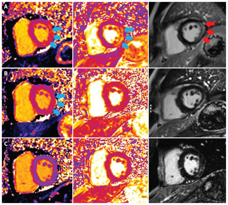
Figure 3:
Cardiac magnetic resonance imaging of a 41-year-old man with myocarditis. (A) Short-axis oblique images showing edema of the inferolateral wall on T1 and T2 mapping (blue arrows) (T1 = 1083 [normal 950–1050] ms and T2 = 71 [normal < 57] ms) and on late gadolinium enhancement (LGE) imaging (red arrows) during hospital admission. (B) Persistent myocardial edema in the inferolateral wall on T1 (blue arrows) but not T2 mapping at 2 months (T1 = 1080 ms; T2 = 48 ms). (C) Myocardial edema resolved on T1 and T2 mapping at 6 months (T1 = 1010 ms; T2 = 46 ms). All images were acquired from the same 1.5 T scanner (Aera, Siemens Healthineers).

