Abstract
Acne vulgaris, the most common form of acne, is characterized by a mixed eruption of inflammatory and noninflammatory skin lesions primarily affecting the face, upper arms, and trunk. The pathogenesis of acne is multifactorial and includes abnormal keratinization and plugging of the hair follicles, increased sebum production, proliferation and activation of Cutibacterium acnes (C. acnes; formerly Propionibacterium acnes, P. acnes), and finally inflammation. Recent studies have found that cannabidiol (CBD) may be beneficial in the treatment of acne. The aim of this study was to explore natural plant extracts that, when combined with CBD, act synergistically to treat acne by targeting different pathogenic factors while minimizing side effects. The first stage of the study investigated the capacity of different plant extracts and plant extract combinations to reduce C. acnes growth and decrease IL-1β and TNFα secretion from U937 cells. The results found that Centella asiatica triterpene (CAT) extract as well as silymarin (from Silybum marianum fruit extract) had significantly superior anti-inflammatory activity when combined with CBD compared to either ingredient alone. In addition, the CAT extract helped potentiate CBD-induced C. acnes growth inhibition. The three ingredients were integrated into a topical formulation and evaluated in ex vivo human skin organ cultures. The formulation was found to be safe and effective, reducing both IL-6 and IL-8 hypersecretion without hampering epidermal viability. Finally, a preliminary clinical study of this formulation conducted on 30 human subjects showed a statistically significant reduction in acne lesions (mainly inflammatory lesions) and porphyrin levels, thereby establishing a tight correlation between in vitro, ex vivo, and clinical results. Further studies must be conducted to verify the results, including placebo-controlled clinical assessment, to exclude any action of the formulation itself.
1. Introduction
Acne vulgaris, one of the most common human skin diseases, reduces the quality of life of millions of people worldwide. It affects almost everyone between the ages of 15 and 17, with 15–20% of affected individuals experiencing moderate to severe disease [1]. Although acne is principally a disorder of adolescence, current research indicates that the prevalence of acne in adult patients, especially women, is increasing. The clinical manifestations of acne include noninflammatory lesions (open and closed comedones) and inflammatory lesions (papules and pustules) [2]. Major factors in the development of acne include abnormal hyperkeratosis and shedding of the follicular epithelium, increased sebum production, proliferation of the bacteria Cutibacterium acnes (C. acnes; formerly known as Propionibacterium acnes, P. acnes), and finally inflammation. Current guidelines recommend a combination of ingredients and treatments aimed at addressing these different pathogenic factors through unique mechanisms of actions. While many options exist, most available acne treatments cause some degree of irritation, leading to low compliance rates [3]. Over the last decade, a rising interest in natural and plant-derived ingredients has led to the discovery and development of new products that provide good efficacy with less irritation, resulting in better compliance and outcomes [4–6].
Due to the important role of the endocannabinoid system in the skin, current research has focused on the role of cannabinoids in the treatment of several skin disorders [7]. Recently, cannabidiol (CBD) has been suggested as a new treatment option for acne [8]. In addition to its known anti-inflammatory activity, Oláh et al. have shown that CBD can reduce lipolysis and sebocyte proliferation in vitro [9]. Several clinical studies are now underway to evaluate the therapeutic potential of these properties [10].
The development of cannabinoids as therapeutic agents in dermatology and their use in cosmetic applications have been challenged by limited efficacy data and unclear regulatory and legal guidelines surrounding their use [11]. To circumvent these challenges, our team explored the use of established active molecules to provide synergistic properties, both in efficacy and tolerability, when combined with CBD in the treatment of acne. In this study, the compatibility of these herbal extracts with CBD was evaluated in vitro, ex vivo, and clinically. A statistically significant correlation of these findings using our final mixture was demonstrated across all stages of development.
2. Materials and Methods
All cell and skin culture mediums and supplements were supplied by Biological Industries (Beit HaEmek Israel). Unless mentioned, all chemical reagents came from Sigma-Aldrich (Israel). Centella asiatica triterpenes (leaf extract, containing 36–44% asiaticoside and 56–64% asiatic acid and madecassic acid) and silymarin from Silybum marianum fruit extract (50–60% silymarin calculated as silibinin: 20–45% silicristin, 40–65% silibinin A and B, and 10–20% isosilibinin A and B) were produced and supplied by Indena Group. Lonicera japonica (honeysuckle) flower extract, Salvia miltiorrhiza root extract (1% danshensu), and Camellia sinensis (green tea) extract (50% caffeine) came from Draco Natural Products. Salix babylonica (white willow) bark extract (25% salicin) was supplied from Botaniex, while hemp oil and CBD came from Echo Pharmaceuticals. U937 (a promonocytic, human myeloid leukemia cell line), as well as C. acnes, was supplied by ATCC.
2.1. Cell, Bacteria, and Skin Organ Cultures
U937 cells were grown and maintained in RPMI-1640 supplemented with 10% fetal calf serum and 1% streptomycin/penicillin. The cells were maintained at 5% CO2 in a humidified incubator at 37°C. For passaging, the cells were diluted in fresh media at a ratio of 1 : 4. Prior to the experiment, the cells were seeded at 300,000 cell/ml (150 μl pr 96-well plates) and differentiated by PMA (phorbol 12-myristate 13-acetate) into a macrophage-like phenotype, an observation similarly reported in other studies [12]. After 24 hours, the adhered cells were treated as indicated. C. acnes was grown in a modified and reinforced clostridial broth medium with reduced levels of oxygen.
Human skin samples were obtained from 40- to 60-year-old healthy women undergoing aesthetic abdominal surgery, after signing an informed consent form. The experiments were conducted with the approval of the IRB Committee of the Soroka Medical Center, Beer Sheva, Israel (#0258-19-SOR, approval protocol scrc20016; 29.06.2020). The tissue was processed and maintained in an air/liquid interface with the dermal side submerged, as previously detailed [13, 14].
2.2. Viability Determination
U937 cells and skin tissue epidermal viability were monitored by the MTT assay. Following treatment, the cells were washed, and the cellular indicator was added (150 μl of 0.5 mg/mL (3-(4,5- dimethylthiazol-2-yl)-2,5-diphenyltetrazolium bromide dissolved in PBS) to the 96-well plates. The plates were then incubated for 1 hour at 37°C, after which the solution was aspirated. One hundred and fifty microliters of isopropanol was then added to the solution to extract the formazan dye. The absorbance was recorded at 570 nm using a plate reader (TECAN, f200 Infinite, Switzerland). Epidermal viability was determined similarly following heat separation of the epidermis (1 min., 56°C) and its transfer to the 96-well plates.
2.3. Inflammation Induction and Cytokine Quantification
The cells were seeded at 300,000 cell/ml (150 μl pr 96-well plates) and differentiated by PMA (phorbol 12-myristate 13-acetate, 10 ng/ml) into a macrophage-like phenotype. Then, the cells were treated without or with 1 μg/mL in the absence or presence of the different treatments listed below. In addition, dexamethasone (10 μM) was used as a positive control. After different treatments, the spent medium from the cell and tissue cultures was centrifuged (to remove particles) and the supernatant (100 μl) was aliquoted and stored at −20°C until used. Human ELISA tests were performed according to the manufacturer's instructions (Biolegend, San Diego, CA).
2.4. MIC (Minimum Inhibitory Concentration) Assay
To evaluate the impact of the different treatments on C. acnes, a stock of frozen bacteria was removed from a −80°C freezer and thawed into a U-shaped falcon tube containing 4 ml of the modified, reinforced clostridial broth medium at 37°C under reduced oxygen levels (anaerobic chamber). After 3 hr, the bacteria were inoculated into agar plates (100 μl/10 cm plate) and grown under reduced oxygen levels. A single colony was grown in 4 ml media to a midlog phase (O.D. 0.5; 600 nm, approx. 3 hr). Then, the bacteria were diluted to O.D. 0.05 in the absence or presence of different treatments. Bacterial growth, determined by absorbance (O.D. 600), was monitored kinetically using a microplate reader (Infinite f200, TECAN), heated to 37°C. Ampicillin, at 10 μg/ml, was used as a positive control.
2.5. Formulation
A topical formulation containing 1% CBD, 1% CAT, and 1% silymarin was prepared by Echo Pharmaceuticals. The commercial formulation was developed as a hydroalcoholic gel using xanthan gum and acrylates/sodium acryloyldimethyl laurate copolymers as rheology modifiers. The formulation also has contained 0.5% of salicylic acid. The active ingredients were added in a step-wise manner at 45°C under constant stirring to increase solubility. A step-by-step preparation can be obtained by the authors, as well as samples of the formula for academic usage, after standardized NDA.
2.6. Clinical Study
The study was performed according to the Declaration of Helsinki principles and subsequent amendments. The study was conducted in the spirit of Good Clinical Practice Guidelines and general principles of Law 46/2004 of August 19th. The protocol and test conditions were reviewed by the Internal Review Board (opinion no. 6776/2021), and the standard protocol was submitted to the ethical commission of PhD trials (from December 27th, 2019). This study was conducted in accordance with the general conditions of PhD trials and was established as a research project involving human subjects, summarised by the protocol (MD.122/02 Protocol PT.06.01/final04).
2.6.1. Inclusion Criteria
Eligible study participants included male or female subjects aged 15–40 years with mild to moderate facial acne (Grade 2 or 3 according to the Investigator's Global Assessment (IGA)) and skin Fitzpatrick phototype II to IV. All enrolled participants and/or their parent/legal guardian (for minor subjects) provided written, informed consent and willingly agreed to comply with study requirements.
2.6.2. Exclusion Criteria
Subjects were excluded from the study if 1 or more of the following treatments were used: oral retinoids (within 6 months prior), topical retinoid treatment or any facial aesthetic or medical treatments (within 1 month prior), and excessive UV exposure (within 1 month prior). Pregnant women, breastfeeding women, or women planning a pregnancy during the clinical study were also excluded. Participants with medical conditions or those receiving treatments that, according to the investigator's judgment, could compromise the safety of the participant or interfere with outcomes of the study were also excluded. Furthermore, subjects with a planned or expected major surgical procedure during the clinical study were not included.
2.6.3. Study Population
It is presented in Table 1. Thirty-three subjects aged 15–40 with mild to moderate acne were enrolled in the study, with three withdrawals (#5 on D0, #19 on D28 and #11 on D56). Data from the remaining 30 subjects were included in our analysis.
Table 1.
Study population, including demographic and skin reactivity.
| Demographic data (subjects who applied the product at least once) | Skin | Subject having mild to moderate facial acne (Grade 2 or 3 according to the Investigator's Global Assessment (IGA)) | Subjects |
|---|---|---|---|
| Number 33 (100%) | Skin reactivity: | 33 (100%) | Included 33 (100%) |
| Females 33 (81.8%) | Reactive 6 (18.2%) | Analyzed 30 (90.9%) | |
| Males 6 (18.2%) | Sensitive 8 (24.2) | Dropped 3 (9.1%) | |
| Adults 25 (75.8%) | Normal 19 (57.6%) | ||
| Adolescents 8 (24.2%) | |||
| Mean age 22.8 | Skin condition: | ||
| Age min 15 | Combined 25 (75.8%) | ||
| Age max 40 | Oily 8 (24.2%) | ||
| Phototype II 8 (24.2%) | |||
| Phototype III 21 (63.6%) | |||
| Phototype IV 4 (12.1%) |
2.6.4. Methods
Study participants were instructed to apply a thin layer of the study cream (ECHO-A-01) to any spot (red or not) and any imperfection on the skin 2 or 3 times a day (morning, noon, and evening (before bedtime)) on clean skin. Instrumental efficacy data were collected on days D0, D28, D42, and D56 and analyzed using the Wilcoxon signed-rank test. All the calculations were performed using SPSS 23 (IBM) with a 95% confidence interval. High-resolution photographs were taken at each visit using the VISIA-CR imaging system, a standardized clinical imaging and image analysis system that uses a standard light (IntelliFlash), a cross-polarized flash, and a parallel, polarized flash under ultraviolet lighting. The software allows a region of interest to be defined and calculated using numbered parameters and quantify porphyrin levels.
3. Results and Discussion
3.1. In Vitro Screening for CBD-Based Herbal Composition
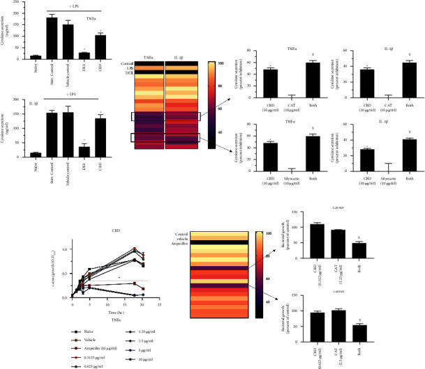
Two initial in vitro screening models were used to evaluate the compatibility and possible synergistic action of herbal mixtures: LPS-induced inflammation of U937 cells and the growth inhibition of C. acnes. First, the impact of CBD (10 μg/ml) alone was evaluated in these systems and is demonstrated in Figures 1(a)–1(c). Exposure to CBD significantly decreased the secretion of both TNFα and IL-1β (Figure 1(a)) in the in vitro LPS-induced inflammatory model. Concentrations of 5 and 10 μg/ml of CBD also reduced C. acnes growth in a comparable manner to ampicillin, which was used as a positive control (Figure 1(c)). These results complement previous findings and reports, suggesting CBD as a potential treatment for acne. Olah et al. demonstrated that CBD inhibits lipogenesis in sebocytes stimulated with either arachidonic acid or a combination of linoleic acid and testosterone. In addition, CBD reduced cell proliferation via the activation of transient receptor potential vanilloid 4 channels [9]. A recent review also shed light on other inflammatory disorders that can be mitigated by CBD-based treatment, including allergic contact dermatitis, psoriasis, acne, scleroderma, and dermatomyositis [15]. In addition, a study dating back to 1976 found that CBD may display antibacterial properties [16], and a more recent, comprehensive study by Blaskovich et al. found that CBD can reduce growth of several bacteria, including highly resistant Staphylococcus aureus, Streptococcus pneumoniae, and Clostridioides difficile. The authors also concluded that membrane impairment is the primary bactericidal mechanism of CBD [17].
Figure 1.

In vitro screening of CBD-based natural mixtures with herbal extracts. (a) U937 cells (300,000 cell/ml) were stimulated with 1 μg/mL LPS and treated for 24 hr. Cytokine levels were evaluated by ELISA. Dexamethasone (DEX) was used as a positive control. (b) The natural mixtures were evaluated similarly, and the compatible blends are presented as a heat map (dark colors depict high inhibition) and selected effective mixture on the right panel. (c) The impact of increasing concentrations of CBD on C. acnes growth was evaluated kinetically by the MIC assay. (d) Bacterial growth after 6 hr was similarly evaluated in the presence of the natural mixtures. n = 3; ∗/$ depicts significance in comparison to the control or CBD group, respectively.
Our studies confirm both the anti-inflammatory and antibacterial properties of CBD, thereby supporting its use in the treatment of acne. However, as acne is a multifactorial skin condition, we sought to create a more comprehensive treatment formulation by adding other already existing chemicals with unique mechanisms of action to act synergistically with CBD to enhance its activity but at a lower dose in order to reduce local side effects and improve tolerability. Therefore, the efficacy of subeffective concentrations of CBD when used alone or combined with seven commercially available herbal extracts was investigated (preliminary screen, data not shown). As shown in Figure 1(b), only two extracts were found to be highly compatible with CBD, the Centella asiatica triterpene (CAT) extract and silymarin extracted from Silybum marianum fruit. The combination of either extract with CBD potentiated its effect in reducing LPS-induced inflammation. Moreover, the CAT extract enhanced the growth arrest of C. acnes when combined with CBD (Figure 1(d)). The CAT extract is well known for its therapeutic effect mainly due to the presence of madecassoside, asiaticoside, madecassic acid, and asiatic acid [18–21]. Our study shows that CAT potentiates the mitigating effect of subtherapeutic concentrations of CBD. Since the combination was active in two separate models, it is possible that it increases the bioavailability of CBD. Further studies are required to understand the mechanism of action underlining this phenomenon. The Silybum marianum fruit extract had been repeatedly shown to modulate the inflammatory system [20, 21] and therefore can be beneficial in the treatment of several skin disorders, including the inflammatory lesions of acne vulgaris. Our study identified a synergistic effect between CBD and silymarin on two cytokines (Figure 1(b)).
3.2. Ex Vivo Validation of the Compounds
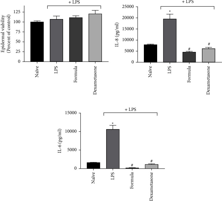
A topical formulation containing 1% CBD, 1% CAT, and 1% silymarin was prepared and evaluated in the ex vivo human skin organ culture (hSOC). The final formulation contained additional 0.5% salicylic acid, which was foreseen to facilitate the therapeutic action of the mixture through several mechanisms [22, 23]. The hSOC system was used repeatedly for both efficacy and safety evaluation, as it emulated the intact tissue [24–26]. The results presented in Figure 2 show that the formulation containing the natural mixture (written as “formula” in Figure 2) was well tolerated by the skin and did not reduce epidermal viability (determined by the MTT method). Although LPS stimulation increased the secretion of IL-6 and IL-8, the cytokines were subsequently blocked by the formula, an effect comparable to that of dexamethasone. Both IL-6 and IL-8 are the main cytokines known to be induced by LPS in the ex vivo system [13]. Importantly, both cytokines have been suggested as possible therapeutic targets to acne vulgaris [27–29]. Therefore, their attenuation, as exhibited in our study, may make our formula clinically relevant as a potent anti-inflammatory formulation.
Figure 2.

The topical formulation containing the natural mixture (“formula”) attenuates LPS-induced inflammation. Inflammation was inflicted in the ex vivo human skin model by 5 µg/ml LPS and treated with the formulated natural mixture for 48 hr. (a) Epidermal viability was evaluated by MTT. (b, c) The quantification of IL-8 and IL-6 by ELISA, respectively. n = 3; ∗/# depicts significance in comparison to the control or LPS-stimulated group, respectively.
3.3. Clinical Evaluation of the Formula
Afterwards, the formula was clinically evaluated on 30 subjects, both male and female aged 15–40 years, with mild to moderate acne (Grade 2 or 3 according to the Investigator's Global Assessment (IGA)). It was applied 2 to 3 times a day directly on spots and any imperfection on the skin (as a spot treatment) for 56 days.
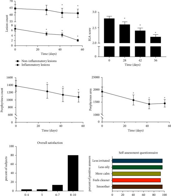
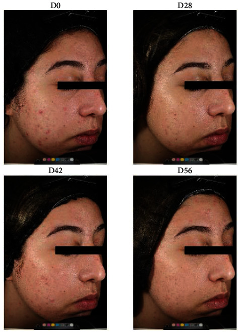
The formula showed a statistically significant reduction in acne lesions, mainly inflammatory. A reduction of 31.8% in total inflammatory lesions was shown following 28 days, 38.2% following 42 days, and 70.9% following 56 days (Figure 3), suggesting that the formula's potent anti-inflammatory activity has significant therapeutic potential in inflammatory acne. The topical application significantly reduced acne severity, shown as a reduction in the Investigator's Global Assessment (IGA) score. In addition, a marked reduction in porphyrin levels (count and area) suggested C. acnes inhibition. Representative images are presented in Figure 4. It should also be noted that the overall rate of volunteer satisfaction was extremely high throughout the trial.
Figure 3.

The topical formulation of the natural mixture attenuates acne lesions. The summary of a 56-day clinical evaluation is presented. (a) Changes in noninflammatory and inflammatory lesions are depicted. (b) The Investigator's Global Assessment (IGA) is presented. (c, d) Porphyrin abundance is depicted. (e, f) The summary of the volunteer questionnaire is shown.
Figure 4.

Representative image of clinical evaluation of the natural mixture.
4. Conclusions
The data presented here demonstrate the efficacy of a newly developed, natural topical formulation in the treatment of acne. Since several pathological pathways underlie the physiology of acne, a treatment that targets several mechanisms of action is advantageous. The topical formulation presented in this study showed significant ability in reducing inflammation and eliminating C. acnes. The ex vivo results show a marked impact of the developed formulation in comparison to untreated tissue. However, the addition of salicylic acid may have further enhanced the efficacy of the formulation, due to its keratolytic activity, and as a result, it may have exhibited positive outcomes in the treatment of acne. Thus, a major limitation of this study is the lack of formulation without CBD and Silybum marianum fruit extract to exclude indirect effects of other compounds. Thus, placebo-controlled evaluation must be performed to validate the study. A significant correlation was shown between in vitro, ex vivo, and clinical tests, where the inhibition of inflammation and C. acnes growth were the main mechanisms of action exhibited by our formulation. The study also demonstrates a transformation factor of 1 : 1000 (∼10 μg/ml to 1%) when converting from in vitro concentrations to ex vivo and clinical ones. The selected concentration in the formulation was determined after a preliminary evaluation determining that the formulation was safe and well tolerated by the tissue on one hand and effective on the other one (data not shown). This finding is also supported by previous findings, transforming data obtained in HaCaT keratinocyte cells to ex vivo experiments [28]. The study has several limitations: in vitro screening was aimed to find CBD-compatible agents, and thus, low levels of the herbal extracts were used. In addition, the significant results obtained clinically were compared to their basal state, as typically performed in cosmetic trials, with no placebo control used. However, the massive reduction in inflammatory acne lesions, as well as porphyrin levels, clearly stands out and was foreseen based on the dual in vitro and ex vivo data.
Acknowledgments
GC acknowledges support provided by the Ministry of Science and Technology (580458776; Israel). The study was funded by Echo Pharmaceutics.
Data Availability
All clinical data are available upon request.
Conflicts of Interest
The authors declare that they have no conflicts of interest.
References
- 1.Dawson A. L., Dellavalle R. P. Acne vulgaris. British Medical Journal . 2013;346(1):p. f2634. doi: 10.1136/BMJ.F2634. [DOI] [PubMed] [Google Scholar]
- 2.Heng A. H. S., Chew F. T. Systematic review of the epidemiology of acne vulgaris. Scientific Reports . 2020;10(1):p. 5754. doi: 10.1038/s41598-020-62715-3. [DOI] [PMC free article] [PubMed] [Google Scholar]
- 3.Zaenglein A. L., Zaenglein A. L. Acne vulgaris. New England Journal of Medicine . 2018;379:1343–1352. doi: 10.1056/NEJMcp1702493. [DOI] [PubMed] [Google Scholar]
- 4.Eichenfield D. Z., Sprague J., Eichenfield L. F. Management of acne vulgaris: a review. The Journal of the American Medical Association . 2021;326(20):p. 2055. doi: 10.1001/jama.2021.17633. [DOI] [PubMed] [Google Scholar]
- 5.del Rosso J. Q. Combination topical therapy in the treatment of acne. Cutis . 2006;78(1):5–12. [PubMed] [Google Scholar]
- 6.Marous M. R., Flaten H. K., Sledge B., et al. Complementary and alternative methods for treatment of acne vulgaris: a systematic review. Current Dermatology Reports . 2018;7(4):359–370. doi: 10.1007/s13671-018-0230-0. [DOI] [Google Scholar]
- 7.Baswan S. M., Klosner A. E., Glynn K., et al. Therapeutic potential of cannabidiol (CBD) for skin health and disorders. Clinical, Cosmetic and Investigational Dermatology . 2020;13:927–942. doi: 10.2147/CCID.S286411. [DOI] [PMC free article] [PubMed] [Google Scholar]
- 8.Dréno B. What is new in the pathophysiology of acne, an overview. Journal of the European Academy of Dermatology and Venereology . 2017;31:8–12. doi: 10.1111/jdv.14374. [DOI] [PubMed] [Google Scholar]
- 9.Oláh A., Tóth B. I., Borbíró I., et al. Cannabidiol exerts sebostatic and antiinflammatory effects on human sebocytes. Journal of Clinical Investigation . 2014;124(9):3713–3724. doi: 10.1172/JCI64628. [DOI] [PMC free article] [PubMed] [Google Scholar]
- 10.Iffland K., Grotenhermen F. An update on safety and side effects of cannabidiol: a review of clinical data and relevant animal studies. Cannabis and Cannabinoid Research . 2017;2(1):139–154. doi: 10.1089/CAN.2016.0034. [DOI] [PMC free article] [PubMed] [Google Scholar]
- 11.Wyse J., Luria G. Trends in intellectual property rights protection for medical cannabis and related products. Journal of Cannabis Research . 2021;3(1):p. 1. doi: 10.1186/s42238-020-00057-7. [DOI] [PMC free article] [PubMed] [Google Scholar]
- 12.Baek Y. S., Haas S., Hackstein H., et al. Identification of novel transcriptional regulators involved in macrophage differentiation and activation in U937 cells. British Medical Journal Immunology . 2009;10(1):p. 18. doi: 10.1186/1471-2172-10-18. [DOI] [PMC free article] [PubMed] [Google Scholar]
- 13.Gvirtz R., Ogen-shtern N., Cohen G. Kinetic cytokine secretion profile of LPS-induced inflammation in the human skin organ culture. Pharmaceutics . 2020;12(4):p. 299. doi: 10.3390/pharmaceutics12040299. [DOI] [PMC free article] [PubMed] [Google Scholar]
- 14.Kahremany S., Hofmann L., Eretz-Kdosha N., Silberstein E., Gruzman A., Cohen G. SH-29 and SK-119 attenuates air-pollution induced damage by activating Nrf2 in HaCaT cells. International Journal of Environmental Research and Public Health . 2021;18(23) doi: 10.3390/ijerph182312371.12371 [DOI] [PMC free article] [PubMed] [Google Scholar]
- 15.Scheau C., Badarau I. A., Mihai L. G., et al. Cannabinoids in the pathophysiology of skin inflammation. Molecules . 2020;25(3):p. 652. doi: 10.3390/molecules25030652. [DOI] [PMC free article] [PubMed] [Google Scholar]
- 16.van Klingeren B., ten Ham M. Antibacterial activity of d9-tetrahydrocannabinol and cannabidiol. Antonie van Leeuwenhoek . 1976;42(1-2):9–12. doi: 10.1007/BF00399444. [DOI] [PubMed] [Google Scholar]
- 17.Blaskovich M. A. T., Kavanagh A. M., Elliott A. G., et al. The antimicrobial potential of cannabidiol. Communications Biology . 2021;4(1):p. 7. doi: 10.1038/s42003-020-01530-y. [DOI] [PMC free article] [PubMed] [Google Scholar]
- 18.Hashim P., Sidek H., Helan M. H. M., Sabery A., Palanisamy U. D., Ilham M. Triterpene composition and bioactivities of Centella asiatica. Molecules . 2011;16(2):1310–1322. doi: 10.3390/MOLECULES16021310. [DOI] [PMC free article] [PubMed] [Google Scholar]
- 19.Sampson J. H., Raman A., Karlsen G., Navsaria H., Leigh I. M. M. O. P. In vitro keratinocyte antiproliferant effect of Centella asiatica extract and triterpenoid saponins. Phytomedicine . 2001;8(3):230–235. doi: 10.1078/0944-7113-00032. [DOI] [PubMed] [Google Scholar]
- 20.Juráňová J., Aury-Landas J., Boumediene K., et al. Modulation of skin inflammatory response by active components of silymarin. Molecules . 2018;24(1):p. 123. doi: 10.3390/molecules24010123. [DOI] [PMC free article] [PubMed] [Google Scholar]
- 21.Amin M. M., Arbid M. S. Estimation of the novel antipyretic, anti-inflammatory, antinociceptive and antihyperlipidemic effects of silymarin in albino rats and mice. Asian Pacific Journal of Tropical Biomedicine . 2015;5(8):619–623. doi: 10.1016/j.apjtb.2015.05.009. [DOI] [Google Scholar]
- 22.Wijayanti T. N. The effectivity of 2% salicylic acid on inflammatory acne vulgaris. Journal of Medical Sciences . 2015;33(2) [Google Scholar]
- 23.Lu J., Cong T., Wen X., et al. Salicylic acid treats acne vulgaris by suppressing AMPK/SREBP1 pathway in sebocytes. Experimental Dermatology . 2019;28(7):786–794. doi: 10.1111/exd.13934. [DOI] [PubMed] [Google Scholar]
- 24.Ogen-Shtern N., Chumin K., Cohen G., Borkow G. Increased pro-collagen 1, elastin, and TGF-ß1 expression by copper ions in an ex-vivo human skin model. Journal of Cosmetic Dermatology . 2020;19(6):1522–1527. doi: 10.1111/jocd.13186. [DOI] [PubMed] [Google Scholar]
- 25.Ogen-Shtern N., Chumin K., Silberstein E., Borkow G. Copper ions ameliorated thermal burn-induced damage in ex vivo human skin organ culture. Skin Pharmacology and Physiology . 2021;34(6):317–327. doi: 10.1159/000517194. [DOI] [PubMed] [Google Scholar]
- 26.Kahremany S., Babaev I., Gvirtz R., et al. Nrf2 activation by SK-119 attenuates oxidative stress, UVB, and LPS-induced damage. Skin Pharmacology and Physiology . 2019;32(4):173–181. doi: 10.1159/000499432. [DOI] [PubMed] [Google Scholar]
- 27.Askari N., Ghazanfari T., Yaraee R., et al. Association between acne and serum pro-inflammatory cytokines (IL-1α, IL-1β, IL-1Ra, IL-6, IL-8, IL-12 and RANTES) in mustard gas-exposed patients: sardasht-Iran cohort study. Archives of Iranian Medicine . 2017;20(2):86–91. [PubMed] [Google Scholar]
- 28.Abd El All H. S., Shoukry N. S., el Maged R. A., Ayada M. M. Immunohistochemical expression of interleukin 8 in skin biopsies from patients with inflammatory acne vulgaris. Diagnostic Pathology . 2007;2(1):p. 4. doi: 10.1186/1746-1596-2-4. [DOI] [PMC free article] [PubMed] [Google Scholar]
- 29.Ragab M., Hassan E. M., Elneily D., Fathallah N. Association of interleukin-6 gene promoter polymorphism with acne vulgaris and its severity. Clinical and Experimental Dermatology . 2019;44(6):637–642. doi: 10.1111/ced.13864. [DOI] [PubMed] [Google Scholar]
Associated Data
This section collects any data citations, data availability statements, or supplementary materials included in this article.
Data Availability Statement
All clinical data are available upon request.


