

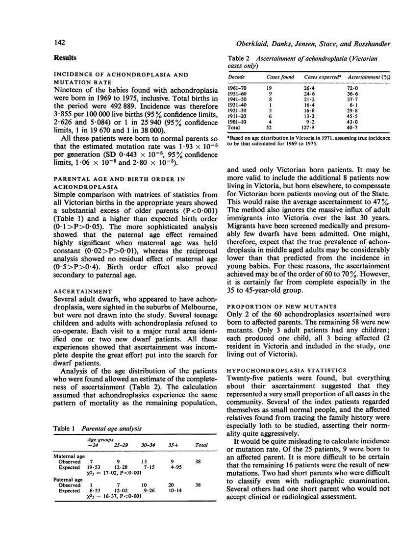
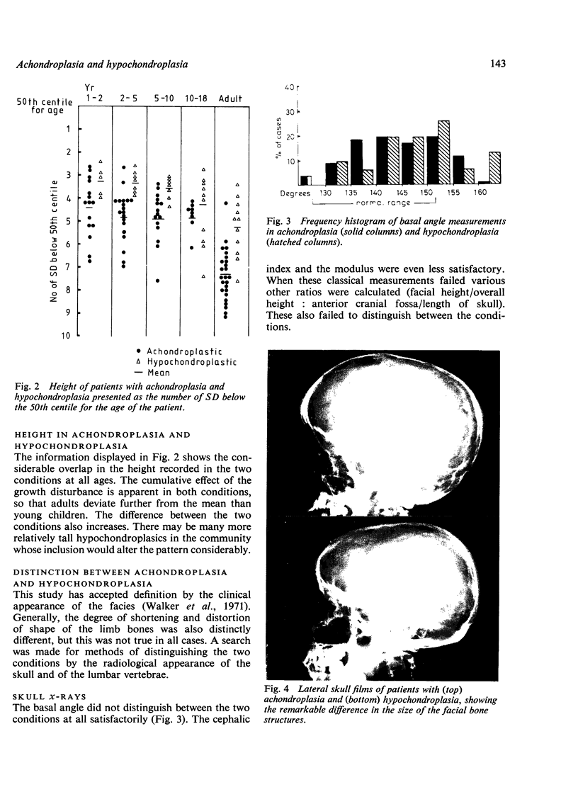
Abstract
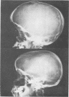
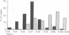
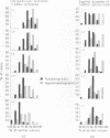
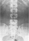
An attempt was made to ascertain all the dwarfs in the State of Victoria. The incidence of achondroplasia proved to be approximately 1 in 26,000 live births in the period 1969 to 1975 when ascertainment was nearly complete. This indicates a mutation rate of 1.93 X 10(-5) per generation in this locus. Paternal age was shown to influence mutation. Ascertainment in earlier years of the study was low despite the very great effort made to find all cases. Patients with hypochondroplasia were particularly difficult to find. However, 25 cases were found for study. Overlap between hypochondroplasia and achondroplasia was found in all features except the facial appearance (which was the basis of definition). Achondroplasia was more severe in all regards, but some individuals with hypochondroplasia were very short and some had extreme degrees of spinal canal stenosis. The classical measurements used to describe the skull changes in acondroplasia failed to distinguish this condition from hypochondroplasia. More efficient indices were devised, but visual assessment of the size of the facial region compared to that of the cranial valult proved more reliable than any index. The clinical distinction based upon facial appearance remains the arbitrary basis of definition.
Full text
PDF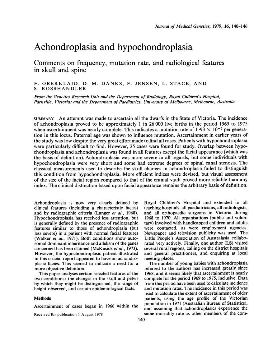
Images in this article
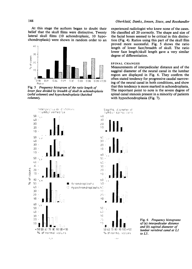
Selected References
These references are in PubMed. This may not be the complete list of references from this article.
- Hinck V. C., Clark W. M., Jr, Hopkins C. E. Normal interpediculate distances (minimum and maximum) in children and adults. Am J Roentgenol Radium Ther Nucl Med. 1966 May;97(1):141–153. doi: 10.2214/ajr.97.1.141. [DOI] [PubMed] [Google Scholar]
- Hinck V. C., Hopkins C. E., Clark W. M. Sagittal diameter of the lumbar spinal canal in children and adults. Radiology. 1965 Nov;85(5):929–937. doi: 10.1148/85.5.929. [DOI] [PubMed] [Google Scholar]
- Langer L. O., Jr, Baumann P. A., Gorlin R. J. Achondroplasia: clinical radiologic features with comment on genetic implications. Clin Pediatr (Phila) 1968 Aug;7(8):474–485. doi: 10.1177/000992286800700809. [DOI] [PubMed] [Google Scholar]
- McKusick V. A., Kelly T. E., Dorst J. P. Observations suggesting allelism of the achondroplasia and hypochondroplasia genes. J Med Genet. 1973 Mar;10(1):11–16. doi: 10.1136/jmg.10.1.11. [DOI] [PMC free article] [PubMed] [Google Scholar]
- Silverman F. N., Brünner S. Errors in the diagnosis of achondroplasia. Acta Radiol Diagn (Stockh) 1967 Jul;6(4):305–321. doi: 10.1177/028418516700600401. [DOI] [PubMed] [Google Scholar]
- Tanner J. M., Whitehouse R. H., Takaishi M. Standards from birth to maturity for height, weight, height velocity, and weight velocity: British children, 1965. I. Arch Dis Child. 1966 Oct;41(219):454–471. doi: 10.1136/adc.41.219.454. [DOI] [PMC free article] [PubMed] [Google Scholar]
- Walker B. A., Murdoch J. L., McKusick V. A., Langer L. O., Beals R. K. Hypochondroplasia. Am J Dis Child. 1971 Aug;122(2):95–104. doi: 10.1001/archpedi.1971.02110020029001. [DOI] [PubMed] [Google Scholar]